Jump to content
In this section we have spot diagnoses posted on a daily basis since June 2010, now over 4000! You can review the archived cases and read the suggested diagnoses by users and the final comment by the contributors.
Case are uploaded each week day by 10 am UK time with the correct diagnosis will generally be posted at 8 pm UK time. Why not view the most recent spot diagnosis and proffer a diagnosis?

Case Number : Case 1513 - 12 April Posted By: Guest

Please read the clinical history and view the images by clicking on them before you proffer your diagnosis.
Submitted Date :
   (0 reviews)

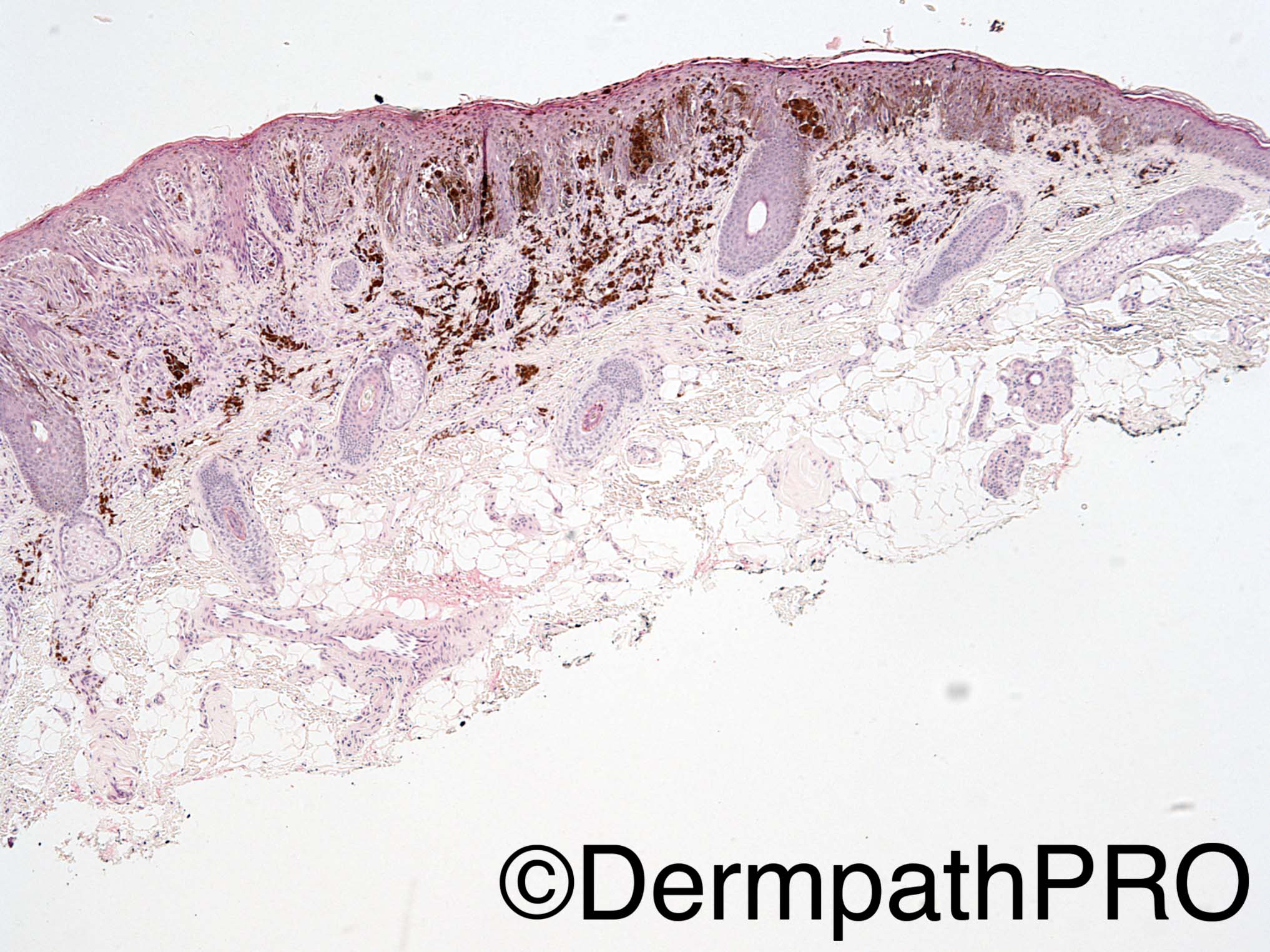
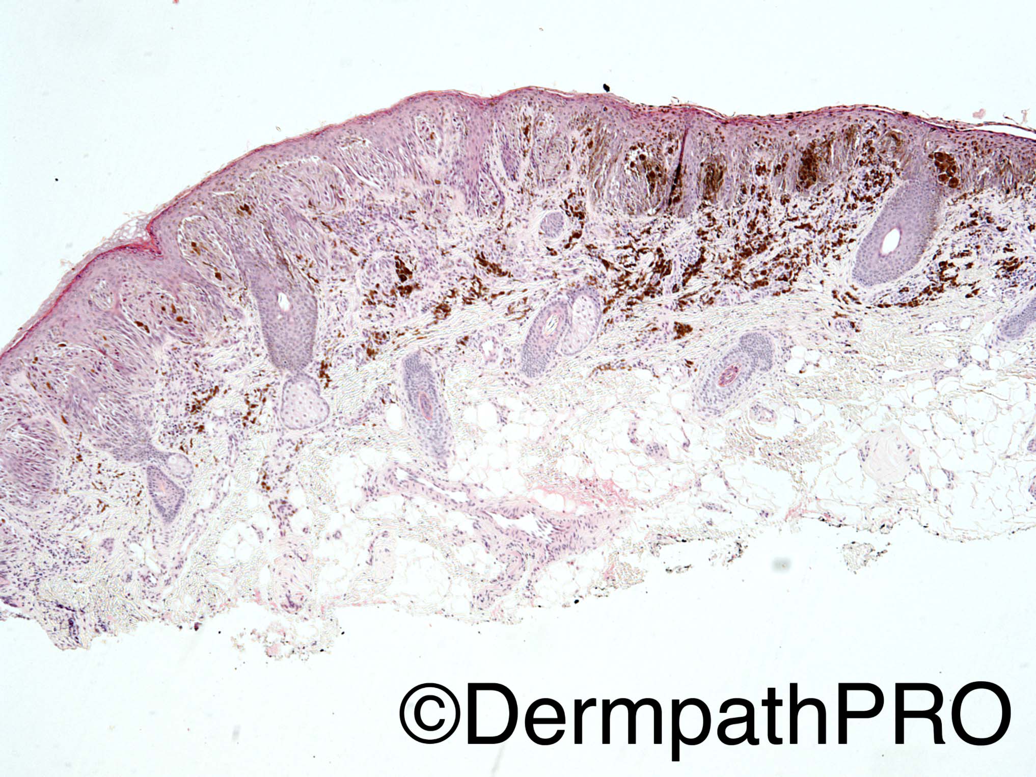
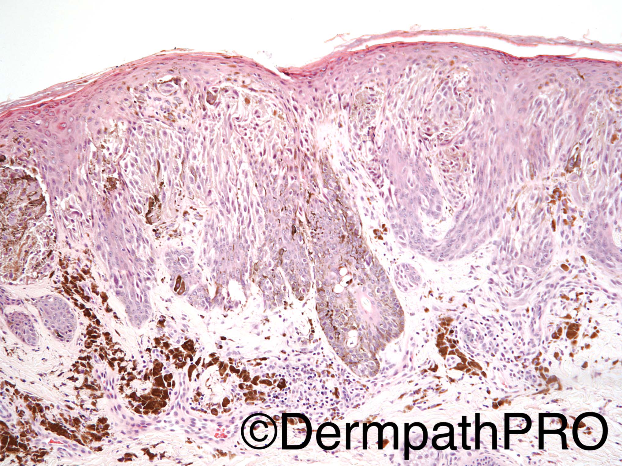
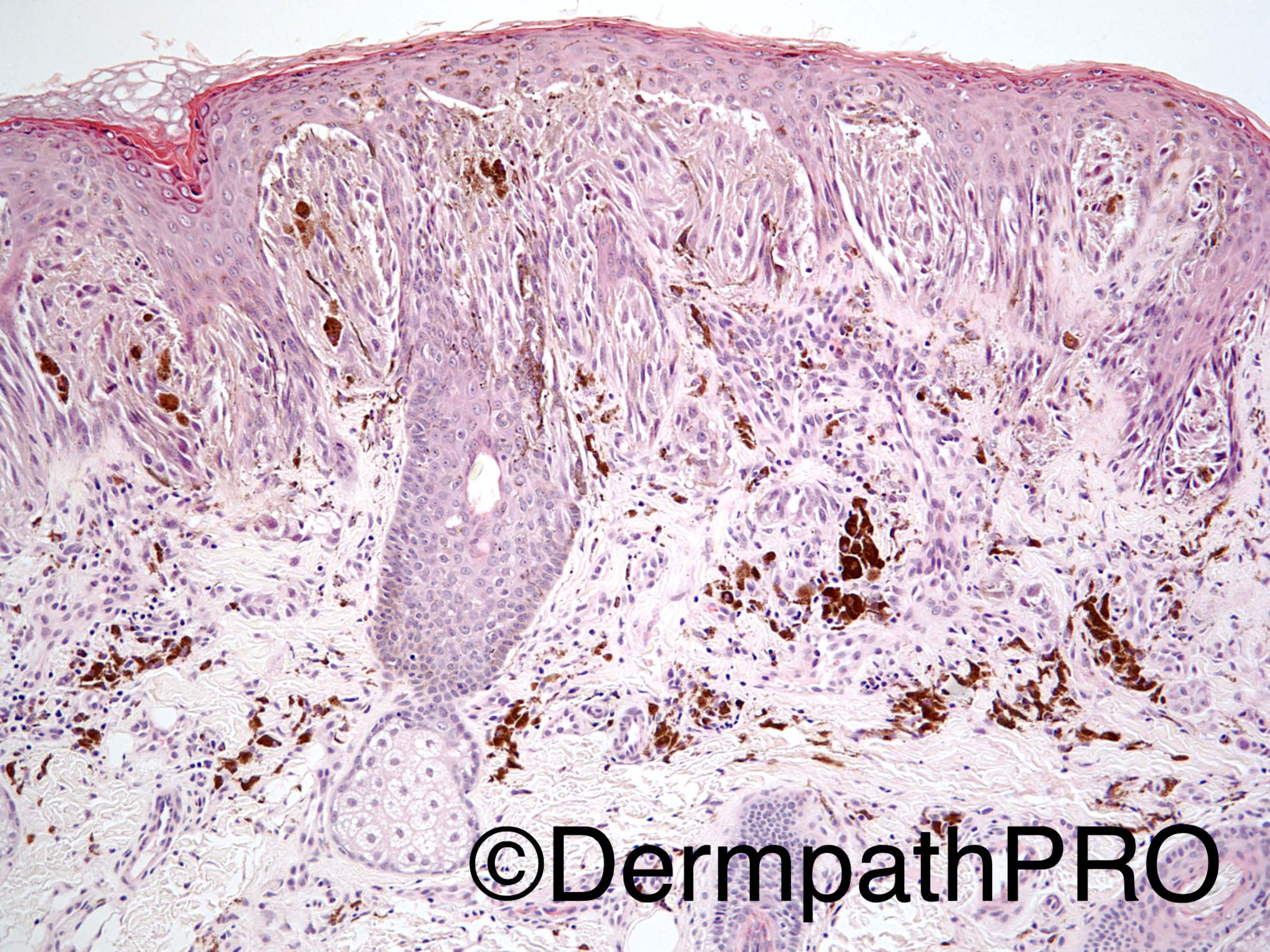
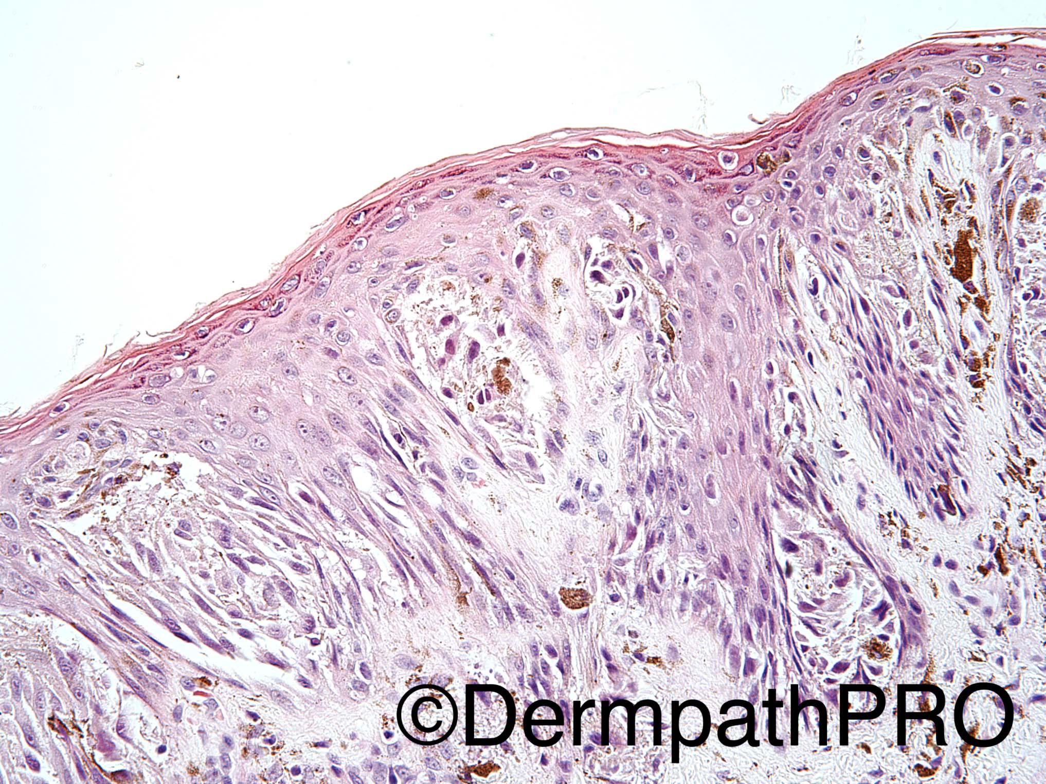
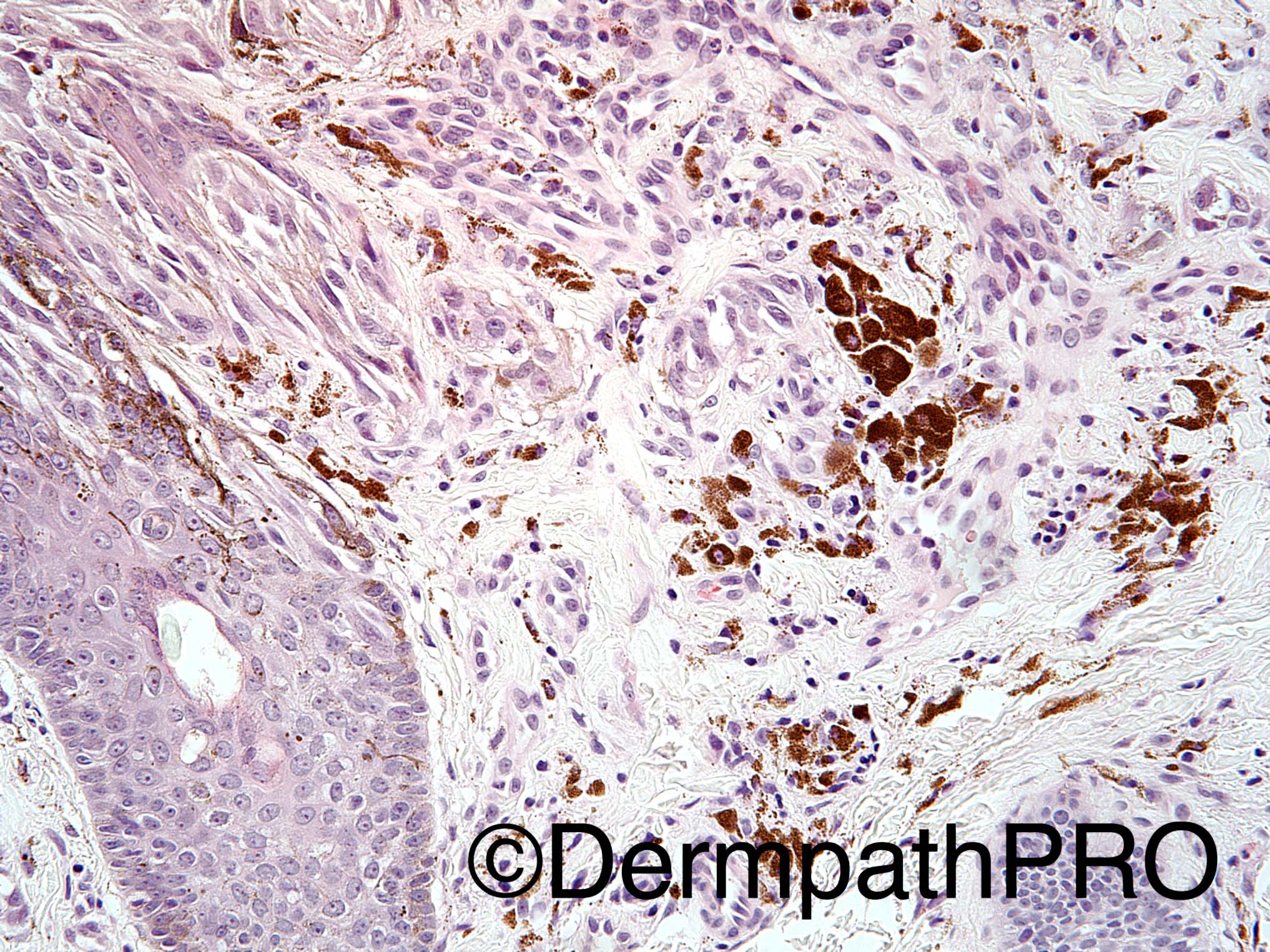
23 year old female with lesion on right upper arm.

Dr Uma Sundram.


  Report Record

User Feedback


I think this is a Spitzoid Melanoma. This could be a good case in which the FISH using 9p21 would be usefull.

Share this comment


Link to comment
share_externally

vincenzo polizzi

Posted

Junctional plaque-like growth pattern of spindled pigmented melanocytes, running parallel to epidermis. Some pagetoid spread but not so worrisome...The cells are the same in all the lesion, without pleomorphism. 

I favor pigmented spindle cell nevus (Reed).

Share this comment


Link to comment
share_externally

Raul Perret

Posted

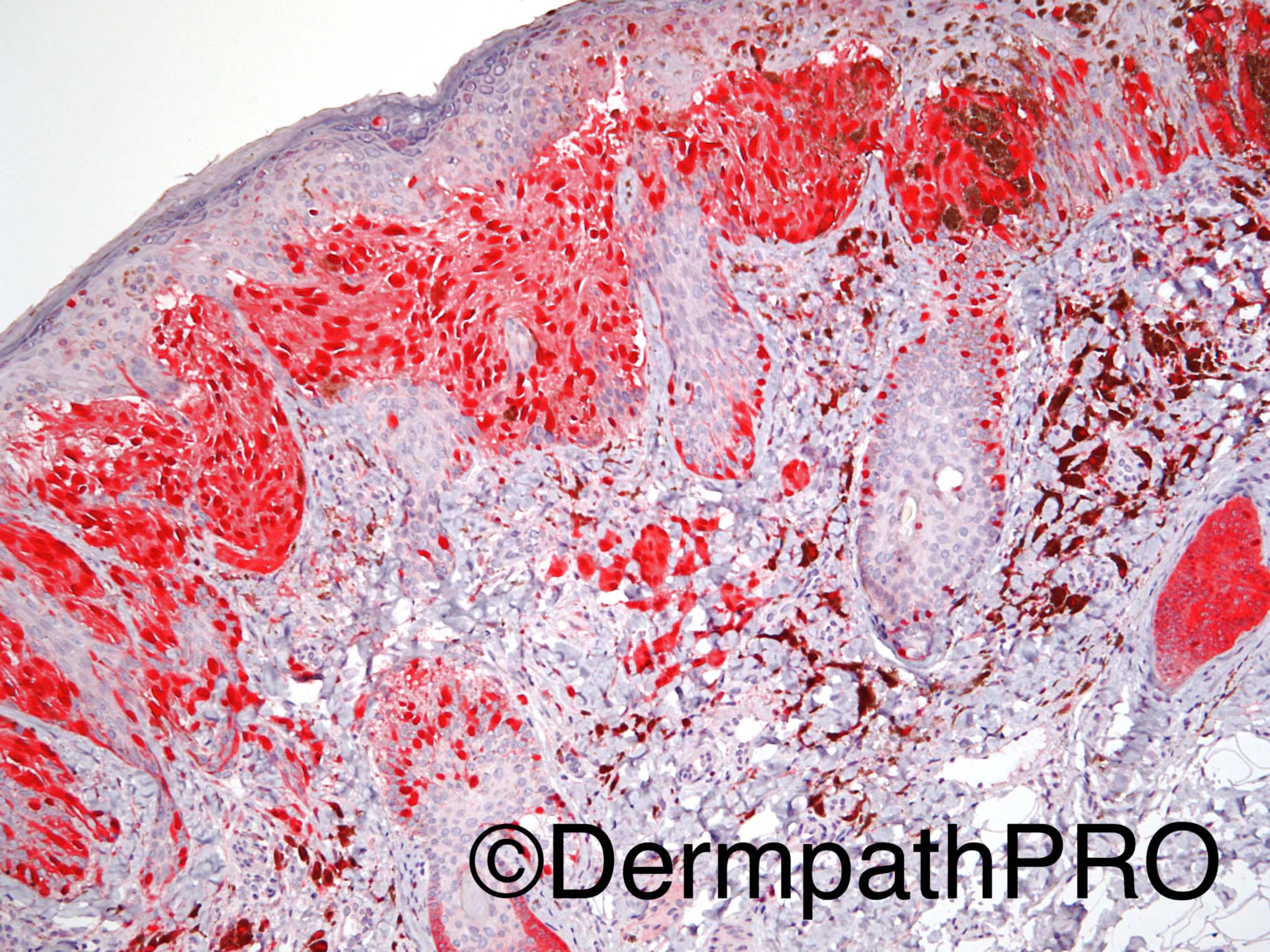
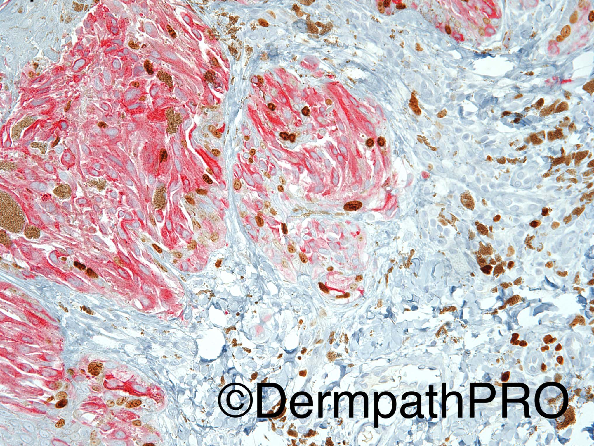
Sincerely I incline more to the benign spectrum of lesions. My impression was Reed nevus/pigmented Spitz. Ki67 could be of some help also. It is known that some spitzoid lesions have high interobserver variability (even between expert dermatopathologists). When I saw this lesion I saw similar aspects to the ones mentioned by Vincenzo (spindle pigmented cells with homogeneous atypia, distribution mainly in nests with parallel orientation, no mitosis and I think some maturation of cells reflected on morphology and Immunohistochemistry in the last picture). The only aspect that bothered me a bit was the assimetrical distribution of pigment. I think it could be interesting to list the features considered to support our diagnosis in melanocytic lesions in order to adjust our criteria when the final diagnosis is posted (if the expert dermatopathologists remark the criteria they considered relevant for the final diagnosis it would be helpful too)

Share this comment


Link to comment
share_externally

Guest Arash Daryakarr

Posted

I favor Reed nevus/Pigmented spitz nevus,too.

Share this comment


Link to comment
share_externally

Dr. Richard Carr

Posted

First word of the report should be "difficult" then Reed-naevus like (thin, compound Spitzoid) lesion that lacks symmetry (two slightly different areas).  Slight uncertainty with respect to behaviour (MELTUMP probably benign behaviour - given thin and Spitzoid and not overtly a melanoma exceedingly unlikely to have metastatic potential).  Correlation with clinical features (not provided) and dermoscopy are obviously essential.  It would be prudent to ensure complete excision.  Given likely 100% good outcome might be overkill to do more but obviously additional IHC and molecular studies may be of academic interest.

Share this comment


Link to comment
share_externally

Pigmented spindle cell nevus of reed/ pigmented spitz
Good stratification sign by immuno guide us toward benign not spitziod melanoma

Share this comment


Link to comment
share_externally

Dr. Mona Abdel-Halim

Posted

Difficult (as Dr Carr said ). Prefer to designate as MELTUMP for further molecular studies especially in the absence of clinical description of the lesion as the lesion is very asymmetrical.

Share this comment


Link to comment
share_externally

Nitin Khirwadkar

Posted

Very Reed-nevus like. Slightly concerned, especially areas where the melanocytic nests appear to efface the rete. As has been mentioned by others, IHC (p16, Ki67), and FISH/CGH may help.

Share this comment


Link to comment
share_externally

Uma Sundram

Posted

Thank you for the wonderful discussion. My apologies for the missing information about the stains: IHC picture #1=MITF; IHC picture #2=p16; IHC picture #3=Mart-1/Ki67. We too thought this was a difficult case. While we favored a pigmented Spitz nevus/nevus of Reed (thin lesion, lack of significant overall pleomorphism or mitotic activity, dermal maturation, low Ki67 rate within the dermis localized to melanocytes), given the asymmetric staining pattern with p16, we recommended full excision and follow up over time. Array CGH analysis of this tumor was negative.

Share this comment


Link to comment
share_externally


Join the conversation

You can post now and register later. If you have an account, sign in now to post with your account.

Guest
Add a comment...

×   Pasted as rich text.   Paste as plain text instead

  Only 75 emoji are allowed.

×   Your link has been automatically embedded.   Display as a link instead

×   Your previous content has been restored.   Clear editor

×   You cannot paste images directly. Upload or insert images from URL.

Loading...

×
×
  • Create New...