Jump to content
In this section we have spot diagnoses posted on a daily basis since June 2010, now over 4000! You can review the archived cases and read the suggested diagnoses by users and the final comment by the contributors.
Case are uploaded each week day by 10 am UK time with the correct diagnosis will generally be posted at 8 pm UK time. Why not view the most recent spot diagnosis and proffer a diagnosis?

Case Number : Case 1515 - 14 April Posted By: Guest

Please read the clinical history and view the images by clicking on them before you proffer your diagnosis.
Submitted Date :
   (0 reviews)

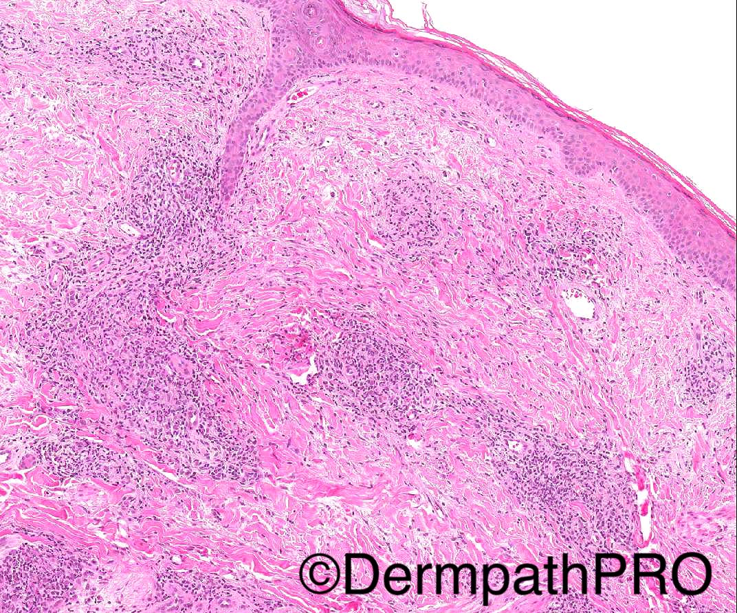
34/M widespread discrete plaques, some indurated.
PMH- recurrent chest infections and bronchiectasis, hepatosplenomegaly (splenectomy done in 2014), lymphadenopathy

- Special stains for AFB and fungi negative.

Case posted by Dr Arti Bakshi


  • Thanks 1
  Report Record

User Feedback


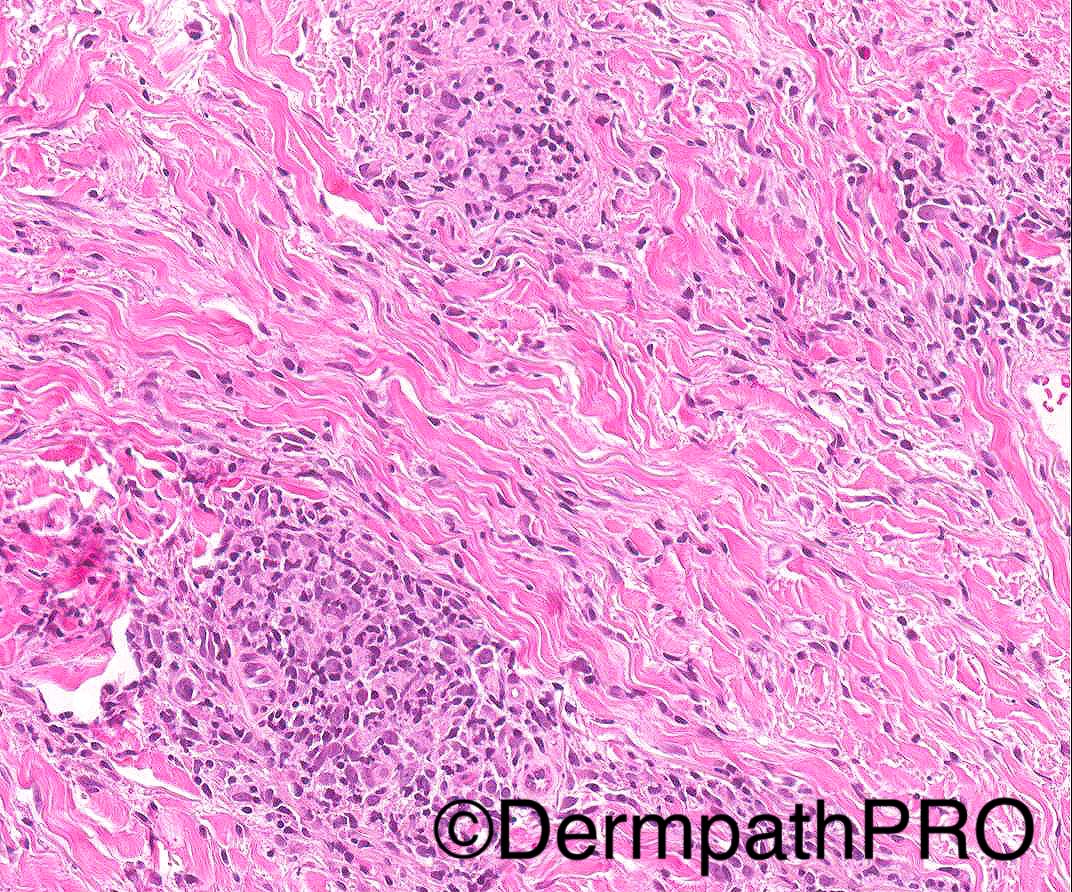
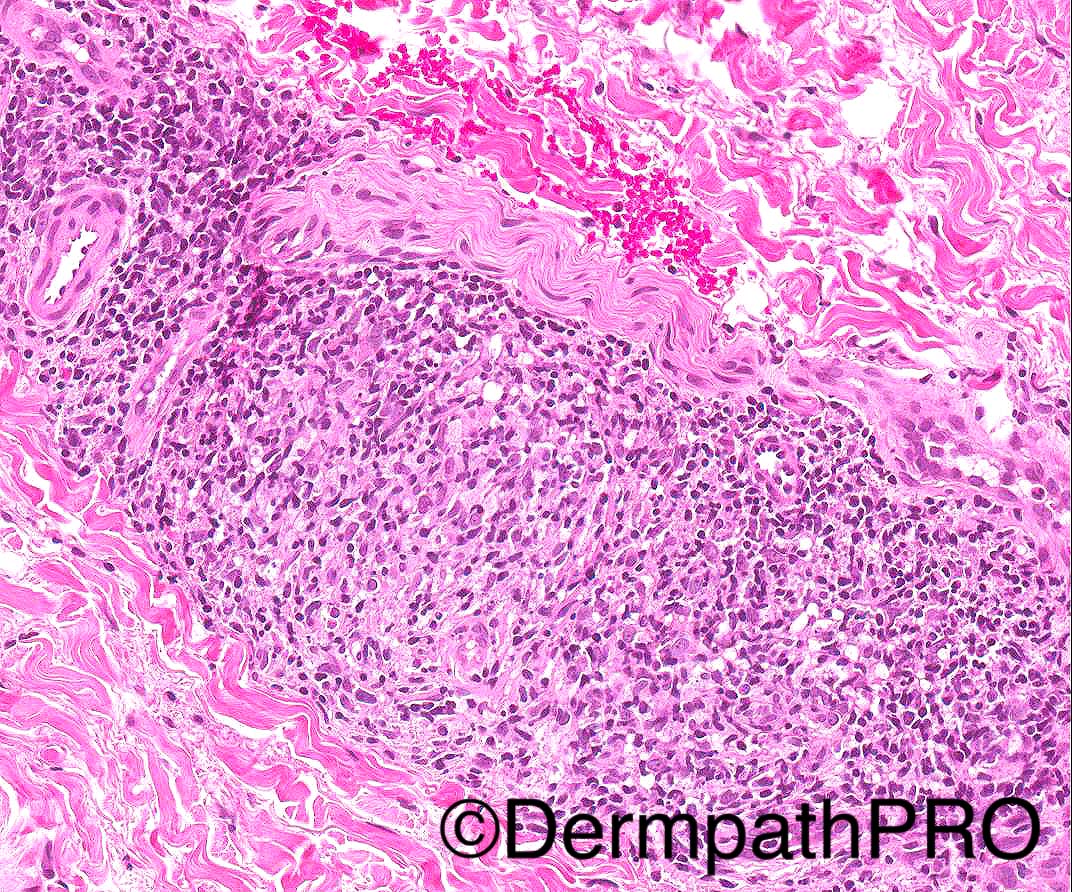
I see some naked granulomas within the dermis with perineural inflammation on last picture. The clinical findings of lung disease and hepatosplenomegaly may also be seen in cases of sistemic Sarcoidosis. So my first thought is Sarcoidosis, but I would do special stains before.

Share this comment


Link to comment
share_externally

Raul Perret

Posted

Non necrotizing dermal granulomatous inflammation with paucity of lymphocytes should make us put sarcoidosis in the differential. Special stains should be perform first of course but I think they will be negative. The clinical history fits with sarcoidosis although I think it would be important to find out if an immunodeficiency syndrome is suspected (like common variable immunodeficiency) in the patient as there can be some granulomatous dermal inflammation in these cases

Share this comment


Link to comment
share_externally

Arti Bakshi

Posted

Sorry, I meant to put in the history...the special stains were all negative.

Share this comment


Link to comment
share_externally

Raul Perret

Posted

Perineural inflammation is rare in sarcoidosis but it can be seen sometimes. I think that to be prudent, we should state that an infectious cause cannot be 100% ruled out even with negative special stains. CPC is of extreme importance in these kind of cases. My preferred diagnosis in this case are: Sarcoidosis/granulomatous inflammation due to immunodeficiency and far down I would put infectious etiology 

Share this comment


Link to comment
share_externally

vincenzo polizzi

Posted

I have many doubts now. May be there are some Leishmania in my brain...

Share this comment


Link to comment
share_externally

Dr. Mona Abdel-Halim

Posted

To me not straight forward sarcoidosis pathologically. There is sarcoidal like granulomas and perivascular lymphohistiocytic aggregates, but I think there is also collagen alteration (necrobiosis). No frank vasculitis, but I am wondering about Wegner's granulomatosis (granulomatosis with polyangitis) presenting with hemphophagocytic lymphohistiocytosis, hence the associated HSM, splenectomy and lymphadenopathy.

Share this comment


Link to comment
share_externally

Dr. Richard Carr

Posted

These are tuberculoid granulomas (lots of lymphocytes), following neurovascular bundles and adnexa.  Funny there is some leishmania-like granularity but suspect that is just a red herring. A high power would not go amiss though. DDx Sarcoid, lymphoma (can present with cutaneous granulomatous process completely mimicking sarcoid), good to consider immunodeficiency too. I've a sneaky suspicion will be a T-cell lymphoma or lymphomatoid granulomatosis.

Share this comment


Link to comment
share_externally

Dr. Mona Abdel-Halim

Posted

Agree with Dr Carr in the possibility of granulomatous lymphoma, also crossed my mind.

Share this comment


Link to comment
share_externally

Sarcoidosis.  In one series, leprosy-like perineural granulomas were present in a quarter of cases.  http://www.ncbi.nlm.nih.gov/pubmed/14690461.  Immunodeficiency diseases are possible, but given that these diseases are uncommon and that granulomas are uncommon in these diseases, I think they are a distant second. 

Share this comment


Link to comment
share_externally

As far as I know, lymphomatoid granulomatosis shows an (1) angiocentric, (2) angiodestructive and (3) lymphocyte predominant infiltrate, none of which we have here.  Plus, lymphomatoid granulomatosis is not a nodal lymphoma and, therefore, lymphadenopathy is not a feature. 

Share this comment


Link to comment
share_externally

Arti Bakshi

Posted

Great discussion!

This is a case of granulomatous disease in association with Common Variable Immunodeficiency.

The patient had severe hypogammaglobulinemia at presentation 3 yrs back and has been on Ig replacement therapy. Biopsies from spleen and lymph node also showed non caseating granuloma.

Infectious aetiologies were ruled out clinically. 

The main differential was sarcoidosis (as there is a known association of sarcoidosis and CVID  and the patient had rasied ACE levels).  The distinction is difficult histologically (although I agree with Richard that the granulomata do show quite a bit of lymphocytic mantle). Based on clinical and radiological correlation, a diagnosis of granulomatous disease in association with CVID was favoured. (apparently raised serum ACE is not specific for sarcoidosis and can be seen with any granulomatous process). Patient was started on oral steroids (the recommended treatment for granulomas in CVID) and at the last visit, the skin lesions were healing well.

Share this comment


Link to comment
share_externally

Dr. Mona Abdel-Halim

Posted

Thanks Arti :-)
Great case :-)
Well done Raul for suggesting this association :-)

Share this comment


Link to comment
share_externally

Arti Bakshi

Posted

Forgot to mention....many thanks to my colleague, Dr Stefanos, for sharing this case and to Dr Darroch for his clinical input.

Share this comment


Link to comment
share_externally


Join the conversation

You can post now and register later. If you have an account, sign in now to post with your account.

Guest
Add a comment...

×   Pasted as rich text.   Paste as plain text instead

  Only 75 emoji are allowed.

×   Your link has been automatically embedded.   Display as a link instead

×   Your previous content has been restored.   Clear editor

×   You cannot paste images directly. Upload or insert images from URL.

Loading...

×
×
  • Create New...