Jump to content
In this section we have spot diagnoses posted on a daily basis since June 2010, now over 4000! You can review the archived cases and read the suggested diagnoses by users and the final comment by the contributors.
Case are uploaded each week day by 10 am UK time with the correct diagnosis will generally be posted at 8 pm UK time. Why not view the most recent spot diagnosis and proffer a diagnosis?

Case Number : Case 1518 - 19 April Posted By: Guest

Please read the clinical history and view the images by clicking on them before you proffer your diagnosis.
Submitted Date :
   (0 reviews)

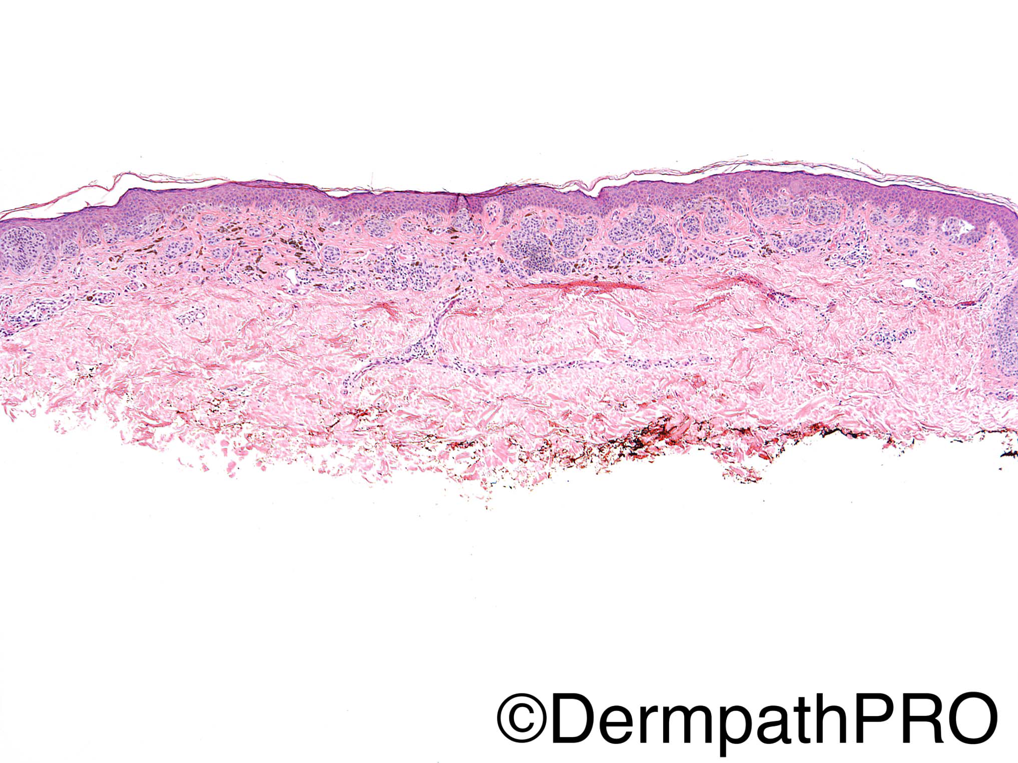
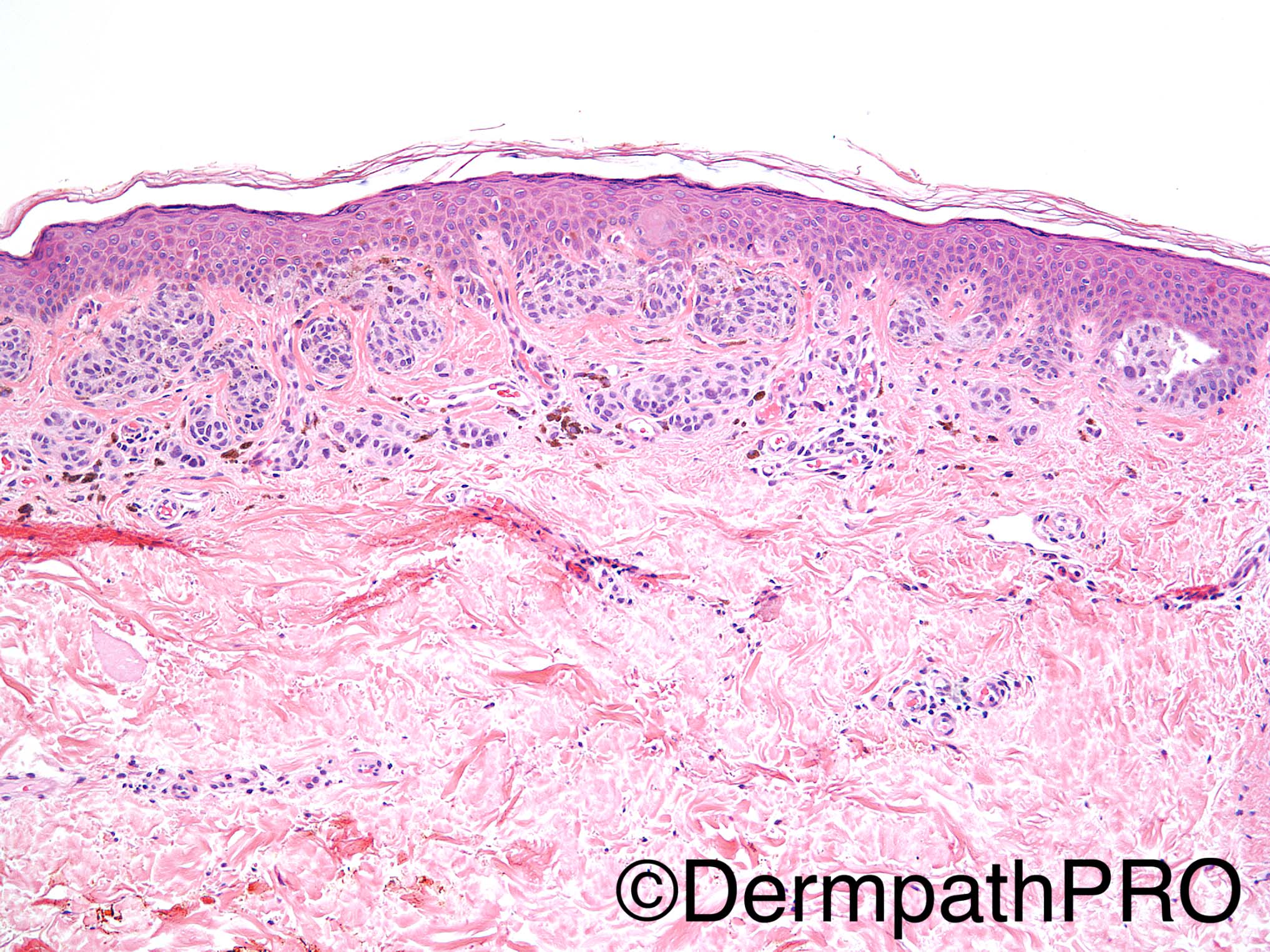
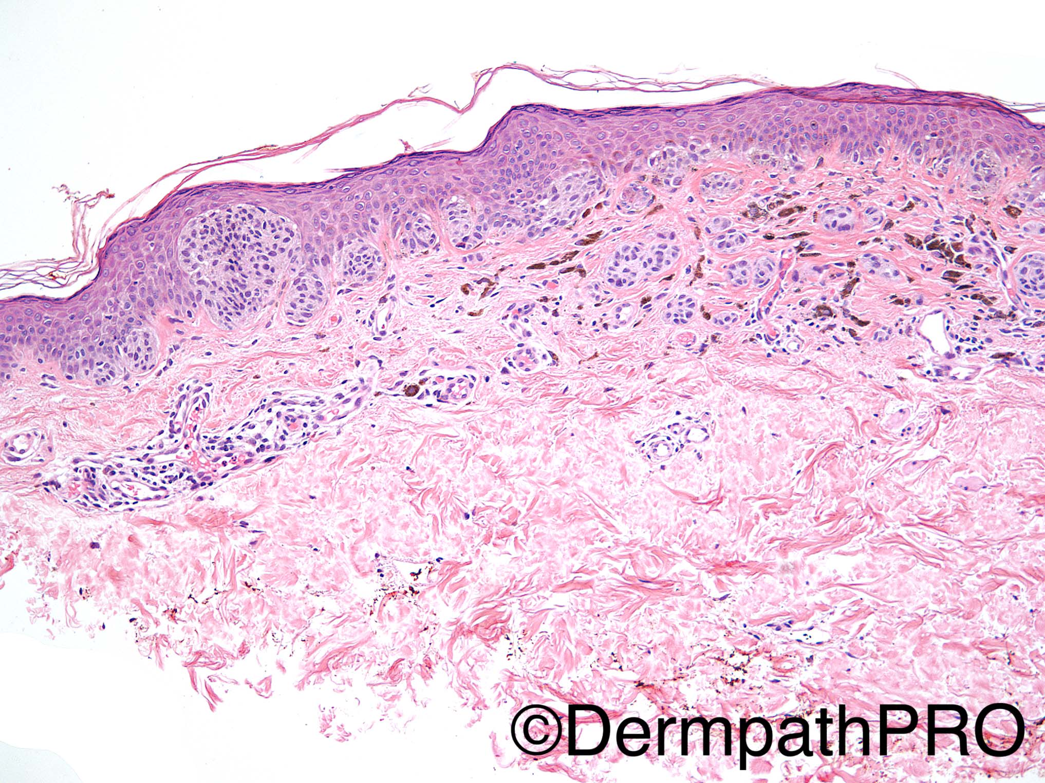
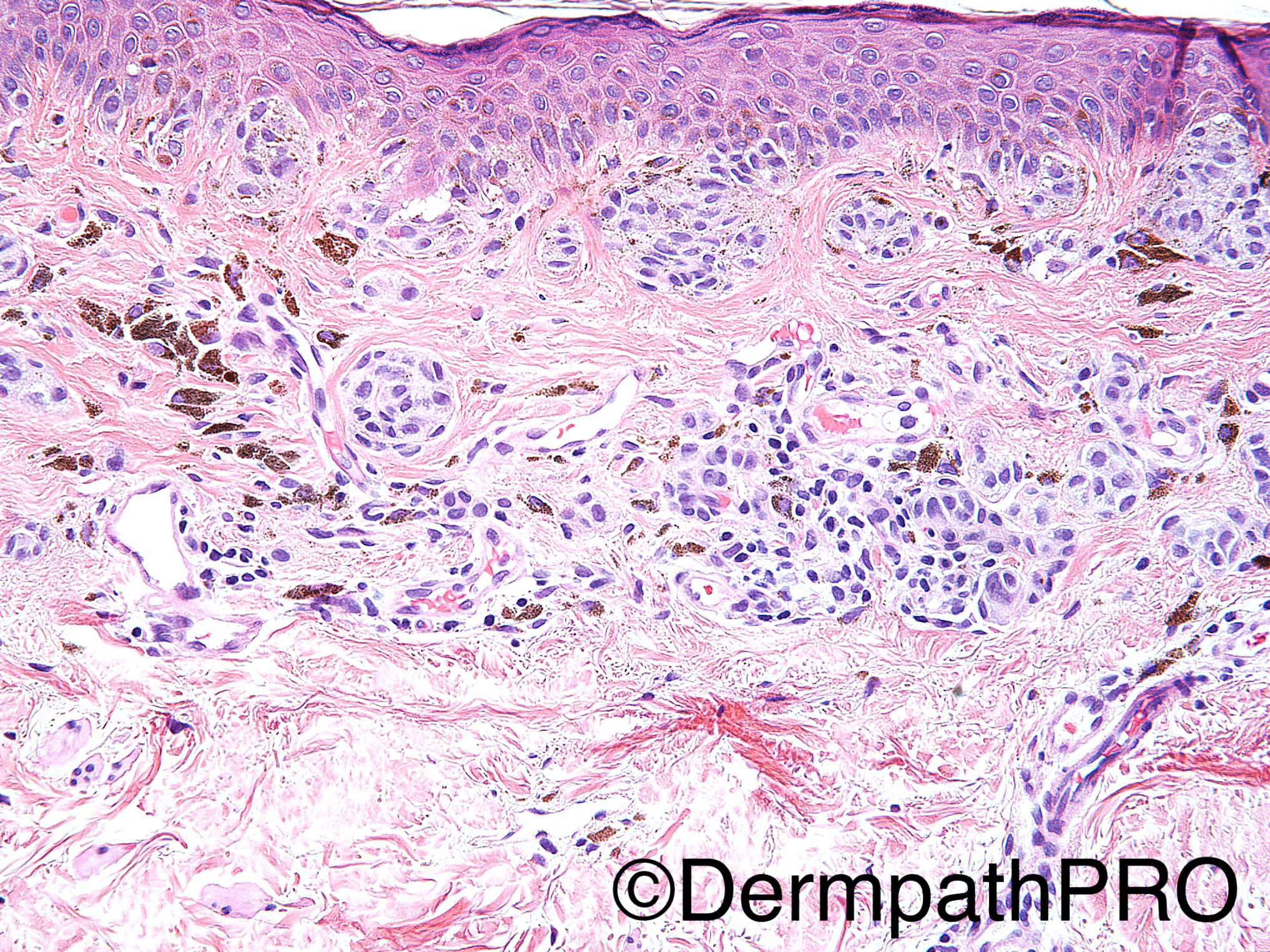
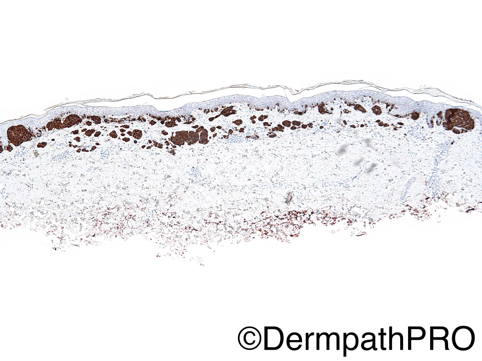
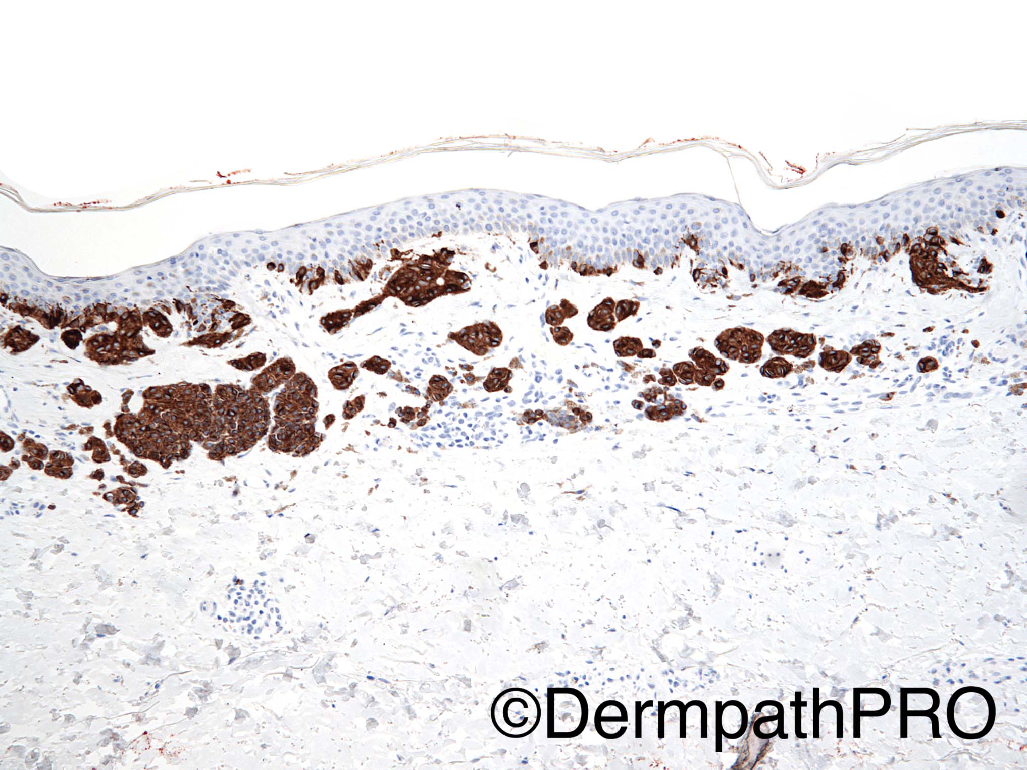
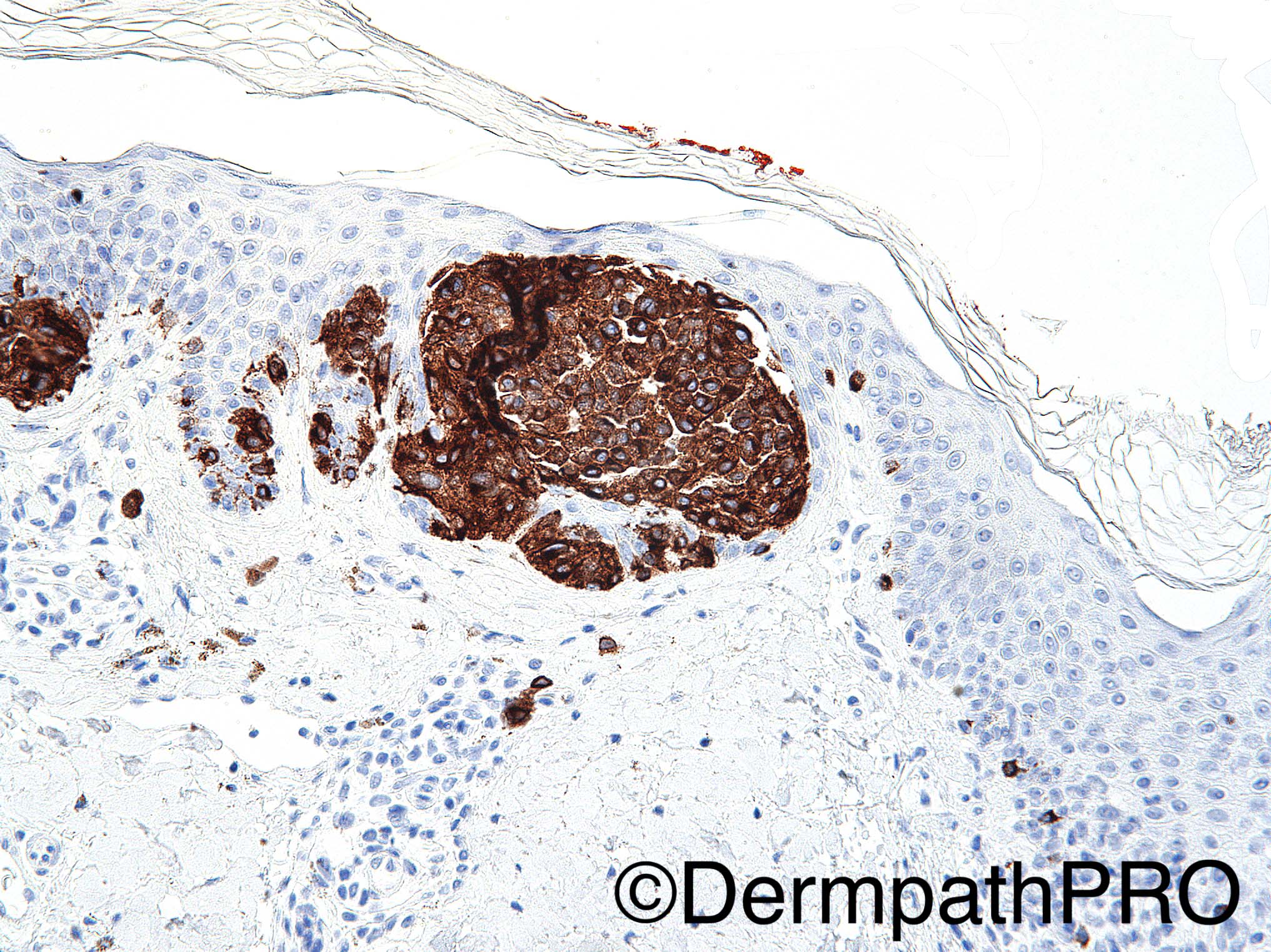
32 year old male with right back brownish black papule. The immunohistochemical stain used is Melan-A/MART-1.

Dr Uma Sundram.


  Report Record

User Feedback


vincenzo polizzi

Posted

Compound nevus, with some random atypical change in the dermal component, and some asymmetric intraepidermal placement of melanocytes...displastic nevus. There is  a maturation gradient in a intrapapillary dermal nest on the center of fig1, with type A melanocytes on the top and type B on the bottom.

Share this comment


Link to comment
share_externally

Raul Perret

Posted

Another not that easy thin melanic lesion. There is some assymmetry, expansile crowded nests, areas of confluence, impairment of clear maturation (demonstrated also with the homogeneous hmb-45 expression) and some cells with conspicuous nucleoli and hypercromasia. On the other hand the lesion starts and ends with nests, there is not a clear lentiginous growth, no pagetoid spread, absence of mitosis (even if I would make a ki67/phh3), extension to adnexae is not clearly seen (would do some levels) and the inflammatory infiltrate is scarce. I would go for MELTUMP in this particular case for further study with FISH/CGH but I would slightly favour a benign lesion. 

Share this comment


Link to comment
share_externally

Arti Bakshi

Posted

Agree with points made by Raul.

Overall, I think there are more reassuring features than concerning ones.The irregular pigment distribution (including a few ?dendritic ones) made me think of a combined naves/naevus with phenotypic heterogeniety.

Share this comment


Link to comment
share_externally

Guest Arash Daryakarr

Posted

this is a melanocytic neoplasm composed of junctional nests and single cells few of nests are fairly large.
focal bridging of junctional nests with surrounding concentric fibroplasia is evident. pigment incontinence & few inflammatory cells are seen in dermis.
Few dermal nests are large and contain large atypical melanocytes at base.
i didn't see any dermal mitosis,any pagetoid spread,diffuse cytologic atypia in favor of melanoma.
i favor more that of dysplastic nevus.

Share this comment


Link to comment
share_externally

My feeling is that this is a benign compound naevus. 

Share this comment


Link to comment
share_externally

Dr. Mona Abdel-Halim

Posted

Favouring dysplastic nevus.

Share this comment


Link to comment
share_externally

Nitin Khirwadkar

Posted

Largely nested, shallow melanocytic lesion. There is mild to moderate architectural atypia, and mild cytonuclear atypia. Difficult to assess maturation in this thin lesion, but some impairment as Raul has suggested is there. No active regression or overt atypia. p16 and PHH3 may not particularly help. If this is a completely excised lesion, I feel would represent a mildly dysplastic naevus.

Share this comment


Link to comment
share_externally

Dr. Richard Carr

Posted

Thin, junctionally "active", compound naevus with minimal atypia. Not overly worried on the H&E. Management based on clinical concern.

Share this comment


Link to comment
share_externally

Uma Sundram

Posted

Agree with all of your assessments. We were not significantly worried about this lesion and ultimately called it a dysplastic nevus with mild atypia. Given the age of the patient, both patient and dermatologist opted for watchful waiting. It was interesting in seeing the range of approaches in this forum--these types of cases tend to be very instructional!

Share this comment


Link to comment
share_externally


Join the conversation

You can post now and register later. If you have an account, sign in now to post with your account.

Guest
Add a comment...

×   Pasted as rich text.   Paste as plain text instead

  Only 75 emoji are allowed.

×   Your link has been automatically embedded.   Display as a link instead

×   Your previous content has been restored.   Clear editor

×   You cannot paste images directly. Upload or insert images from URL.

Loading...

×
×
  • Create New...