Jump to content
In this section we have spot diagnoses posted on a daily basis since June 2010, now over 4000! You can review the archived cases and read the suggested diagnoses by users and the final comment by the contributors.
Case are uploaded each week day by 10 am UK time with the correct diagnosis will generally be posted at 8 pm UK time. Why not view the most recent spot diagnosis and proffer a diagnosis?

Case Number : Case 1521 - 22 April Posted By: Guest

Please read the clinical history and view the images by clicking on them before you proffer your diagnosis.
Submitted Date :
   (0 reviews)

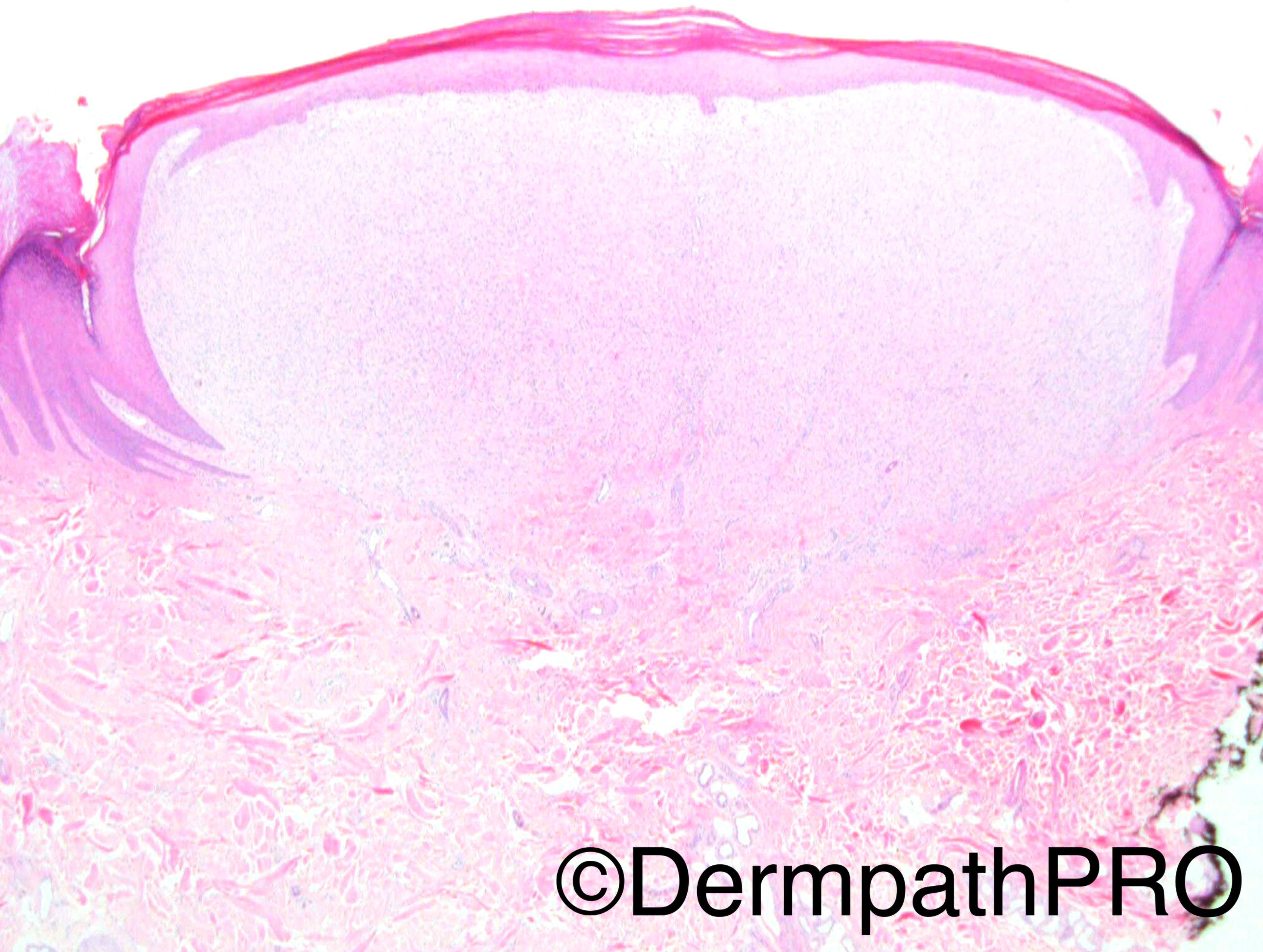
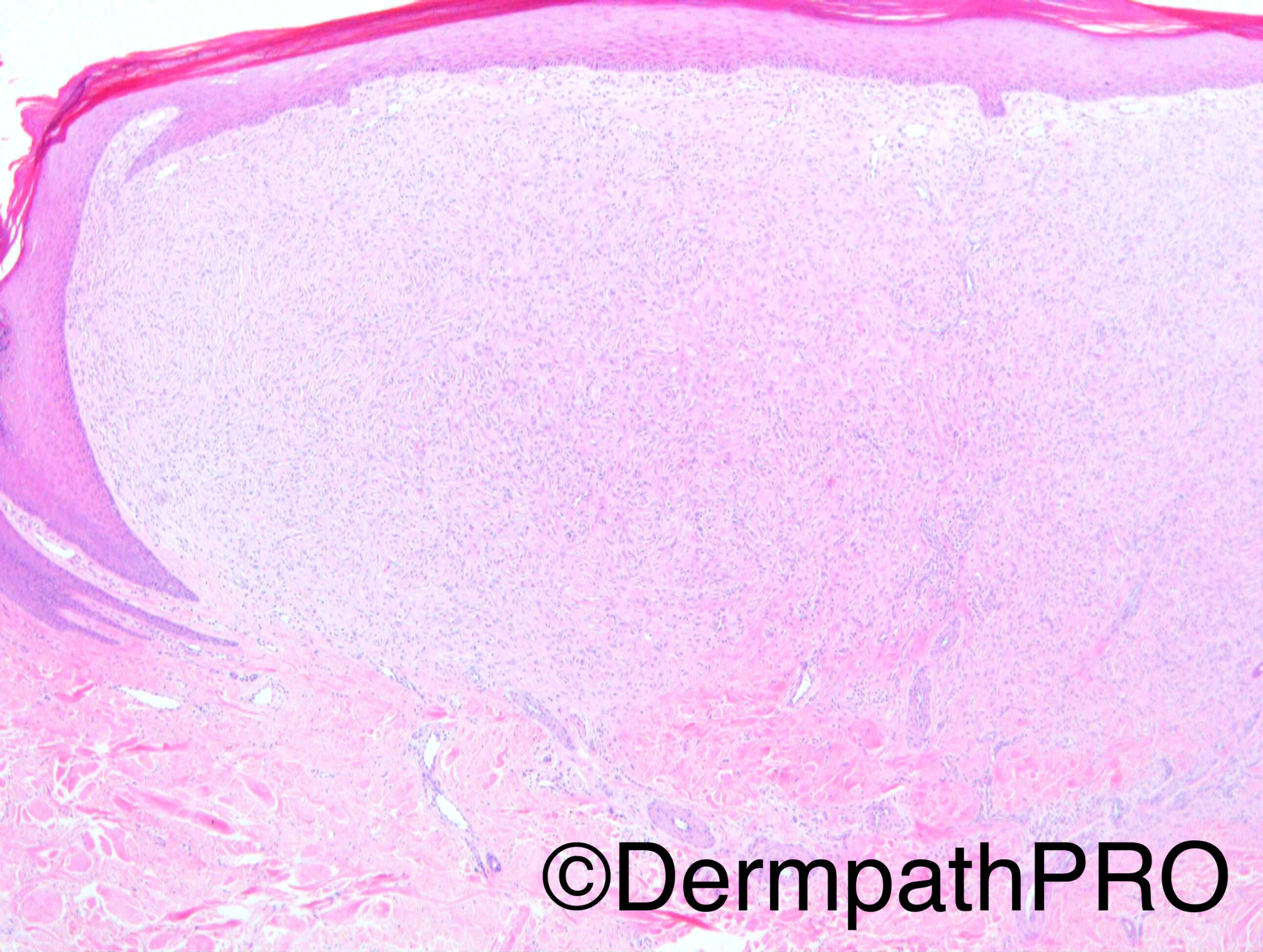
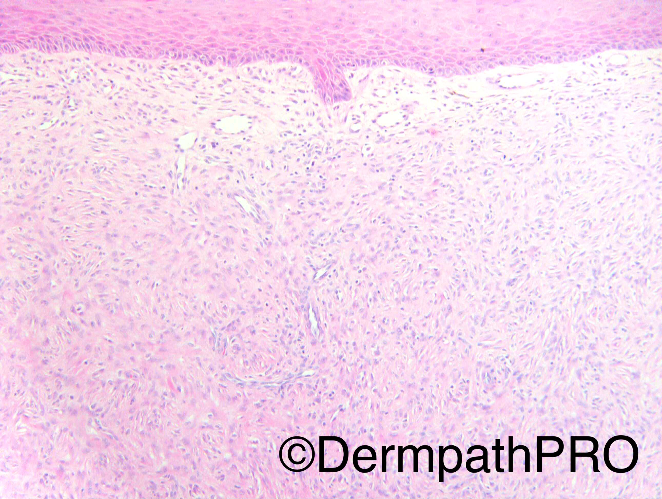
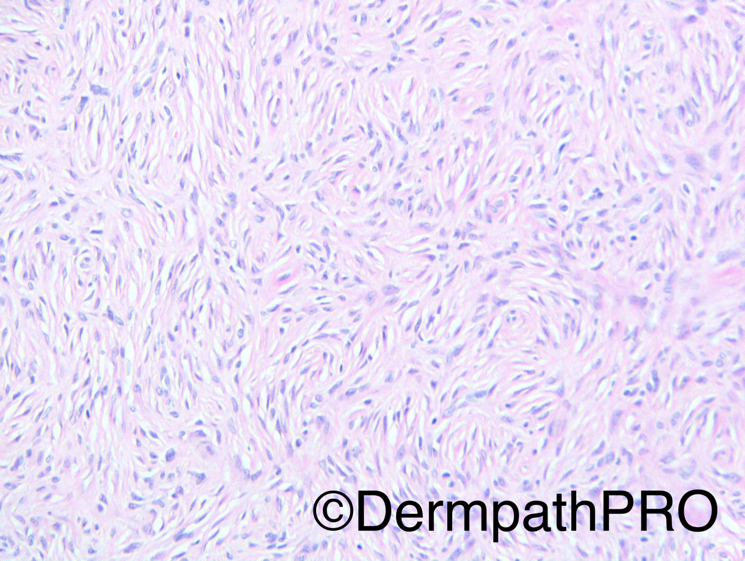
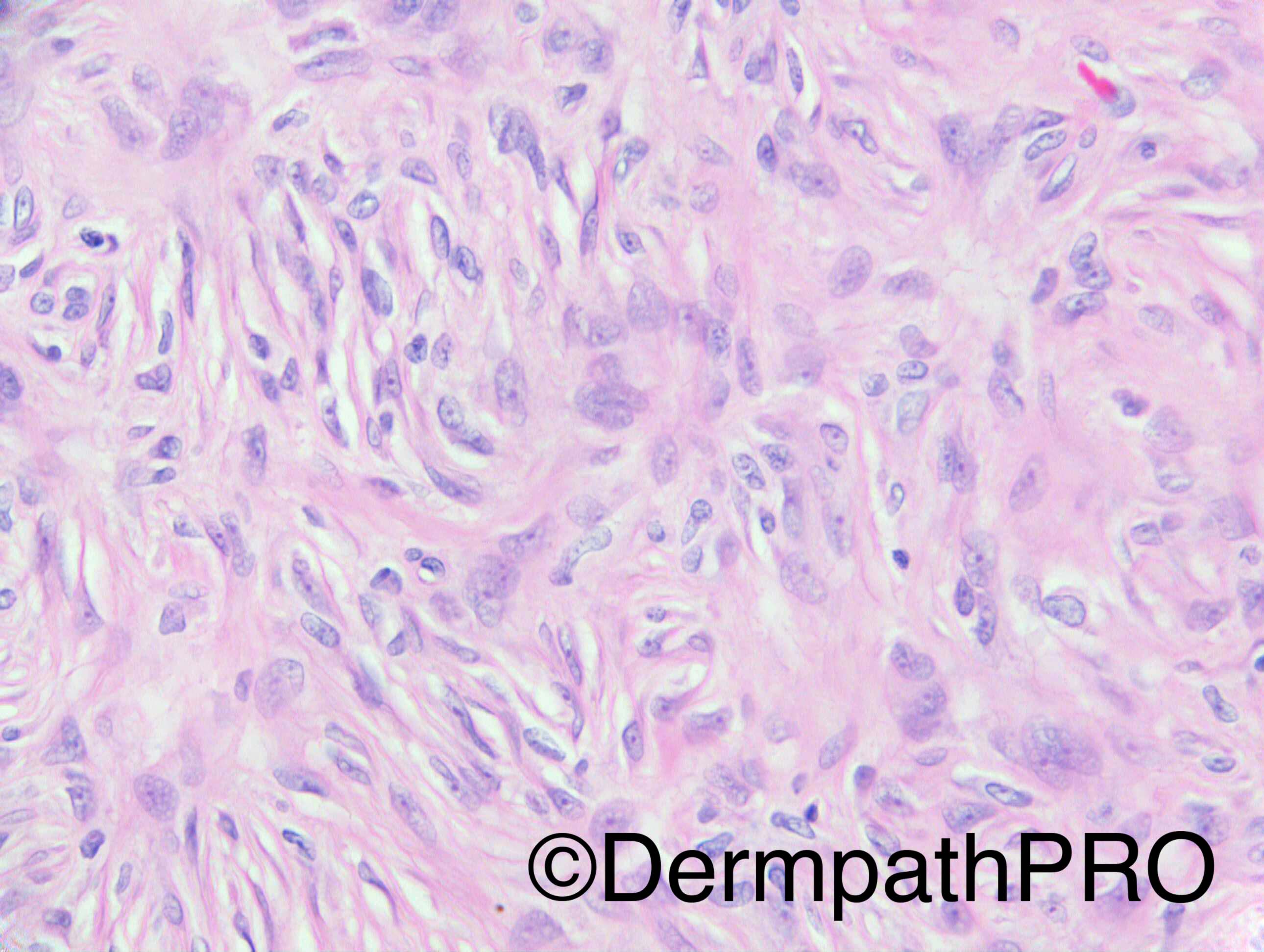
M60. Lesion right knee, 6-7 years. Recent episode of bleeding. ?Angioma


Dr Richard Carr.


  Report Record

User Feedback


vincenzo polizzi

Posted

Epithelioid dermatofibroma. Nice epidermal collarette.

Share this comment


Link to comment
share_externally

Dr. Mona Abdel-Halim

Posted

Perineurioma first thought. Agree with epithelioid fibrous histiocytoma as a ddx

Share this comment


Link to comment
share_externally

vincenzo polizzi

Posted

I favor ED aka epithelioid cell histiocytoma, because fits better on CPC. It is a red polypoid nodule ( clinically angioma ) and shows ectatic vessels in keeping With recent bleeding. Histologically looks like more cellular that epithelioid but this is in keeping with Epithelioid Dermatofibroma.

Share this comment


Link to comment
share_externally

Nitin Khirwadkar

Posted

Agree with the differentials.

Share this comment


Link to comment
share_externally

Guest Arash Daryakarr

Posted

I favor epithelioid fibrous histiocytoma due to clinical presentation (usually presents as a red nodule which usually confused with pyogenic granuloma) and histomorphologic charactristics.
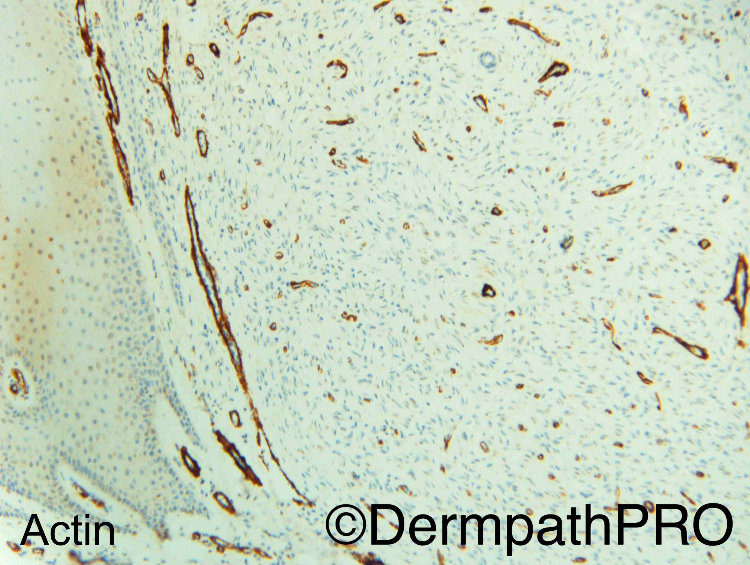
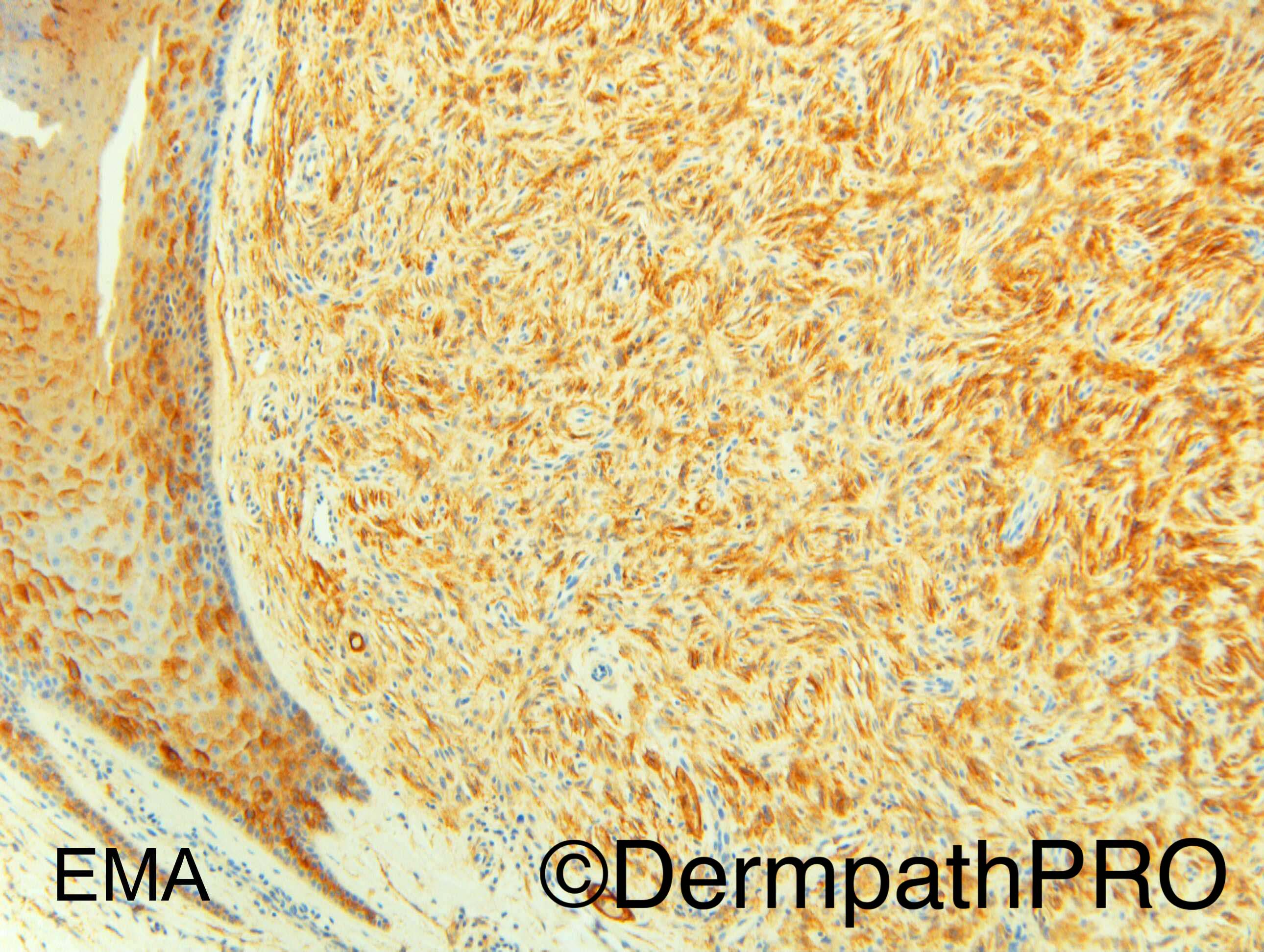
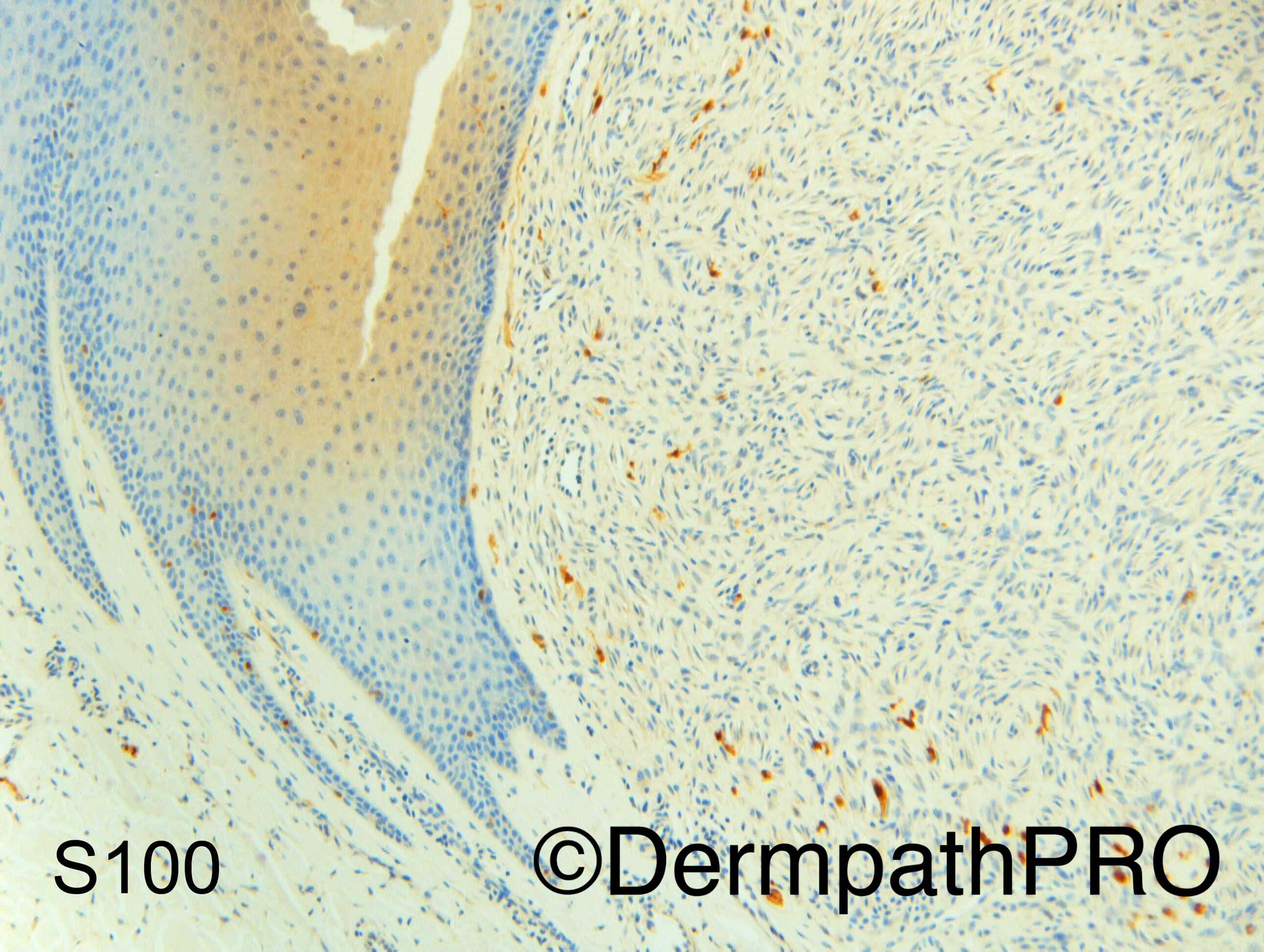
Distinction from epithelioid perineurioma can be made by IHC for EMA and claudin-1.

Share this comment


Link to comment
share_externally

Arti Bakshi

Posted

Yes, epithelioid fibrous histiocytoma vs perineurioma

Share this comment


Link to comment
share_externally

Dr. Mona Abdel-Halim

Posted

So it is perineurioma :-)

Share this comment


Link to comment
share_externally

Dr. Richard Carr

Posted

Good discussion. I reported it as a perineurioma.

Share this comment


Link to comment
share_externally


Join the conversation

You can post now and register later. If you have an account, sign in now to post with your account.

Guest
Add a comment...

×   Pasted as rich text.   Paste as plain text instead

  Only 75 emoji are allowed.

×   Your link has been automatically embedded.   Display as a link instead

×   Your previous content has been restored.   Clear editor

×   You cannot paste images directly. Upload or insert images from URL.

Loading...

×
×
  • Create New...