Jump to content
In this section we have spot diagnoses posted on a daily basis since June 2010, now over 4000! You can review the archived cases and read the suggested diagnoses by users and the final comment by the contributors.
Case are uploaded each week day by 10 am UK time with the correct diagnosis will generally be posted at 8 pm UK time. Why not view the most recent spot diagnosis and proffer a diagnosis?

Case Number : Case 1593 - 03 August Posted By: Guest

Please read the clinical history and view the images by clicking on them before you proffer your diagnosis.
Submitted Date :
   (0 reviews)

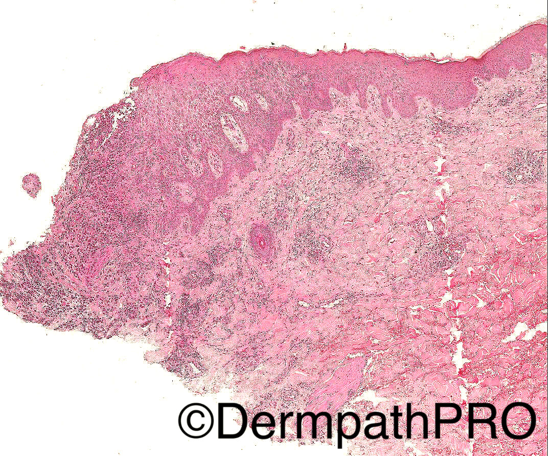
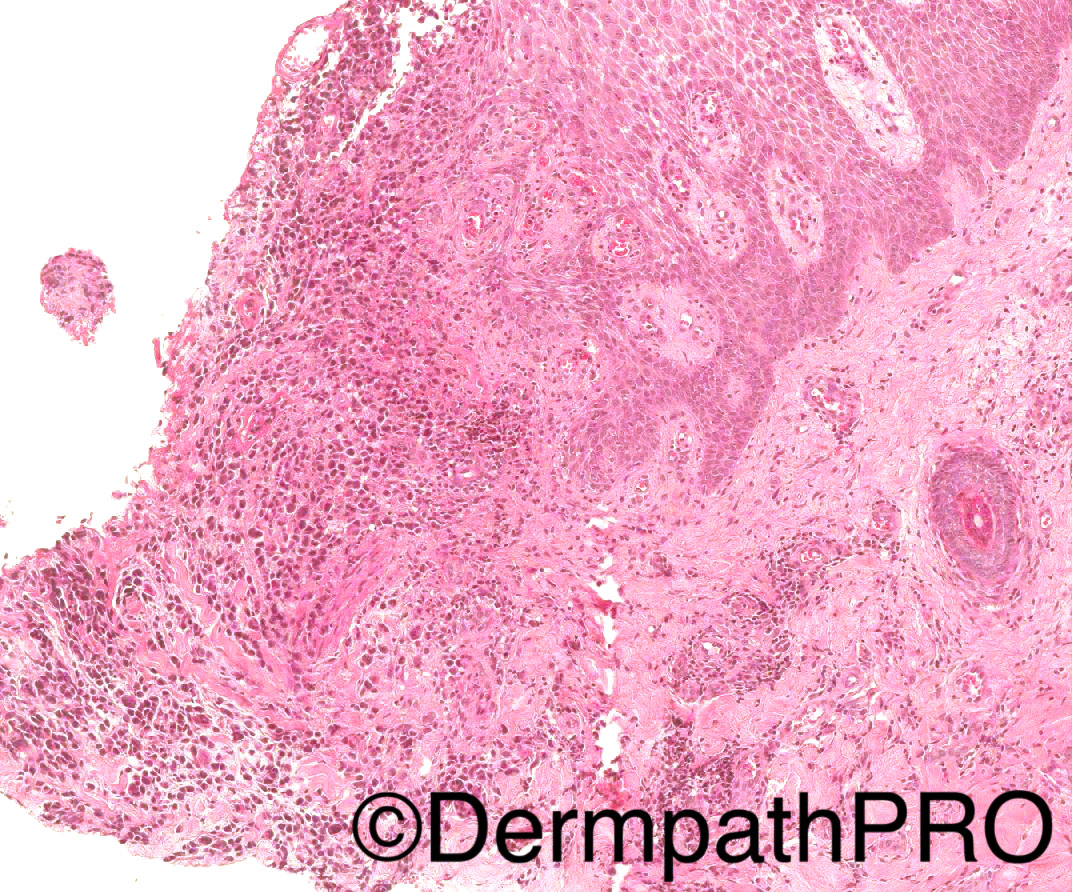
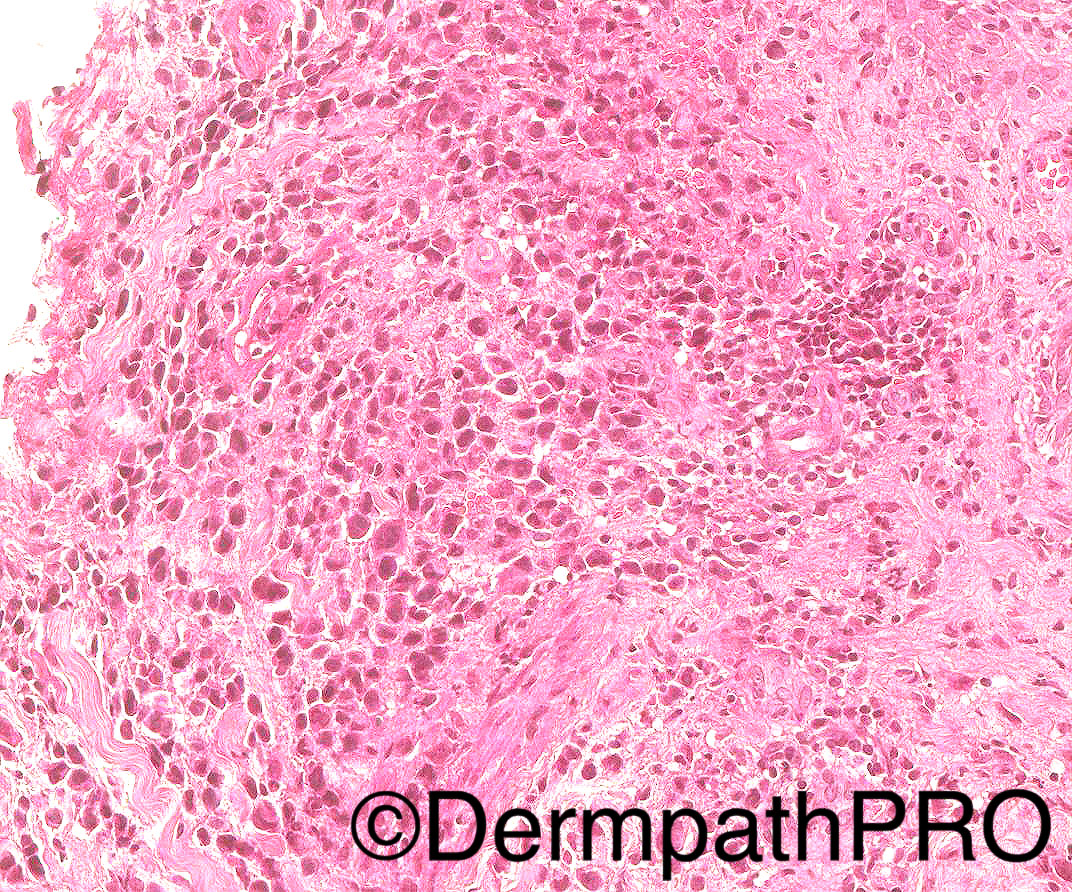
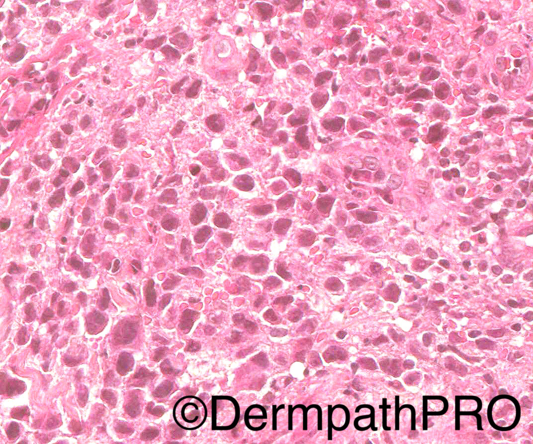
68/M widespread purpuric telangiectatic rash on trunk and extremities, diagnosed as purpuric Mycosis fungoides on a previous bx. T cell clone present. Now, developed 3 new lesions on leg, 4- 5mm papules. Another biopsy done and images below are from the second bx. Clonality studies reveal identical T cell clone as previous biopsy. (same distribution and size of monoclonal peaks)

Case Posted by Dr Arti Bakshi


  Report Record

User Feedback


vincenzo polizzi

Posted

Maybe a MF associated type D LyP!

Lymphomatoid papulosis associated with mycosis fungoides: a study of 21 patients including analyses for clonality.
Zackheim HS, et al. J Am Acad Dermatol. 2003.

Share this comment


Link to comment
share_externally

Dr. Mona Abdel-Halim

Posted

Yes, LyP developing in MF patient

Share this comment


Link to comment
share_externally

Raul Perret

Posted

I thought of large cell transformation in MF and I think that the fact that the same clone was demonstrated actually favours large cell transformation over LyP. Clnicopathologic correlation really important also....

Share this comment


Link to comment
share_externally

Raul Perret

Posted

Actually I read a bit more and same clone can be present in both entities, the most important is the clinical setting. If clinically is more suggestive of LyP it should be LyP but if patient is clinically in bad shape and worsening should be called transformed MF. By the way, correct me if im wrong but I think if the clinical is rather benign we could call it CD30+ lymphoproliferative disorder as it could also be primary cutaneous anaplastic large cell lymphoma (also indolent as LyP); the most specific feature for favouring LyP would be the classic recurrent self-healing papules.

Share this comment


Link to comment
share_externally

vincenzo polizzi

Posted

I think that there are many large anaplastic cells in between small and medium size ones and the infiltrate is predominantly destroying intra epidermal and in superficial dermis, otherwise this is a deep band-like intradermal monotonous population of medium size anaplastic cells in tumoral stage of MF with typical persistent scattered or grouped in nests intra epidermal component. But this is not absolutely true, so agree with above first Raul's comment and with the second one also.

Share this comment


Link to comment
share_externally

Arti Bakshi

Posted

Raul's second comment summarises this case very well!

Yes, the distinction is heavily dependent on the clinical presentation and course of lesions. Purely from a histological point of view, this is basically a CD30 rich T cell lymphproliferation which could be compatible with a transformed MF, primary cutaneous ALCL or LyP, type C... ( @Vincenzo, the infiltrate was quite deep in another section and the surface is ulcerated rather than showing epidermotropism, so would classify as type C rather than Type B LyP).

At our MDT, I was informed that 2 of the lesions have started showing spontaneous regression, the third is still persistent. Remember, Lyp may not always present with crops of lesions but may be oligolesional and the regression period may be over weeks to months. There was some concern amongst our haematologists that an identical clone implies transformed MF, but as you all have rightly commented LyP often shows the same clone as the associated MF. (thanks for the reference Vincenzo, I quoted the same one in my report and to the concerned haematologists!). 

Share this comment


Link to comment
share_externally


Join the conversation

You can post now and register later. If you have an account, sign in now to post with your account.

Guest
Add a comment...

×   Pasted as rich text.   Paste as plain text instead

  Only 75 emoji are allowed.

×   Your link has been automatically embedded.   Display as a link instead

×   Your previous content has been restored.   Clear editor

×   You cannot paste images directly. Upload or insert images from URL.

Loading...

×
×
  • Create New...