Jump to content
In this section we have spot diagnoses posted on a daily basis since June 2010, now over 4000! You can review the archived cases and read the suggested diagnoses by users and the final comment by the contributors.
Case are uploaded each week day by 10 am UK time with the correct diagnosis will generally be posted at 8 pm UK time. Why not view the most recent spot diagnosis and proffer a diagnosis?

Case Number : Case 1600 - 12 August Posted By: Guest

Please read the clinical history and view the images by clicking on them before you proffer your diagnosis.
Submitted Date :
   (0 reviews)

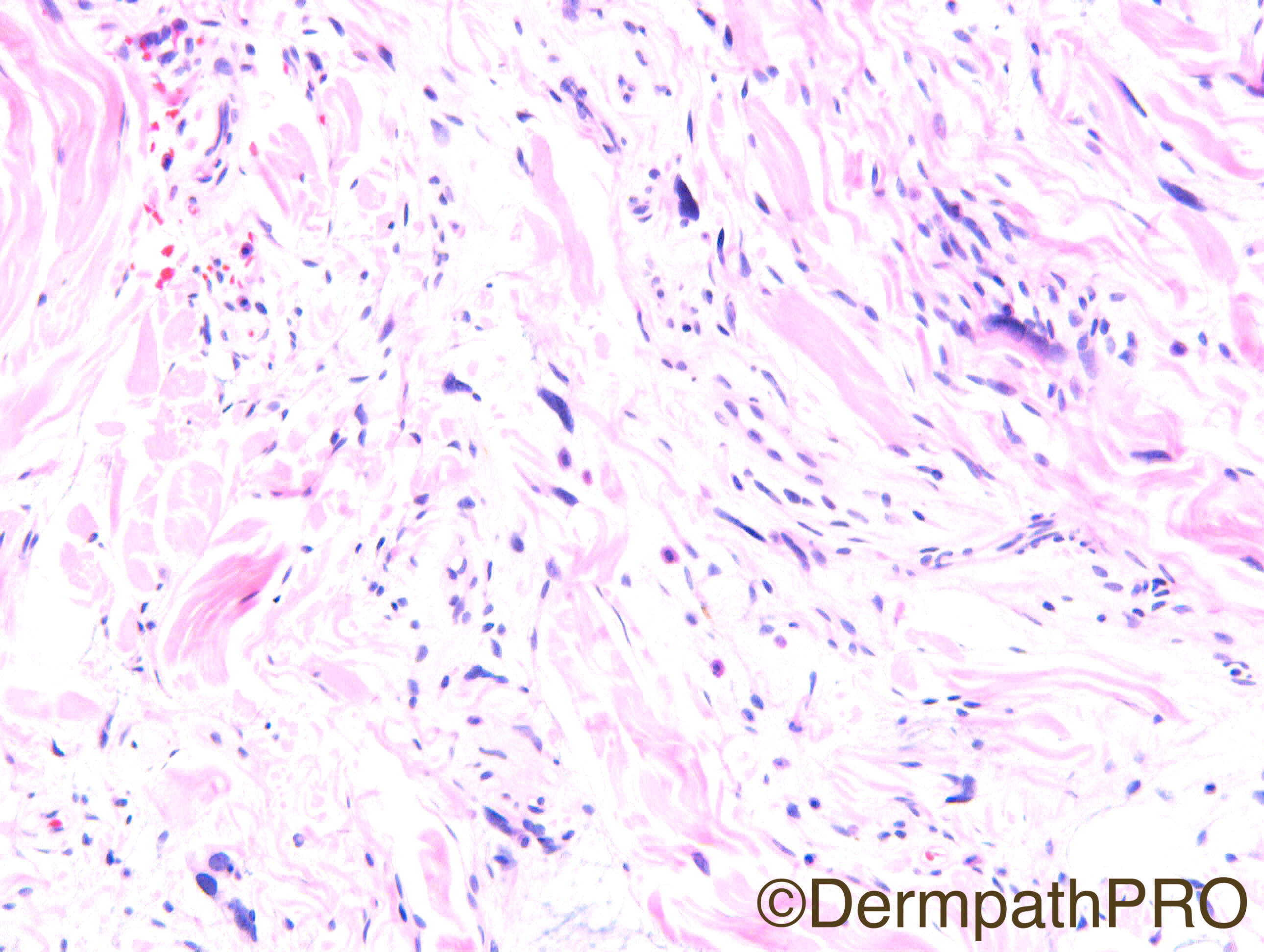
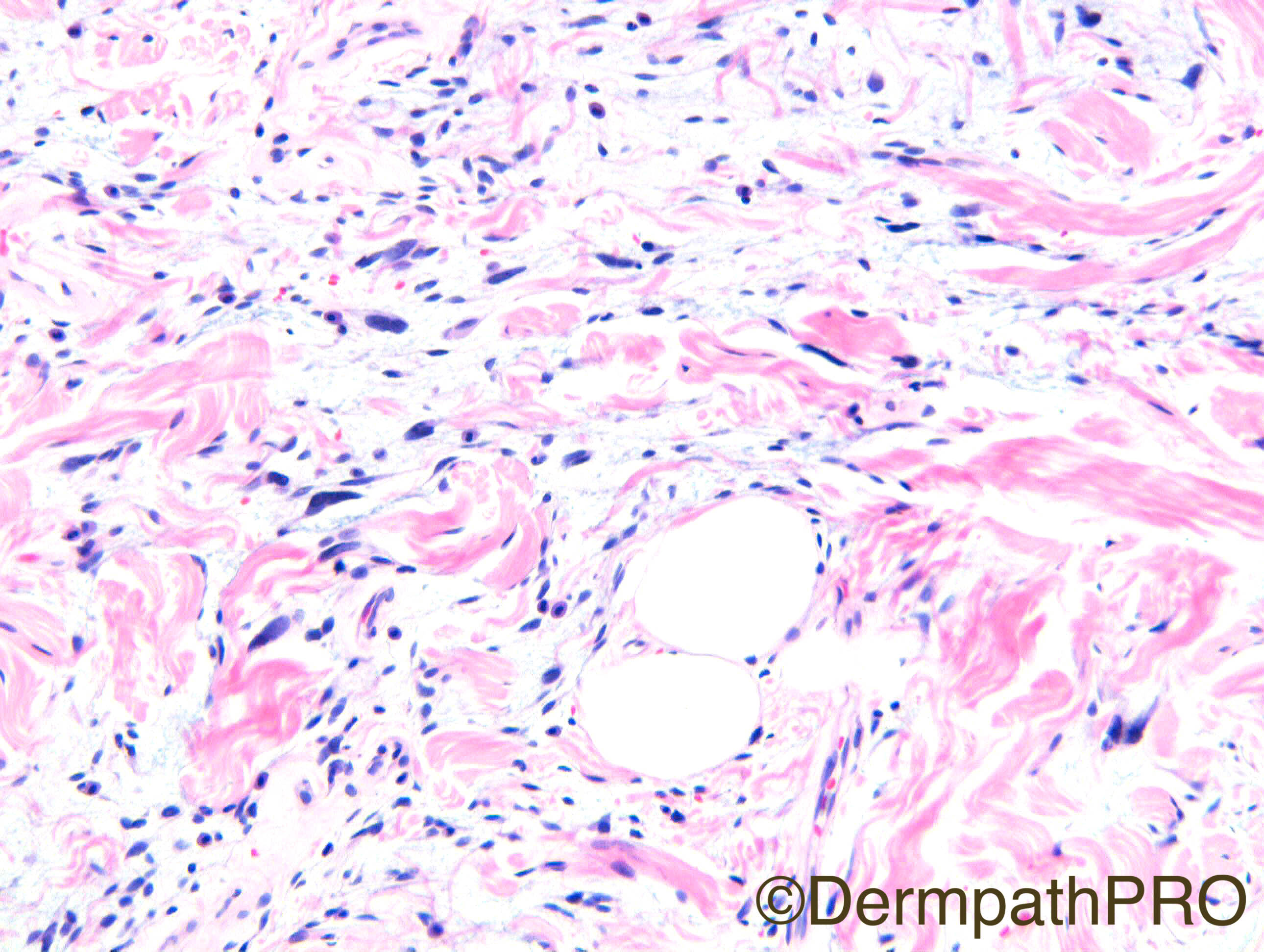
Clinical Details: F80. Calf mass. Referral c/o Dr Terry Jones

Case Posted by Dr Richard Carr

  Report Record

User Feedback


vincenzo polizzi

Posted

Pleomorphic Fibroma.

It is usually described as a polypoid or dome shaped lesion, rather than a mass, but location and morphology fit well!

Share this comment


Link to comment
share_externally

Raul Perret

Posted

There is a lot of mucin, with this location we should ask the fellow clinician how is the thyroid biology of the patient to rule out a tumoral form of pretibial myxedema. Fibroblasts are increased and reactive so another option is lichen myxedematosus (I assume there aren't any other signs or symptons in the patient to consider scleromyxedema). But I think it is a must asking the lab of the patient to discard thyroid pathology first. Another thing that crossed my mind if the lesion is unilateral was a massive form of lymphoedema but I think this is not the typical morphology. To sum up, not tumoral for me but rather a mucinosis

Share this comment


Link to comment
share_externally

Dr. Mona Abdel-Halim

Posted

I am thinking of angiofibroblastoma of the skin although not enough capillaries in the stroma as described for this entity, but we have spindled and stellate fibroblasts in myxoid and fibromyxoid areas.. Not sure though :-) waiting to know the correct answer..

Share this comment


Link to comment
share_externally

Guest Romualdo

Posted

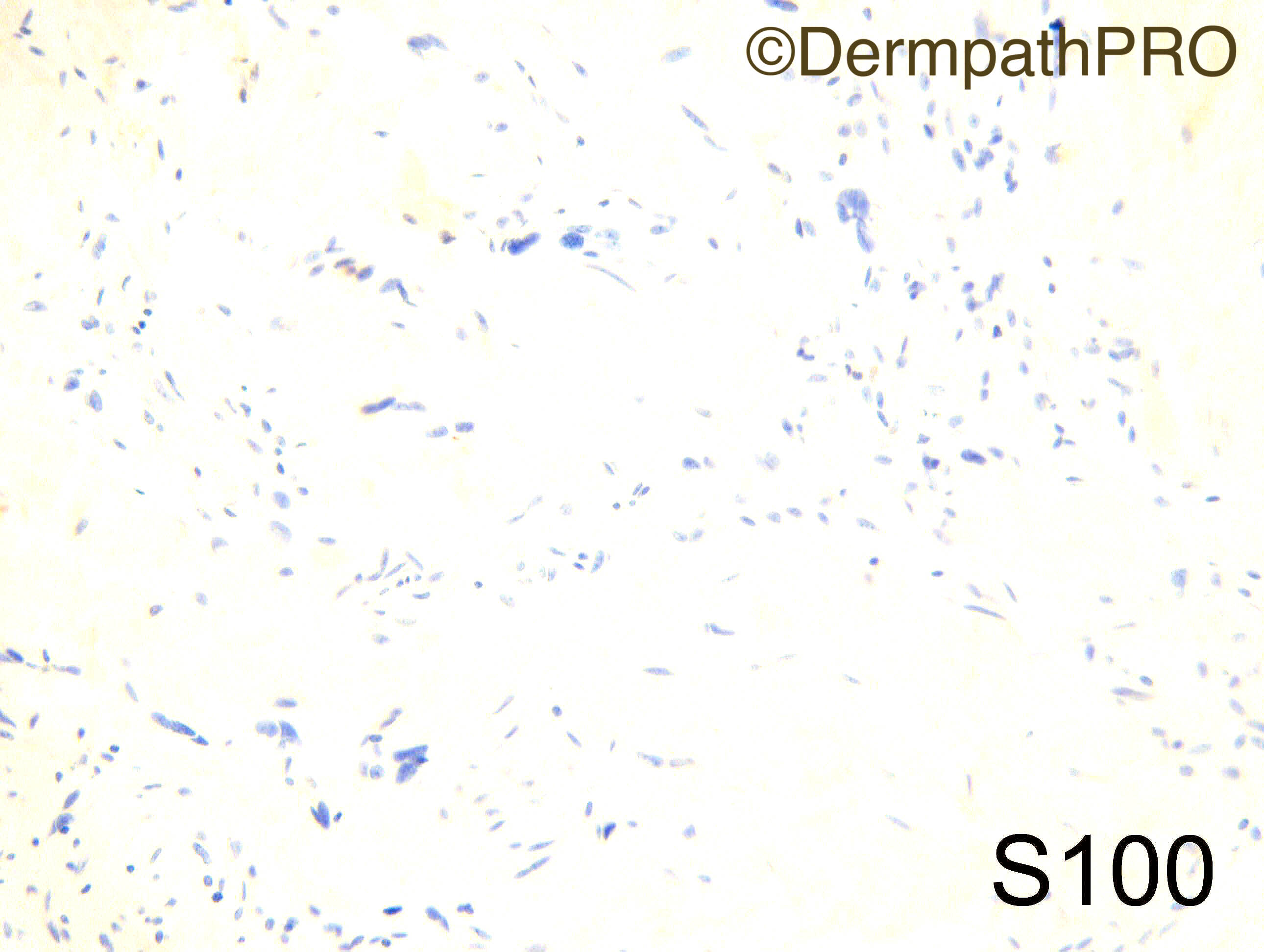
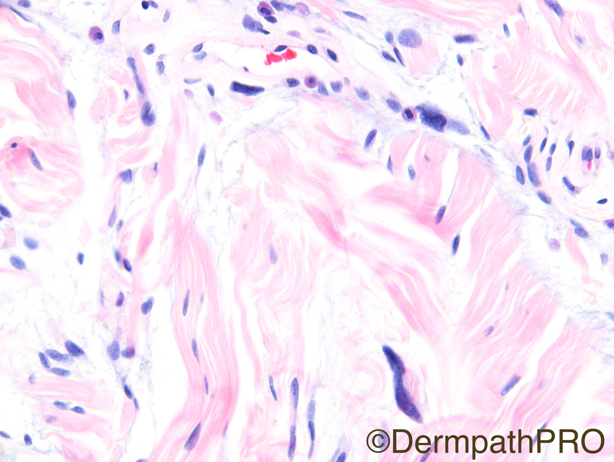
In contrast to much more common subcutaneous lesions, cutaneous spindle cell/ pleomorphic lipomas are more frequent in women, are more infiltrative  and have a wide anatomic distribution. Spindle cells, pleomorphic cells, mucin, mast cells, thick collagen fibers, rare adipocytes and CD34 positivity and S100 negativity fit well.

Share this comment


Link to comment
share_externally

Raul Perret

Posted

Yes you are right Romualdo I should have payed more attention to the case. Always forget to check for the nice ropey collagen seen in picture seven and as you very well remarked there are also evident floret cells.

Share this comment


Link to comment
share_externally

Eman El-Nabarawy

Posted

Also thought of spindle cell lipoma.

Share this comment


Link to comment
share_externally

Nitin Khirwadkar

Posted

Would go for a pleomorphic lipoma. 

Share this comment


Link to comment
share_externally

Dr. Richard Carr

Posted

Yes I thought this was a nice example of a dermally located spindle cell lipoma (overlaps morphologically and cytogenetically with pleomorphic lipoma). 

Share this comment


Link to comment
share_externally


Join the conversation

You can post now and register later. If you have an account, sign in now to post with your account.

Guest
Add a comment...

×   Pasted as rich text.   Paste as plain text instead

  Only 75 emoji are allowed.

×   Your link has been automatically embedded.   Display as a link instead

×   Your previous content has been restored.   Clear editor

×   You cannot paste images directly. Upload or insert images from URL.

Loading...

×
×
  • Create New...