Jump to content
In this section we have spot diagnoses posted on a daily basis since June 2010, now over 4000! You can review the archived cases and read the suggested diagnoses by users and the final comment by the contributors.
Case are uploaded each week day by 10 am UK time with the correct diagnosis will generally be posted at 8 pm UK time. Why not view the most recent spot diagnosis and proffer a diagnosis?

Case Number : Case 1606 - 22 August Posted By: Guest

Please read the clinical history and view the images by clicking on them before you proffer your diagnosis.
Submitted Date :
   (0 reviews)

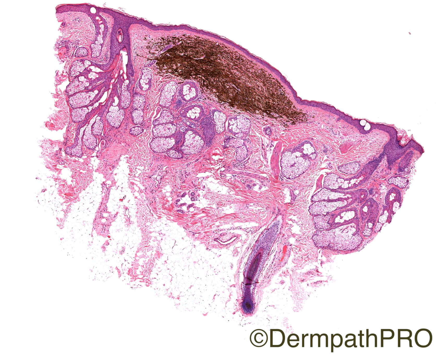
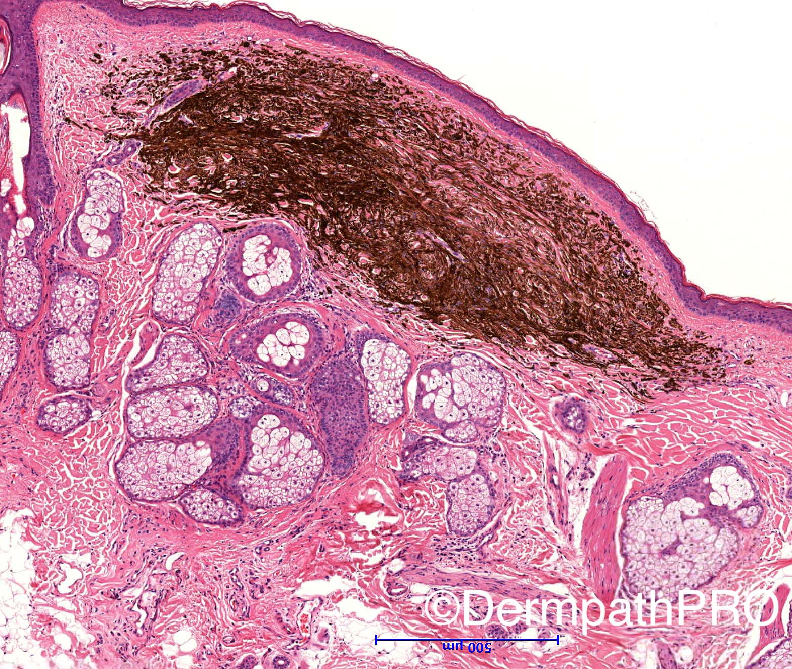
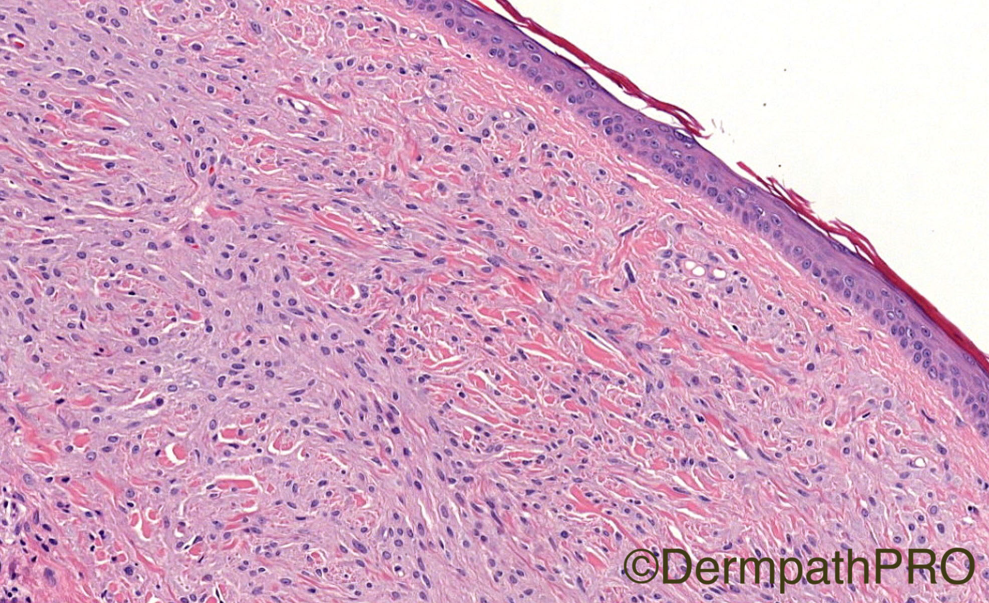
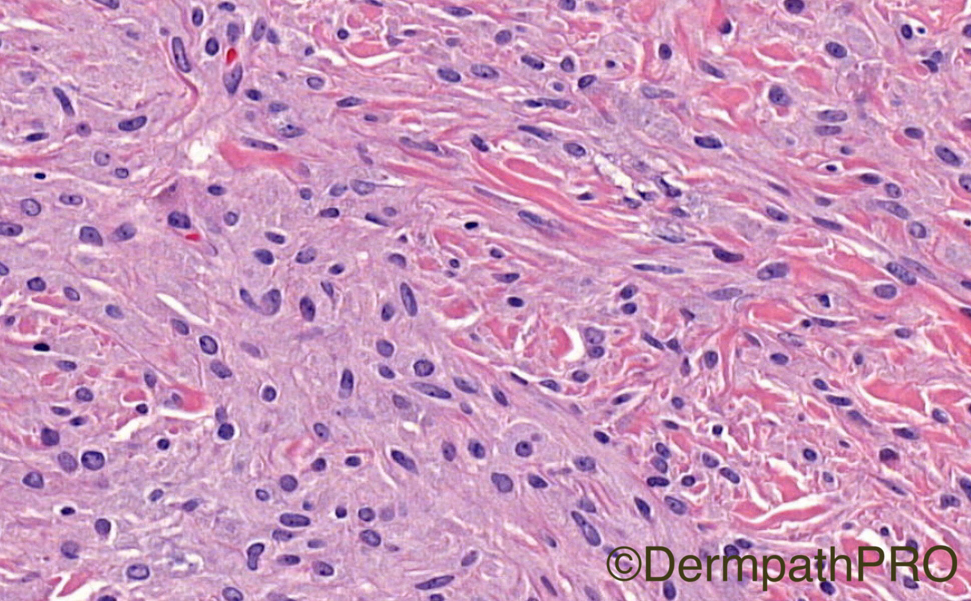
55 year old lesion on scalp. Past history of MM.

Case posted by Dr Iskander Chaudhry


  Report Record

User Feedback


vincenzo polizzi

Posted

Agree. Satellite nodule of previous lesion?

Tumoral melanosis as a manifestation of a completely regressed primary melanoma with metastases.

Ng SH, Chave TA.

Share this comment


Link to comment
share_externally

Iskander H. Chaudhry

Posted

Many thanks for the opinions. Before I tell you the diagnosis the previous was a SSMM minimally invasive, RGP on the leg 5 years ago. So any other differentials? 

Share this comment


Link to comment
share_externally

vincenzo polizzi

Posted

Ipilimumab-associated TM? But I don't think so, because the patient wasn't on advanced stage.
Post trauma TM?
Pigmented Epithelioid Melanocytoma? But can't see any bona-fide melanocyte, epithelioid nor spindled...It lseems just a tumoral melanosis...
Found this art: DOI: http://dx.doi.org/10.5935/scd1984-8773.201681708

Share this comment


Link to comment
share_externally

Dr. Mona Abdel-Halim

Posted

[quote name="Iskander H. Chaudhry" timestamp="1471983266"]Many thanks for the opinions. Before I tell you the diagnosis the previous was a SSMM minimally invasive, RGP on the leg 5 years ago. So any other differentials? [/quote] ? Metastatic melanoma.

Share this comment


Link to comment
share_externally

Iskander H. Chaudhry

Posted

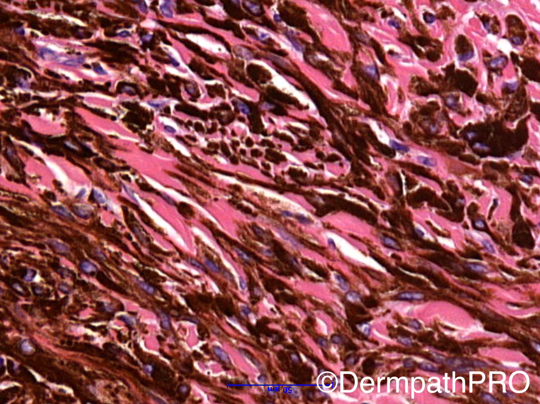
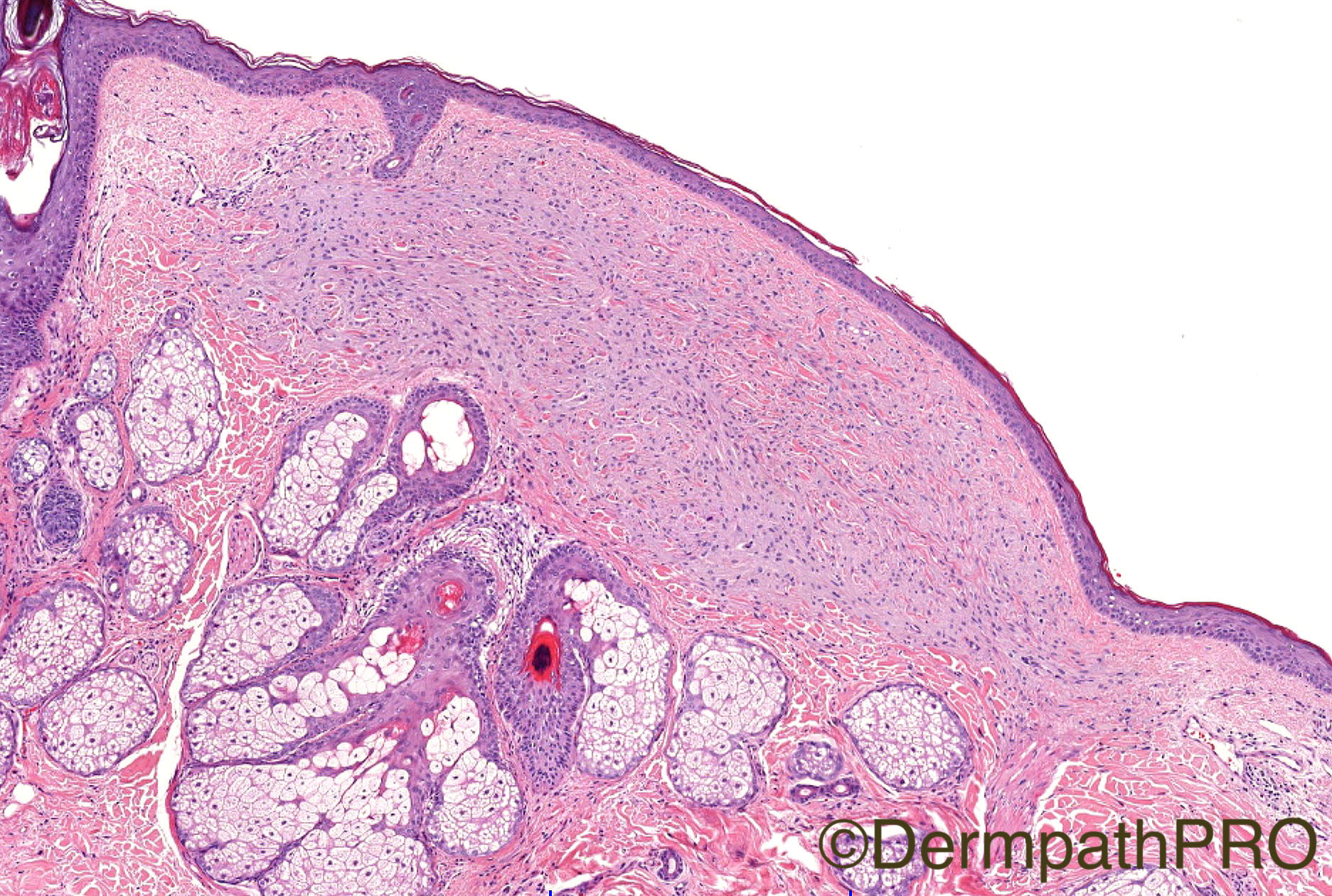
Dear All thank you for your comments; the final diagnosis was epithelioid blue naevus. A pigment synthesizing melanoma (PSM) was considered but this is a small and superficial intradermal lesion with a small focus of lymphocytic infiltrate suggesting partial regression. The lesion is not expansile, there is preservation of the dermal structures, including the papillary dermis and of the epidermis. While pigment obscures view, where visible the cells are bland and have features in keeping with cells of epithelioid blue naevus. Small size, upper dermal location, no involvement of subcutis, non-infiltrating margins and non-expansile mass are all reassuring features and are not those of PSM.
 
My colleague Dr Corrado D'Arrigo agreed with the diagnosis and he bleached the specimen (see below). The cells are melanocytes as apposed to macrophages and are bland! 

Share this comment


Link to comment
share_externally

Admin_Dermpath

Posted

Dr Corrado D'Arrigo's images have been added above, they are the final three.


Cheers, Geoff

Share this comment


Link to comment
share_externally


Join the conversation

You can post now and register later. If you have an account, sign in now to post with your account.

Guest
Add a comment...

×   Pasted as rich text.   Paste as plain text instead

  Only 75 emoji are allowed.

×   Your link has been automatically embedded.   Display as a link instead

×   Your previous content has been restored.   Clear editor

×   You cannot paste images directly. Upload or insert images from URL.

Loading...

×
×
  • Create New...