Jump to content
In this section we have spot diagnoses posted on a daily basis since June 2010, now over 4000! You can review the archived cases and read the suggested diagnoses by users and the final comment by the contributors.
Case are uploaded each week day by 10 am UK time with the correct diagnosis will generally be posted at 8 pm UK time. Why not view the most recent spot diagnosis and proffer a diagnosis?

Case Number : Case 1464 -03 February Posted By: Guest

Please read the clinical history and view the images by clicking on them before you proffer your diagnosis.
Submitted Date :
   (0 reviews)

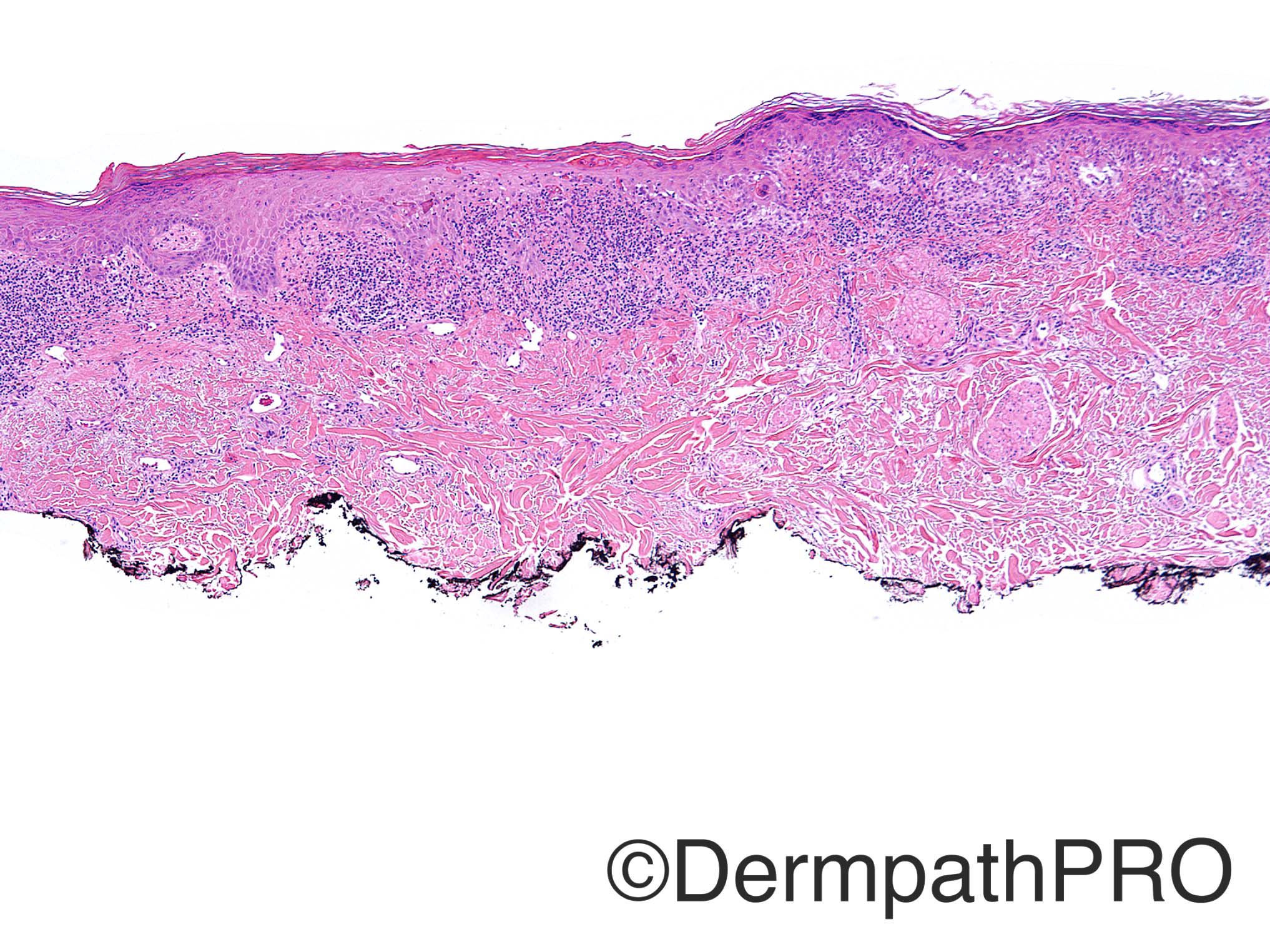
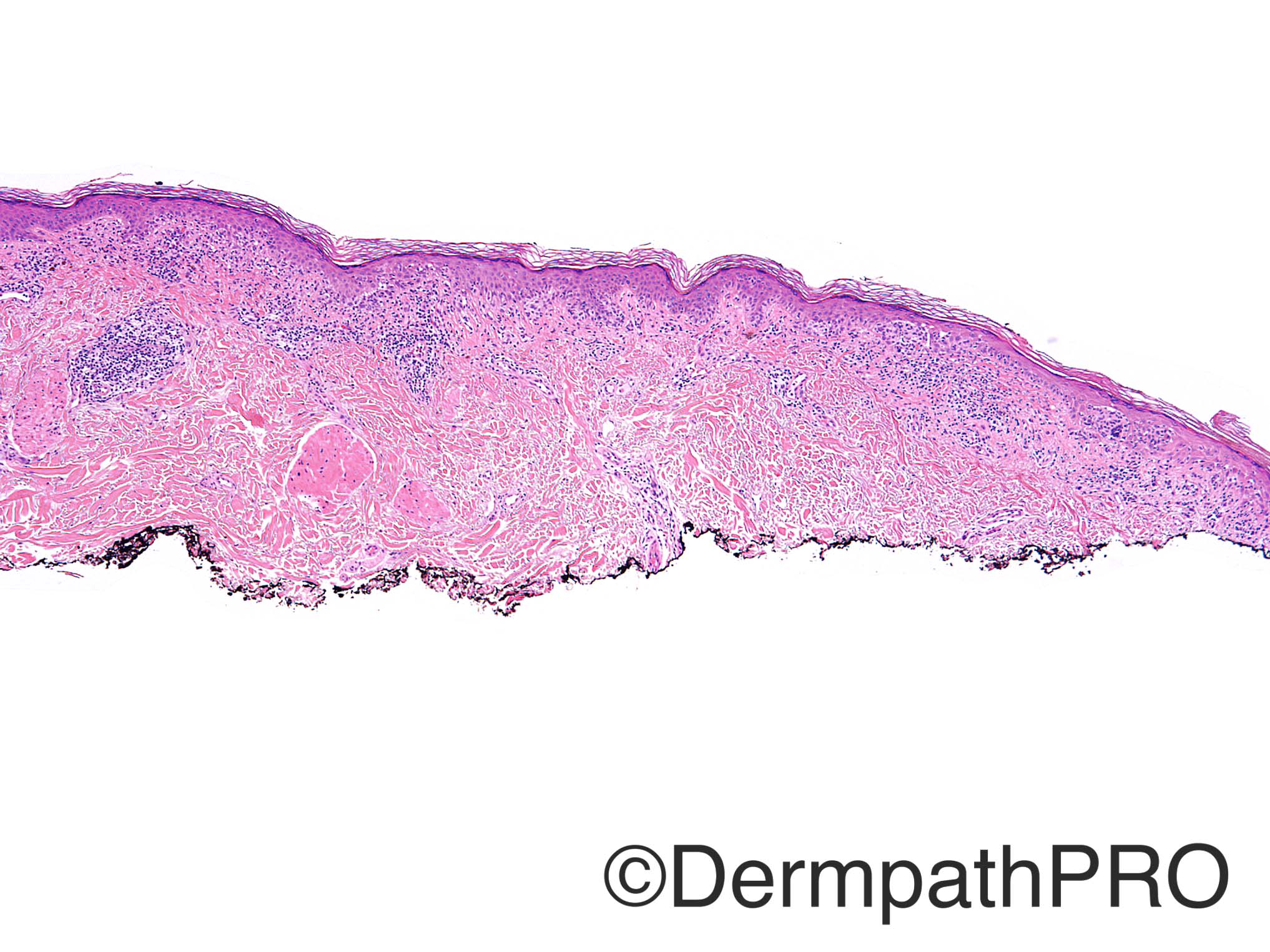
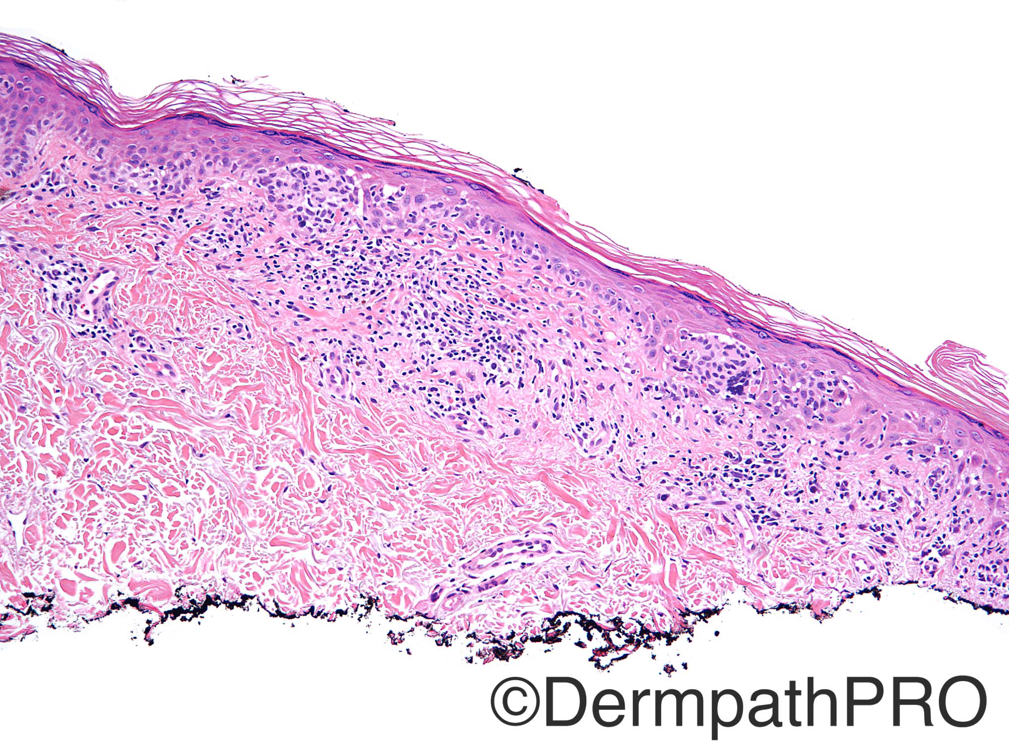
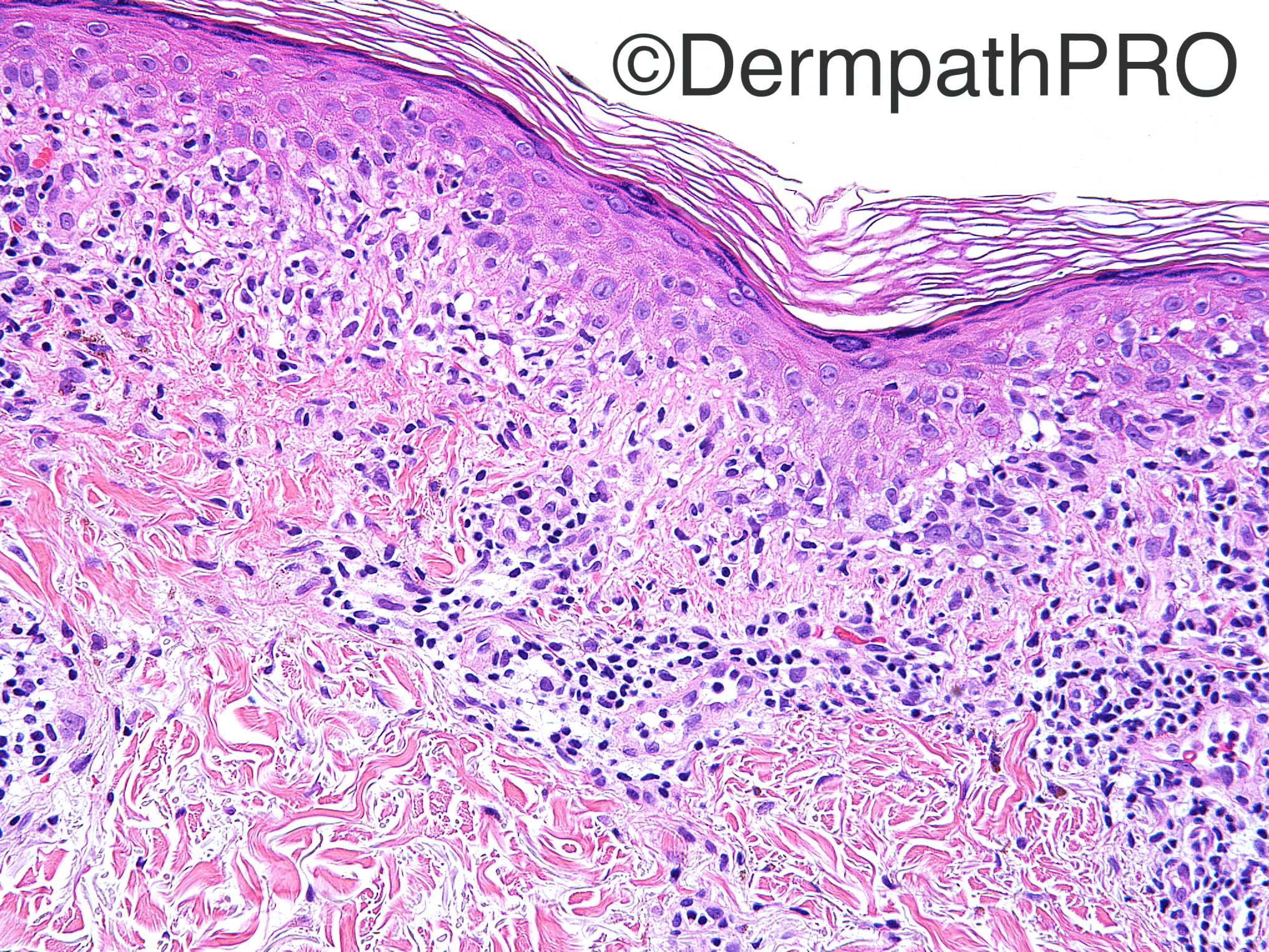
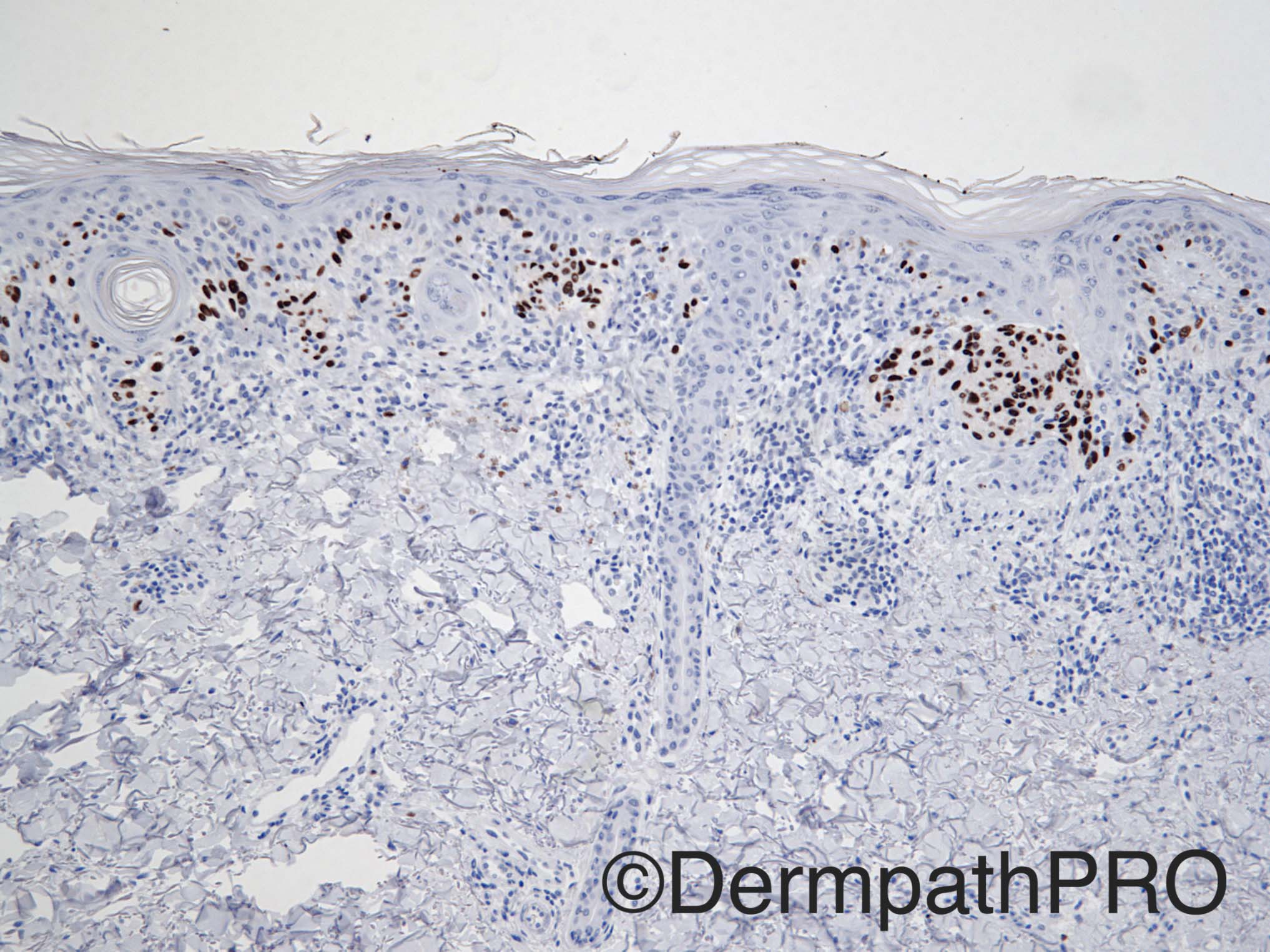
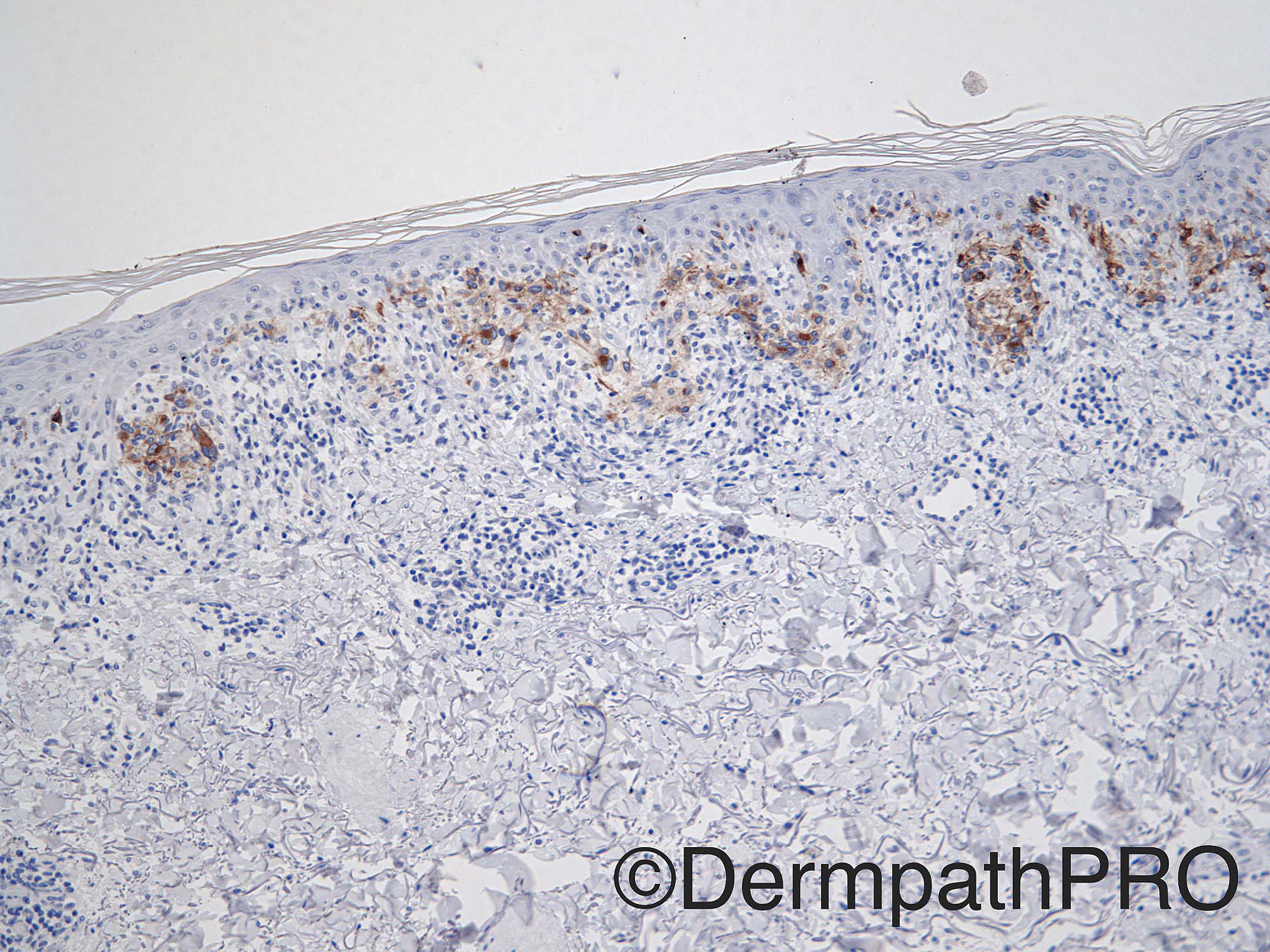
Case History: 66 year old male with 1.9 x 1.3 cm lesion on right mid back. Figure 8=Sox 10; Figure 9=Melan A.

Case posted by Dr Uma Sundram

  • Like 1
  Report Record

User Feedback


Raul Perret

Posted

The lesion seems to be quite extensive, both with nested and individual mélanocytes. Slight confluence of nests and focal extension to the adnexae. The neoplastic cells are moderately atypical with presence of small nucleoli. A conspicuous lichenoid lymphocytic infiltrate altogether with the clinical setting makes me favour melanoma in situ.

Share this comment


Link to comment
share_externally

vincenzo polizzi

Posted

I see a lichenoid regression adiacent to malignant melanoma in situ and a  transition of malignant melanoma to lichenoid tissue reaction with loss of melanocytes at the dermal-epidermal junction in the lichenoid reaction. I favour a regressing early invasive melanoma

Share this comment


Link to comment
share_externally

Raul Perret

Posted

I agree that some of the criteria of regression are present but to my eyes the lesion is completely in situ. I would like to know how do the expert dermatopathologists diagnose lesions like this one, do you add a comment stating that areas of regression cannot be ruled out?

Share this comment


Link to comment
share_externally

vincenzo polizzi

Posted

i'm a general pathologist, not at all an expert dermatipathologist, but i see a dermal scarring in first image. 

Share this comment


Link to comment
share_externally

Nitin Khirwadkar

Posted

Atypical junctional melanocytic proliferation, with focal regression (dermal fibrosis, vascular proliferation and inflammation). Overall, in-situ malignant melanoma. Is this an incisional biopsy?

Share this comment


Link to comment
share_externally

Raul Perret

Posted

In a lesion like this in your usual practice do you stage it as a pTx or a pTis?. Based on the CAP we should stage it as pTx right? (and the surgeon and patient will hate us)

Share this comment


Link to comment
share_externally

Dr. Richard Carr

Posted

It looks like a shave biopsy.  I think you are right to consider the possibility of a lichenoid reaction to melanoma in situ but the location and predominant nesting suggest we should also consider a junctional dysplastic naevus.  The small focus of scarring could be secondary (e.g. to follicle rupture, previous non-diagnostic biopsy, treatments or local trauma etc. etc.  As an aside, in addition to the features mentioned by Nitin I like to see a break in the dermal component (not relevant here). I would examine multiple levels but assuming this is representative leave the diagnosis slightly open and advise complete excision with clear margins.

Share this comment


Link to comment
share_externally

Uma Sundram

Posted

This was a tough case but we thought we could not exclude at least melanoma in situ with a lichenoid background.  The lichenoid nature of the lesion precluded estimation of true biological potential; therefore, early invasive melanoma could not be excluded. As this was a shave biopsy, we recommended complete excision with margins appropriate for melanoma in situ, and a caveat that the lesion may act in a more aggressive fashion than the typical MMIS. We would stage as at least pTis and clarify in the body of the report that the presence of dense inflammation complicates our ability to accurately determine depth.

Share this comment


Link to comment
share_externally

Raul Perret

Posted

Thank you Doctors for you useful comments. Great case

Share this comment


Link to comment
share_externally


Join the conversation

You can post now and register later. If you have an account, sign in now to post with your account.

Guest
Add a comment...

×   Pasted as rich text.   Paste as plain text instead

  Only 75 emoji are allowed.

×   Your link has been automatically embedded.   Display as a link instead

×   Your previous content has been restored.   Clear editor

×   You cannot paste images directly. Upload or insert images from URL.

Loading...

×
×
  • Create New...