Jump to content
In this section we have spot diagnoses posted on a daily basis since June 2010, now over 4000! You can review the archived cases and read the suggested diagnoses by users and the final comment by the contributors.
Case are uploaded each week day by 10 am UK time with the correct diagnosis will generally be posted at 8 pm UK time. Why not view the most recent spot diagnosis and proffer a diagnosis?

Case Number : Case 1475 -18 February Posted By: Guest

Please read the clinical history and view the images by clicking on them before you proffer your diagnosis.
Submitted Date :
   (0 reviews)

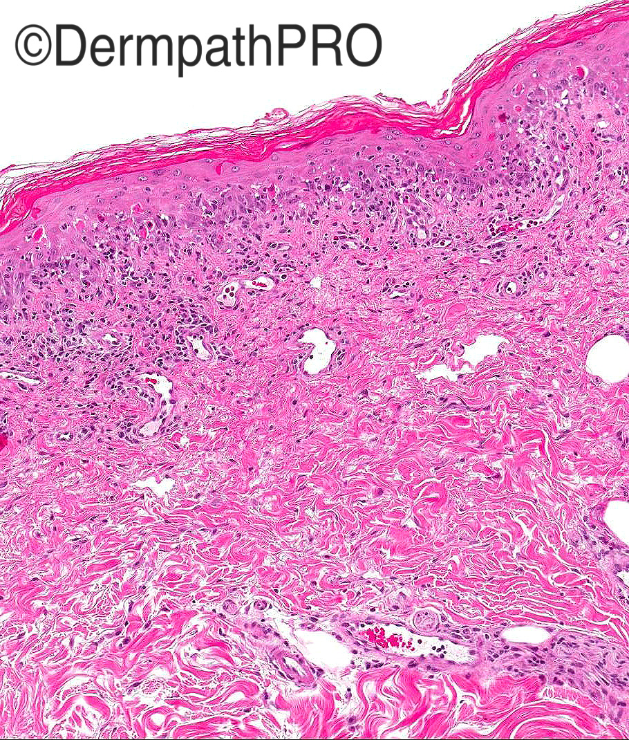
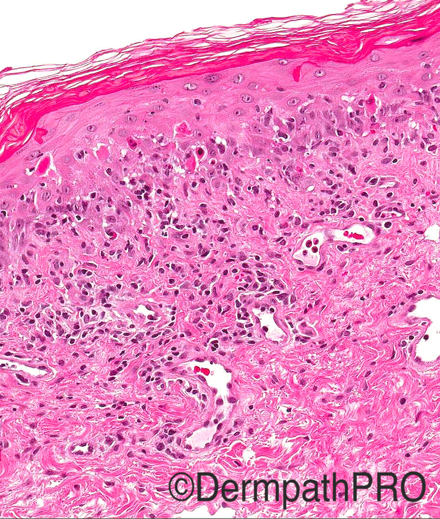
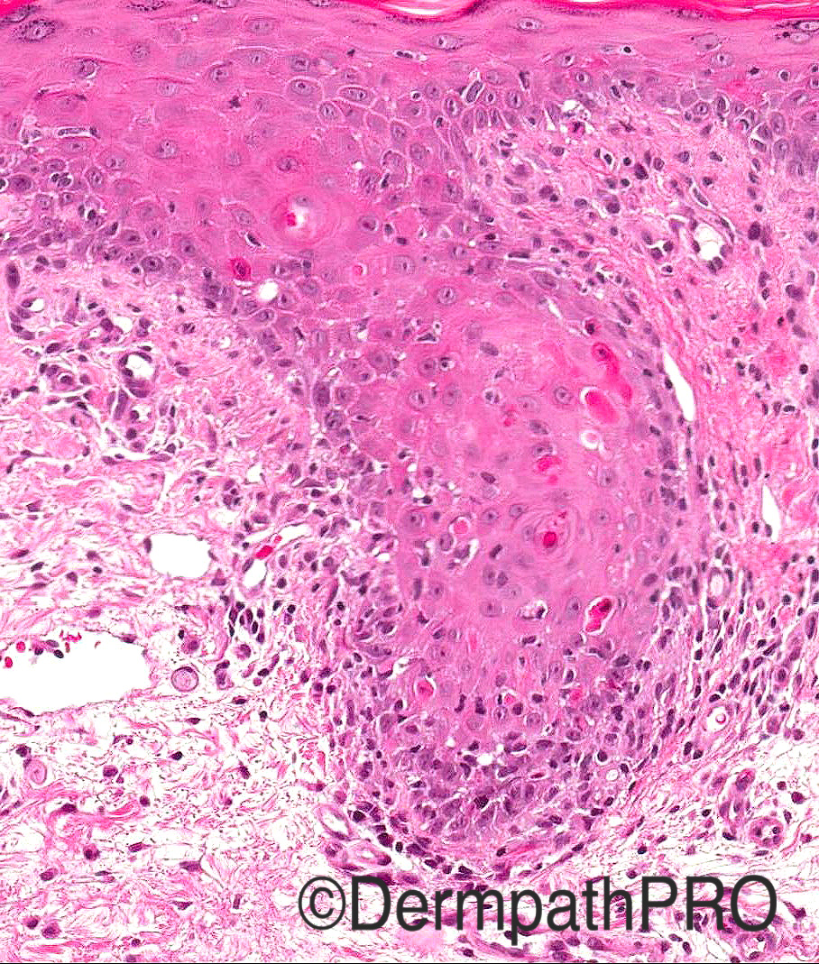
Case History: 70/F with a history of retroperitoneal mass and rapidly progressing, polymorphic blistering rash on limbs and trunk. Oral erosions present

Case posted by Dr Arti Bakshi


  Report Record

User Feedback


Raul Perret

Posted

Early toxic epidermal necrolysis. To my eyes there is an overlap in this photos of TEN with EM but i favour the first one.Probably the retroperitoneal mass is a lymphoma, there are associations between TEN and lymphomas, tough it would be important to know if the patient recieved medications at the moment of the rash including chemotherapy.

Share this comment


Link to comment
share_externally

Dr. Mona Abdel-Halim

Posted

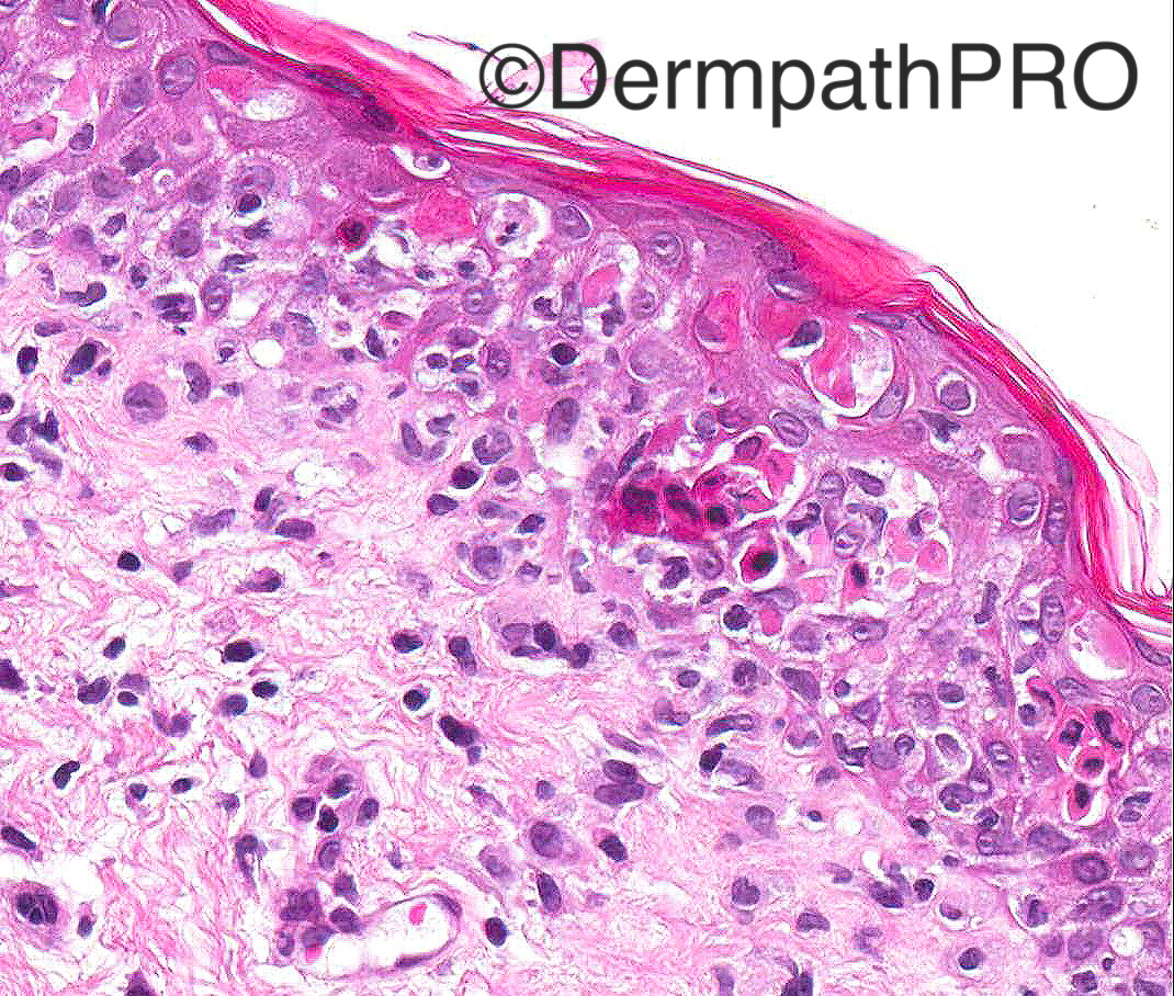
Ok looks EM like, but clinically due to the presence of retriperitoneal mass, one should not forget paraneoplastic pemphigus, may present with EM like pucture. The suprabasal acantholysis can be very focal. DIF is needed as well as serum detection of auto antibodies against multiple antigens in the same patient. According to Weedon, one can not find the suprabasal acantholysis even on multiple sectioning..

Share this comment


Link to comment
share_externally

Raul Perret

Posted

That is a good suggestion Mona. On morphology alone we would not be able to differentiate these lesions

Share this comment


Link to comment
share_externally

vincenzo polizzi

Posted

Agree, although i favour Erythema Multiforme actually, because of focal, not confluent epidermal necrosis. 

Share this comment


Link to comment
share_externally

Guest Romualdo

Posted

I think hyperkeratosis speaks against erythema multiforme and TEN. I go with paraneoplastic pemphigus.

Share this comment


Link to comment
share_externally

biswanath behera

Posted

in Figure 4, probably single acantholytic cell present. 


From the history, probably this is a case of Paraneoplastic pemphigus.


IF will help.

Share this comment


Link to comment
share_externally

Arti Bakshi

Posted

Yes, this is a case of Paraneoplastic Pemphigus.

The Direct IMF showed intercellular IgG and linear C3 along dermo-epidermal junction. (this combination being typical of PNP). The retroperitoneal mass was a sarcoma with lung mets.

Mona has already summarised the key features well, in particular that one may not find any acantholysis at all. It is important to keep this entity in the back of your mind when faced with EM like histology as PNP may precede diagnosis of malignancy. Unfortunately, the disease is multi systemic and has a rapid downhill course with high mortality. (as was also the case in this patient).

Share this comment


Link to comment
share_externally


Join the conversation

You can post now and register later. If you have an account, sign in now to post with your account.

Guest
Add a comment...

×   Pasted as rich text.   Paste as plain text instead

  Only 75 emoji are allowed.

×   Your link has been automatically embedded.   Display as a link instead

×   Your previous content has been restored.   Clear editor

×   You cannot paste images directly. Upload or insert images from URL.

Loading...

×
×
  • Create New...