Jump to content
In this section we have spot diagnoses posted on a daily basis since June 2010, now over 4000! You can review the archived cases and read the suggested diagnoses by users and the final comment by the contributors.
Case are uploaded each week day by 10 am UK time with the correct diagnosis will generally be posted at 8 pm UK time. Why not view the most recent spot diagnosis and proffer a diagnosis?

Case Number : Case 1477 -22 February Posted By: Guest

Please read the clinical history and view the images by clicking on them before you proffer your diagnosis.
Submitted Date :
   (0 reviews)

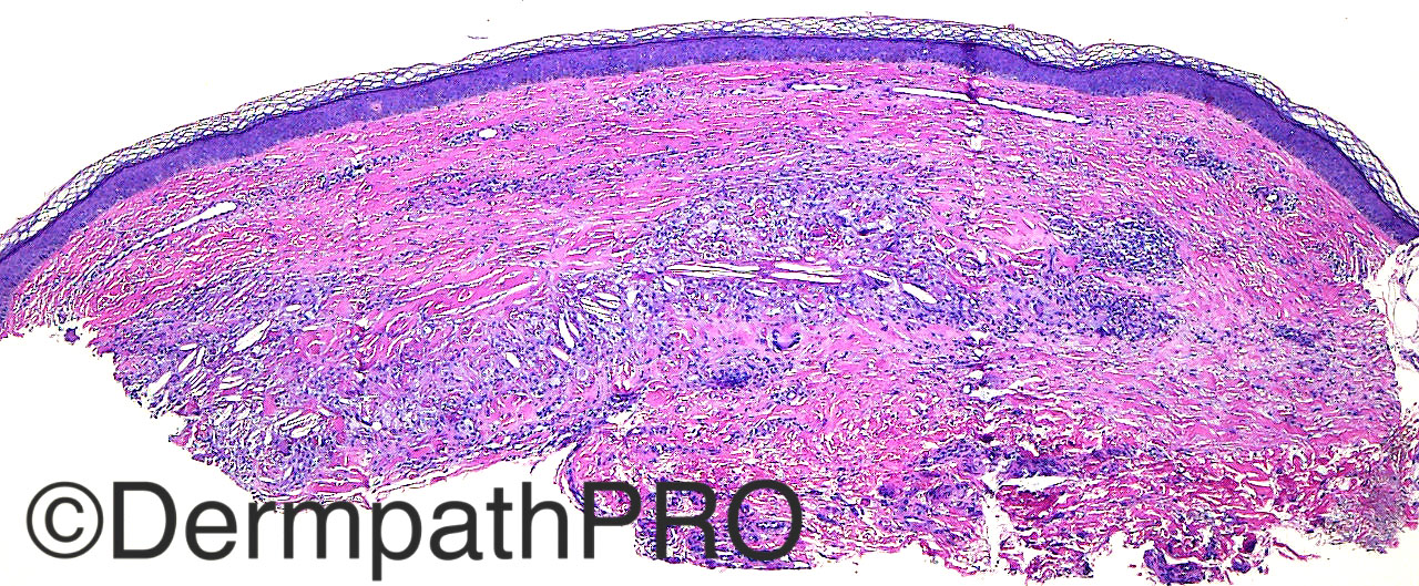
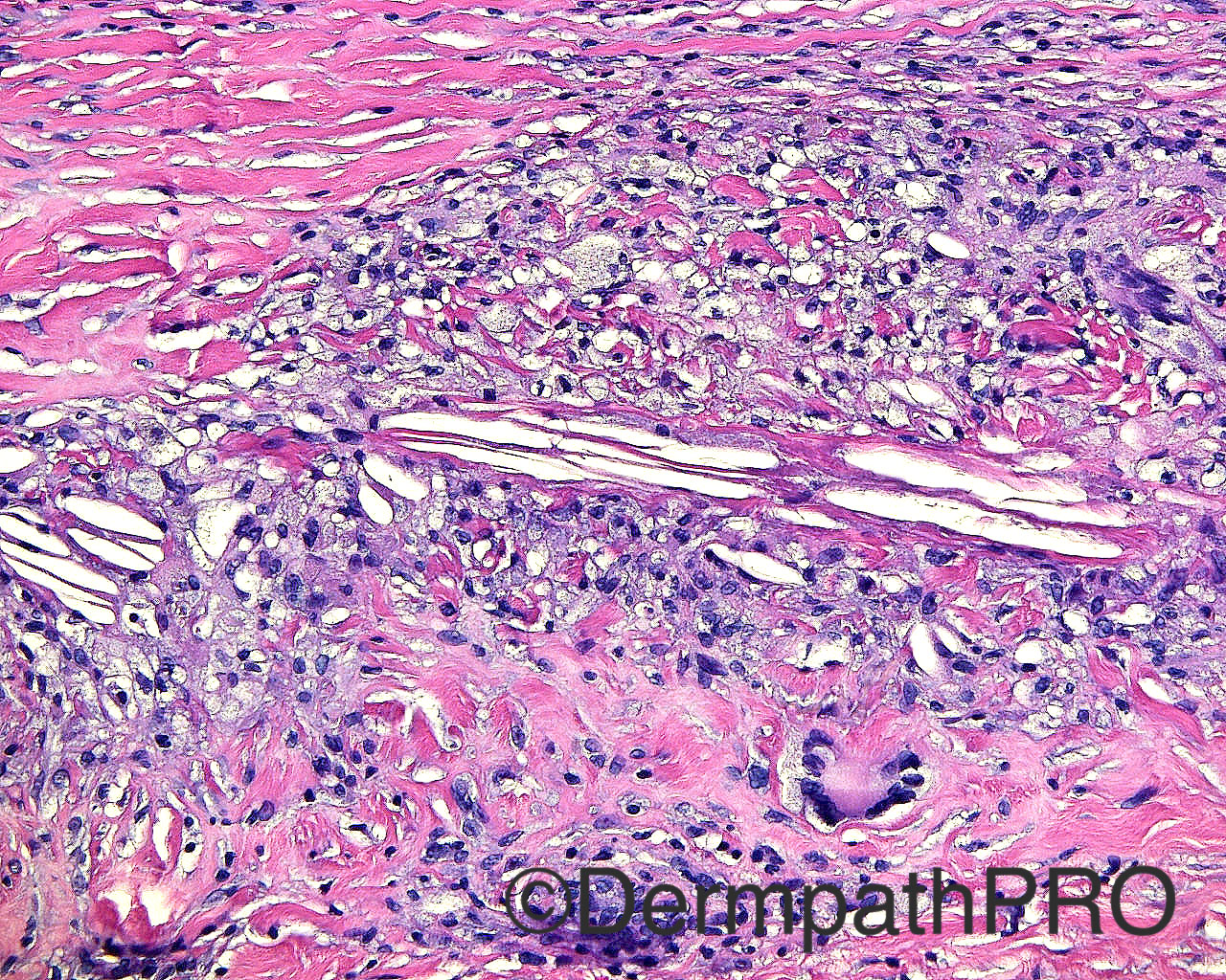
Case History: The patient is a 47 year old woman with a punch biopsy of a glistening, white, atrophic papule with telangiectasia taken from the left medial calf.

Case posted by Dr Mark Hurt


  Report Record

User Feedback


Raul Perret

Posted

xanthoma (flat xanthoma?) Dyslipidemia should be ruled out.

Share this comment


Link to comment
share_externally

Dr. Mona Abdel-Halim

Posted

Necrobiotic xanthogranuloma, atypical site though. Cholesterol clefts are characteristic together with the foamy cells and the Touton giant cells.

Share this comment


Link to comment
share_externally

vincenzo polizzi

Posted

Palisaded granulomatous collection of  Touton, foamy-cells and cholesterol clefting, with necrobiotic collagen. I favour Necrobiotic Xanthogranuloma. Paraproteinemia?

Share this comment


Link to comment
share_externally

Nitin Khirwadkar

Posted

Xanthogranuloma. Pondered about a lipidised DF (although not typical) and a cholesterol granuloma.

Share this comment


Link to comment
share_externally

Raul Perret

Posted

To be honest necrobiotic collagen is not readily apparent to my eyes and the biopsy is quite superficial (we dont see the hypodermis). I would be scared to diagnose necrobiotic xanthogranuloma in this case only based on histology

Share this comment


Link to comment
share_externally

Sasi Attili

Posted

Site typical for necrobiosis lipoidica. Histo more suggestive of NXG

Share this comment


Link to comment
share_externally

Arti Bakshi

Posted

Agree with Sasi that the site is suggestive of necrobiosis lipoidica. (as also presence of dermal fibrosis in a layered pattern).

Apparently one can get cholestrol clefts in necrobiosis lipoidica rarely http://www.ncbi.nlm.nih.gov/pubmed/3351060

Ofcourse clinical correlation is essential

Share this comment


Link to comment
share_externally

Dr. Mona Abdel-Halim

Posted

Sure the ddx should be NLD with the rare occurance of cholesterol clefts...

Share this comment


Link to comment
share_externally

vincenzo polizzi

Posted

Yes, the cholesterol clefts aren't strictly clues to NXG, because of rare occurrence in NLD, implicating diabetes mellitus with complications...but in img2 and img3 there are atypical and Touton type giant cells, so typical for NXG!

Share this comment


Link to comment
share_externally

Dr. Richard Carr

Posted

Mark,

Do you think the adipophilin in this case is following pattern 2 (both intracellular and extracellular after the distribution of palisaded or scattered histiocytes), according to Schulman & Leboit Am J Dermatopathol 2015; 37(3):203-9 (i.e. GA pattern), rather then the pattern 1 (extracellular, within zones of altered collagen) seen in NL.

Share this comment


Link to comment
share_externally


Join the conversation

You can post now and register later. If you have an account, sign in now to post with your account.

Guest
Add a comment...

×   Pasted as rich text.   Paste as plain text instead

  Only 75 emoji are allowed.

×   Your link has been automatically embedded.   Display as a link instead

×   Your previous content has been restored.   Clear editor

×   You cannot paste images directly. Upload or insert images from URL.

Loading...

×
×
  • Create New...