Jump to content
In this section we have spot diagnoses posted on a daily basis since June 2010, now over 4000! You can review the archived cases and read the suggested diagnoses by users and the final comment by the contributors.
Case are uploaded each week day by 10 am UK time with the correct diagnosis will generally be posted at 8 pm UK time. Why not view the most recent spot diagnosis and proffer a diagnosis?

Case Number : Case 1478 -23 February Posted By: Guest

Please read the clinical history and view the images by clicking on them before you proffer your diagnosis.
Submitted Date :
   (0 reviews)

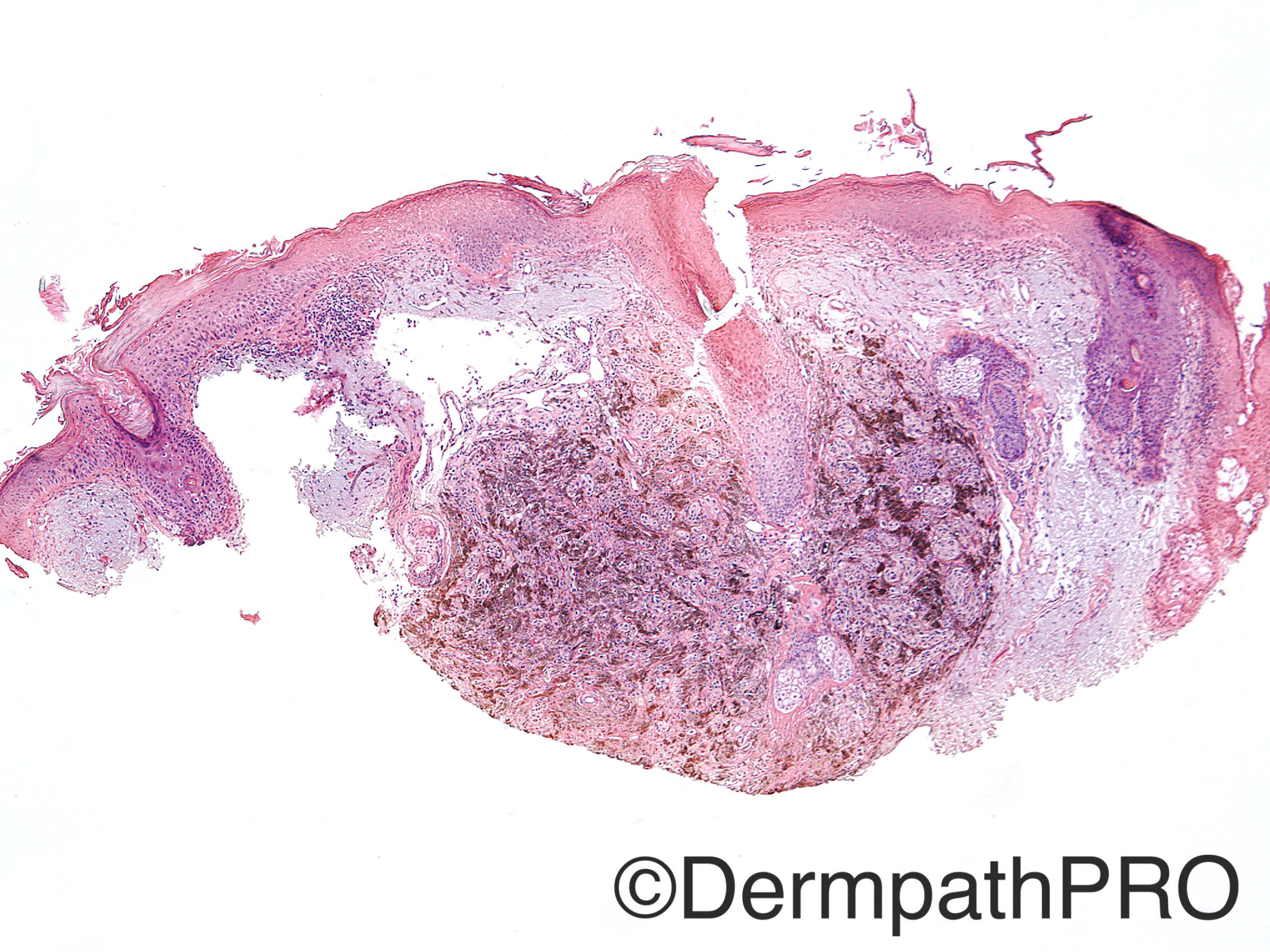
Case History: 9 year-old female with biopsy of scalp lesion.

Case posted by Dr Uma Sundram


  Report Record

User Feedback


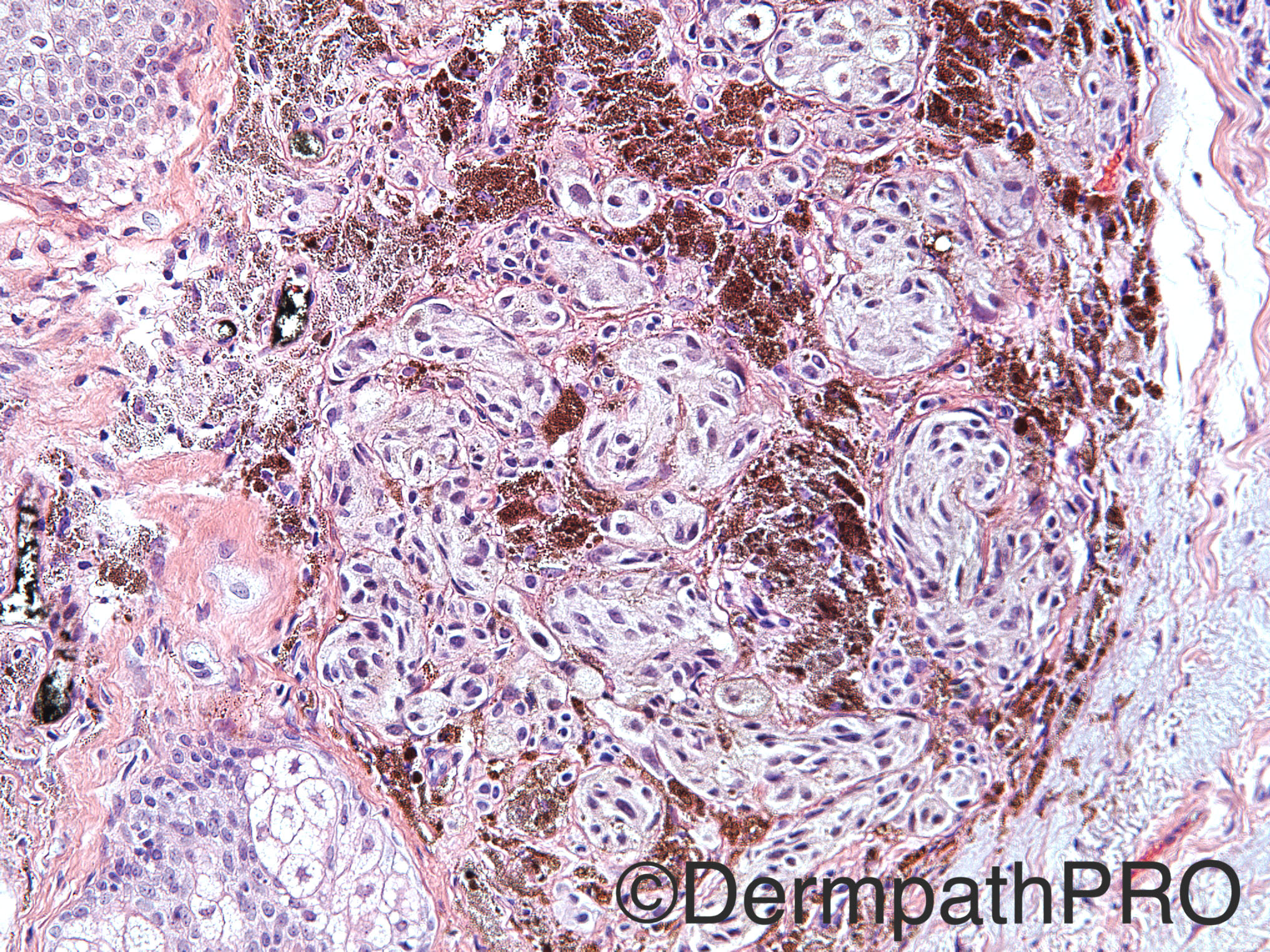
Epithelioid blue nevus. There´s a odd basophilic colagen that looks like solar elastosis. If this is a solar elastosos, maybe the age of the patient is wrong or the slides are from other patient. If this blue colagen in fact is not colagen, but a myxoid matrix, this may be an associated myxoma in the context of the Carney complex.

Share this comment


Link to comment
share_externally

Arti Bakshi

Posted

Cellular Blue naevus.

Agree with Igor's comment about solar elastosis.

Share this comment


Link to comment
share_externally

Nitin Khirwadkar

Posted

Cellular blue naevus. Yes, the amount of solar elastosis is certainly odd for the age!

Share this comment


Link to comment
share_externally

Guest Romualdo

Posted

In an epithelioid blue nevus I would expect marked pigmented epithelioid melanocytes. I think this biphasic pattern, with nests of pale cells compartmentalized by septa rich in melanophages is more in keeping with a cellular blue nevus.

Share this comment


Link to comment
share_externally

Raul Perret

Posted

Agree with the blue nevus type of lesions, if someone called this either deep penetrating nevus or cellular blue nevus I would accept it, I think there is morphological support for either of these lesions. From my point of view it is myxoid change of the stroma that we see. Bothers me that the deep limits are compromised....

Share this comment


Link to comment
share_externally

vincenzo polizzi

Posted

I see an entirely intradermal melanocytic lesion below an elastotic actinic wrap, with two cellular populations: one with non nested heavy pigmented spindle or round cells; and the other with nested less pigmented, epithelioid melanocytes with clear large cytoplasm. Some cell show a egg-fried appearance...I favor an  Epithelioid Blue Nevus.

Share this comment


Link to comment
share_externally

Guest Tiberiu Tebeica

Posted

Cellular blue nevus. I also found the blueish look of the dermis odd...

Share this comment


Link to comment
share_externally

Uma Sundram

Posted

Hi everyone, for some reason the wrong history was uploaded. Here is the correct history:

74 year old man with a pigmented lesion of the left nose clinically thought to be a blue nevus. I'll leave it up for one more day to see if anyone has any new thoughts before posting our diagnosis.

My apologies for the tardy update--bad winter storm in Michigan has everyone going crazy!

Share this comment


Link to comment
share_externally

Dr. Richard Carr

Posted

I was thinking DPN on the original history (I was not sure it was elastosis or mucin!) now I want to see a melanA to rule out lentigo maligna. I would express a some uncertainty and have it fully excised.  Did not think it had the classical cytology of epithelioid blue / pigmented epithelioid melanocytoma / pigment synthesising melanoma / low grade hypermelanotic dermal melanoma etc. so I would probably say DPN-like lesion of uncertain malignant potential but favouring benign behaviour.

Share this comment


Link to comment
share_externally

Uma Sundram

Posted

Given the age of the patient, we too were very cautious about the diagnosis. Our final diagnosis was atypical melanocytic proliferation with deep penetrating nevus-like features; we asked that the lesion be fully excised. No residual was present on the re-excision and the patient is currently being followed. We did not do any further stains as the biopsy was very small.

Share this comment


Link to comment
share_externally


Join the conversation

You can post now and register later. If you have an account, sign in now to post with your account.

Guest
Add a comment...

×   Pasted as rich text.   Paste as plain text instead

  Only 75 emoji are allowed.

×   Your link has been automatically embedded.   Display as a link instead

×   Your previous content has been restored.   Clear editor

×   You cannot paste images directly. Upload or insert images from URL.

Loading...

×
×
  • Create New...