Jump to content
In this section we have spot diagnoses posted on a daily basis since June 2010, now over 4000! You can review the archived cases and read the suggested diagnoses by users and the final comment by the contributors.
Case are uploaded each week day by 10 am UK time with the correct diagnosis will generally be posted at 8 pm UK time. Why not view the most recent spot diagnosis and proffer a diagnosis?

Case Number : Case 1481-26 February Posted By: Guest

Please read the clinical history and view the images by clicking on them before you proffer your diagnosis.
Submitted Date :
   (0 reviews)

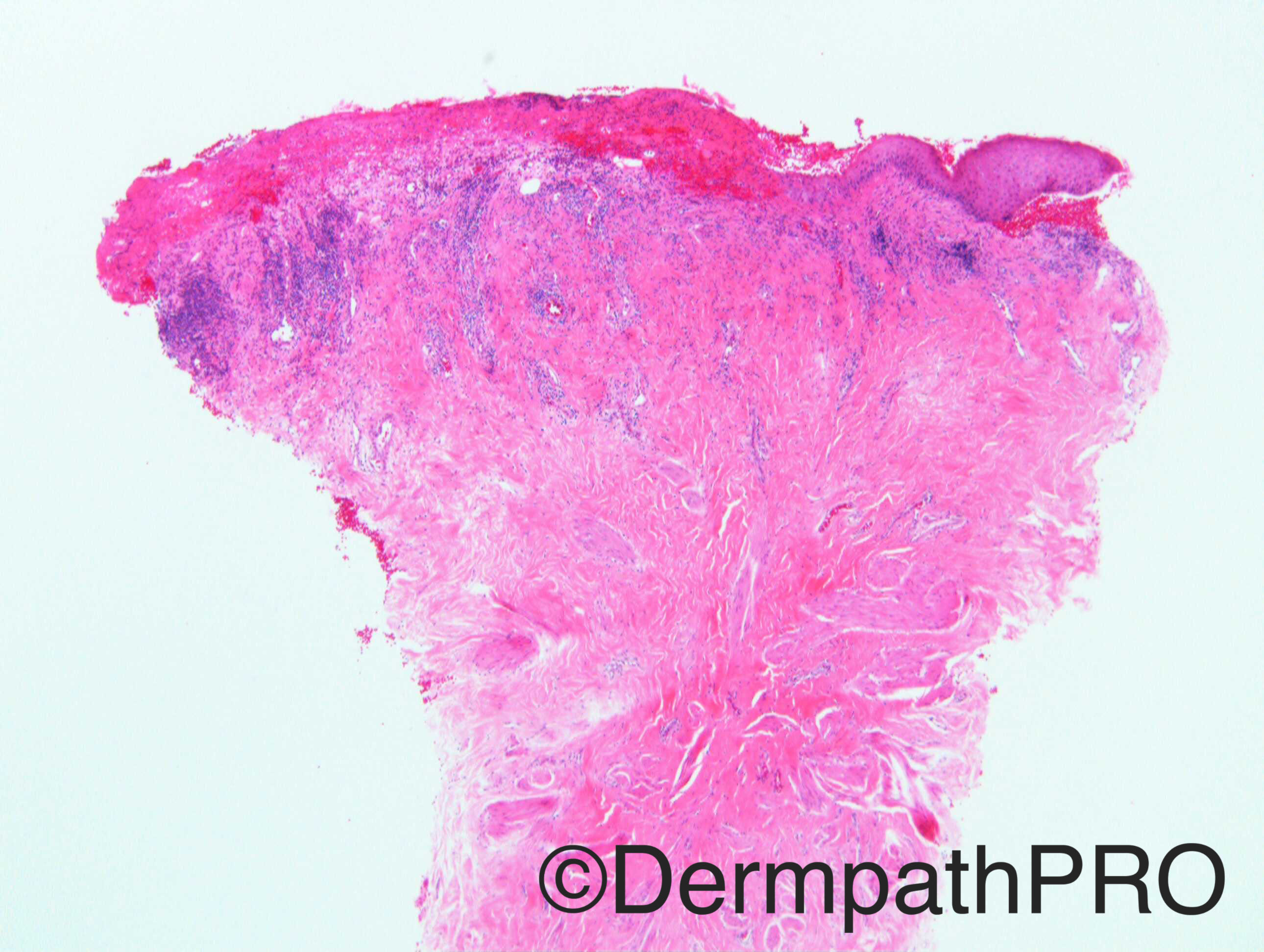
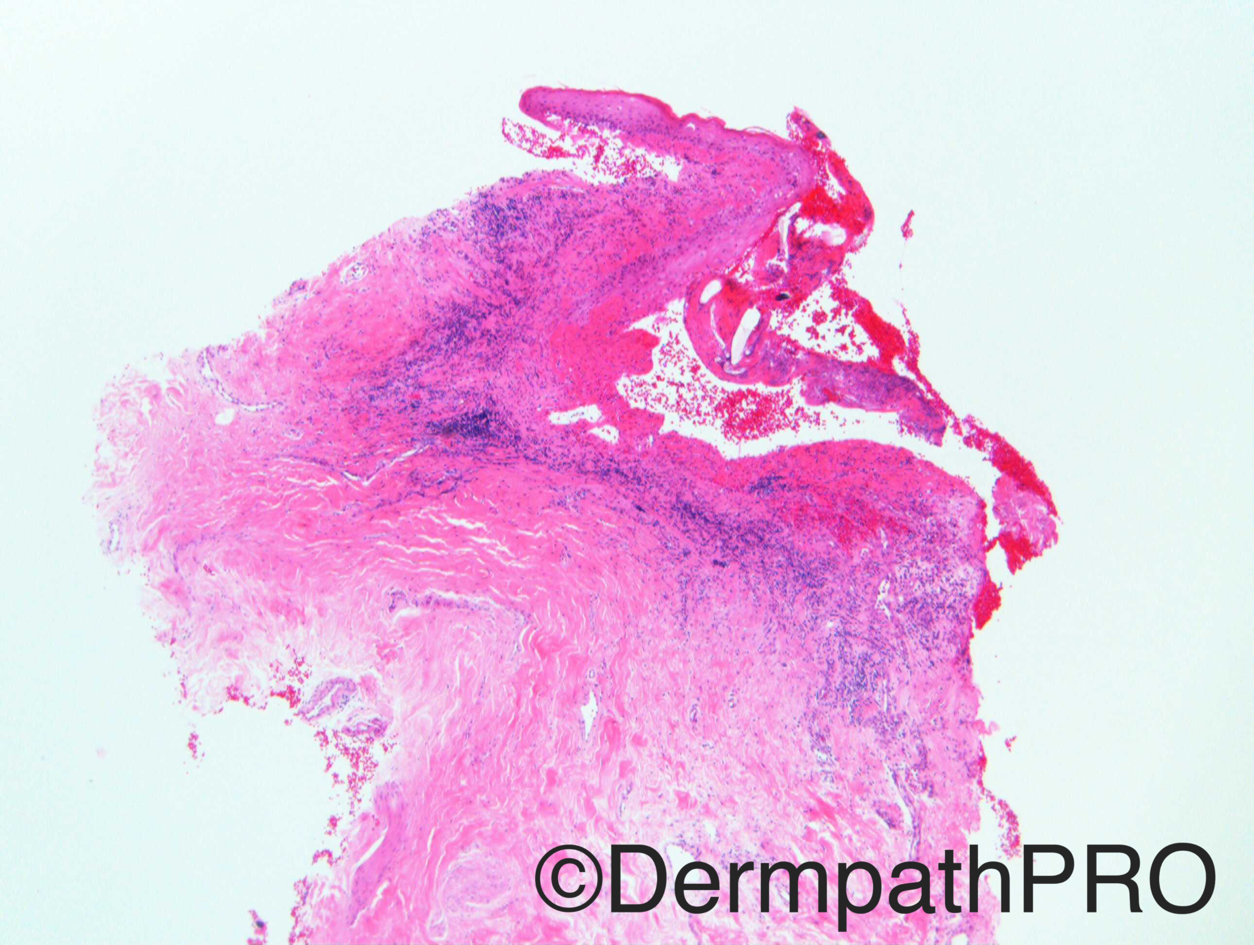
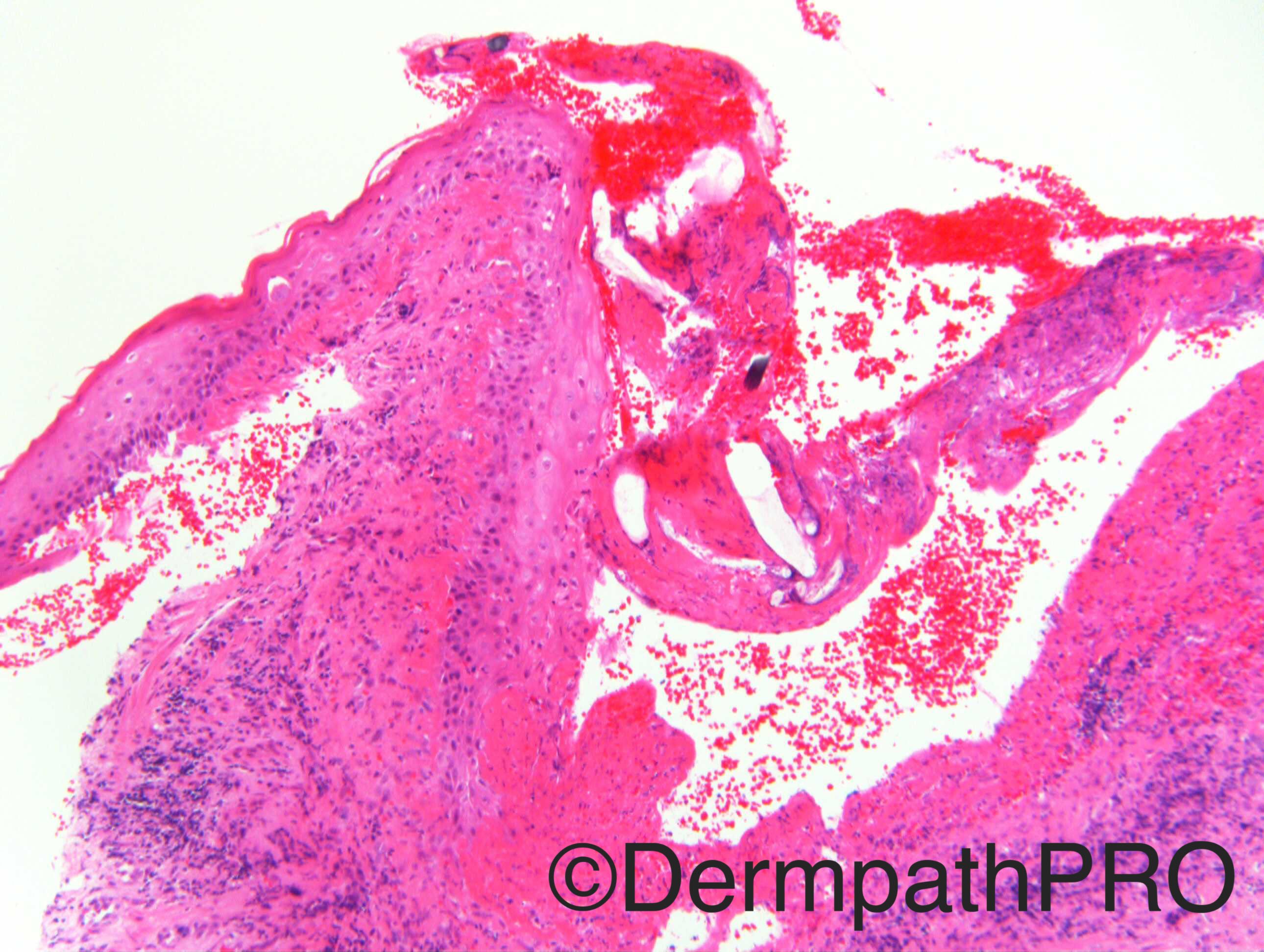
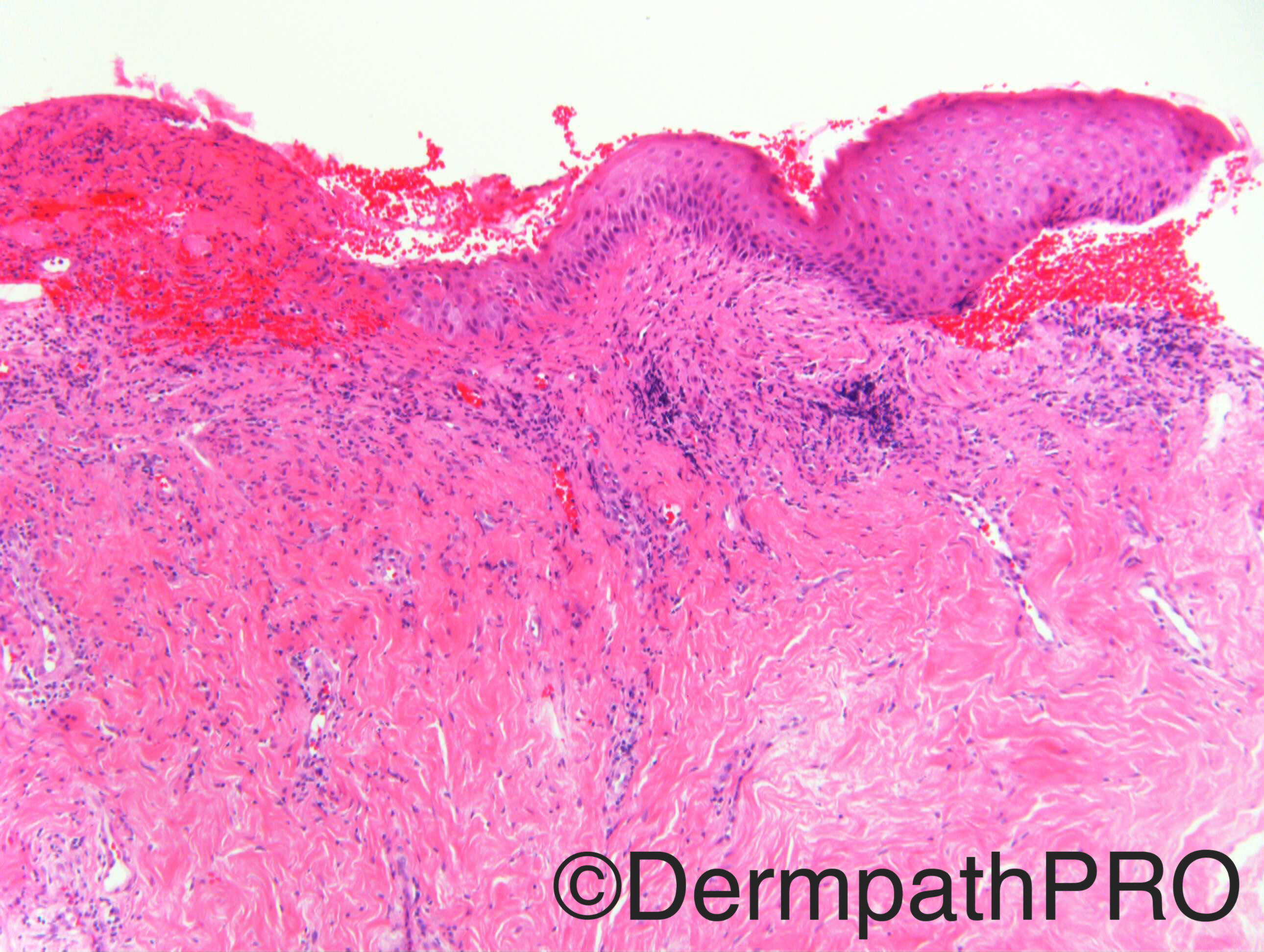
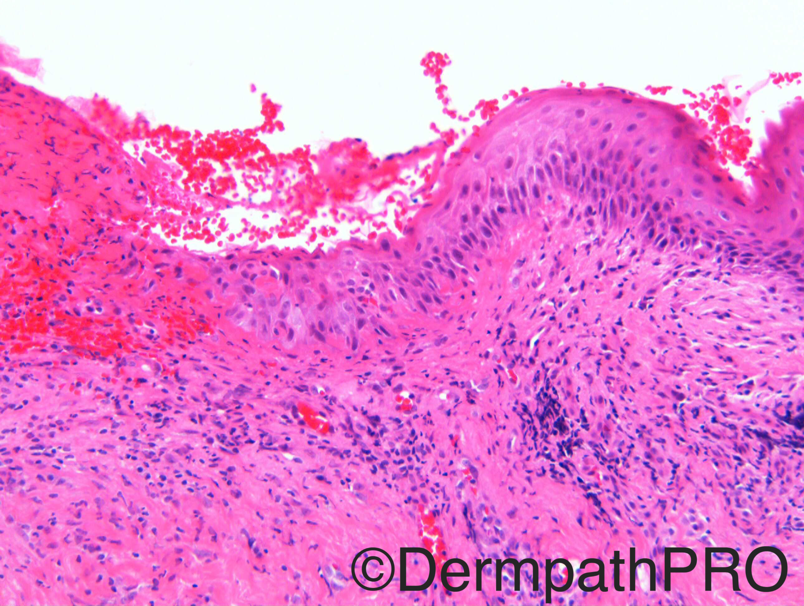
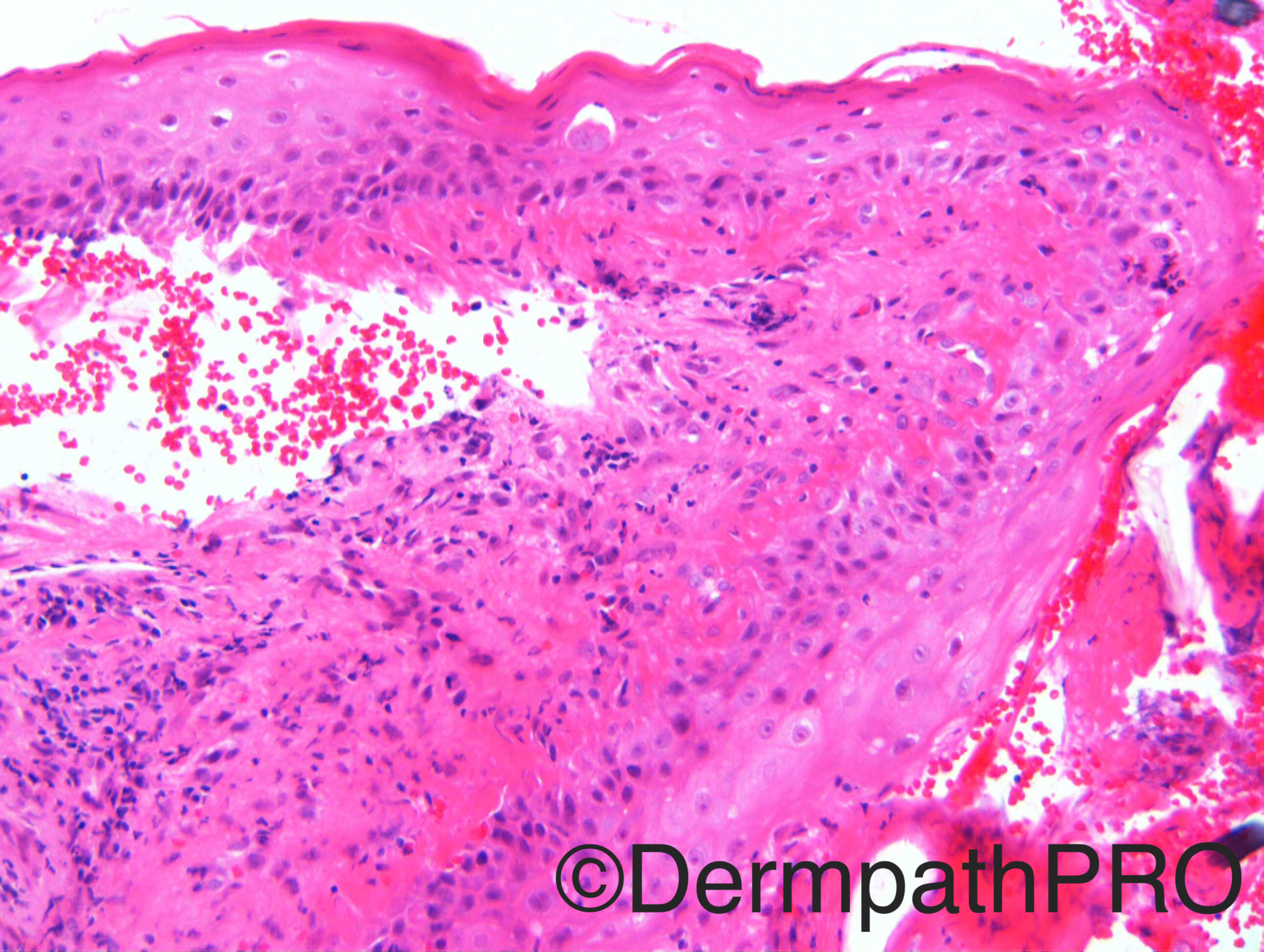
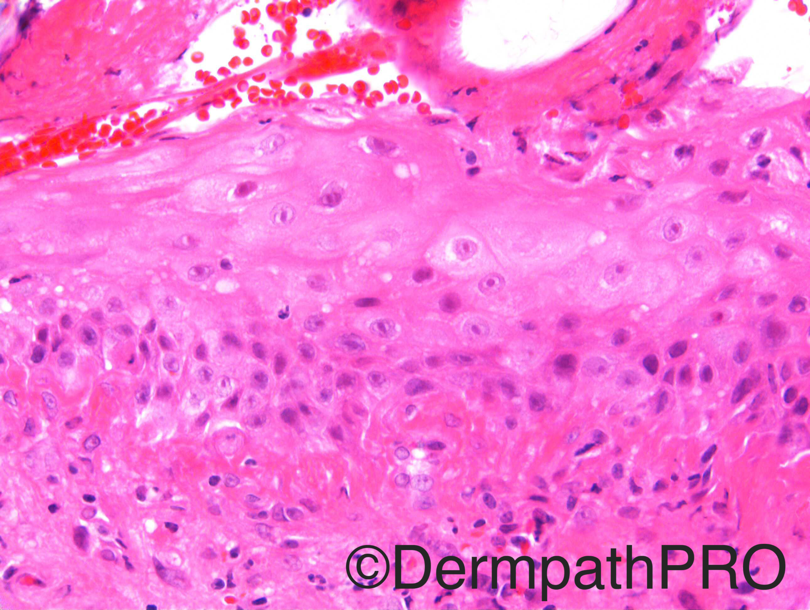
Case History: F60. Bleeding left nipple. ?Paget’s

Case posted by Dr Richard Carr

  Report Record

User Feedback


vincenzo polizzi

Posted

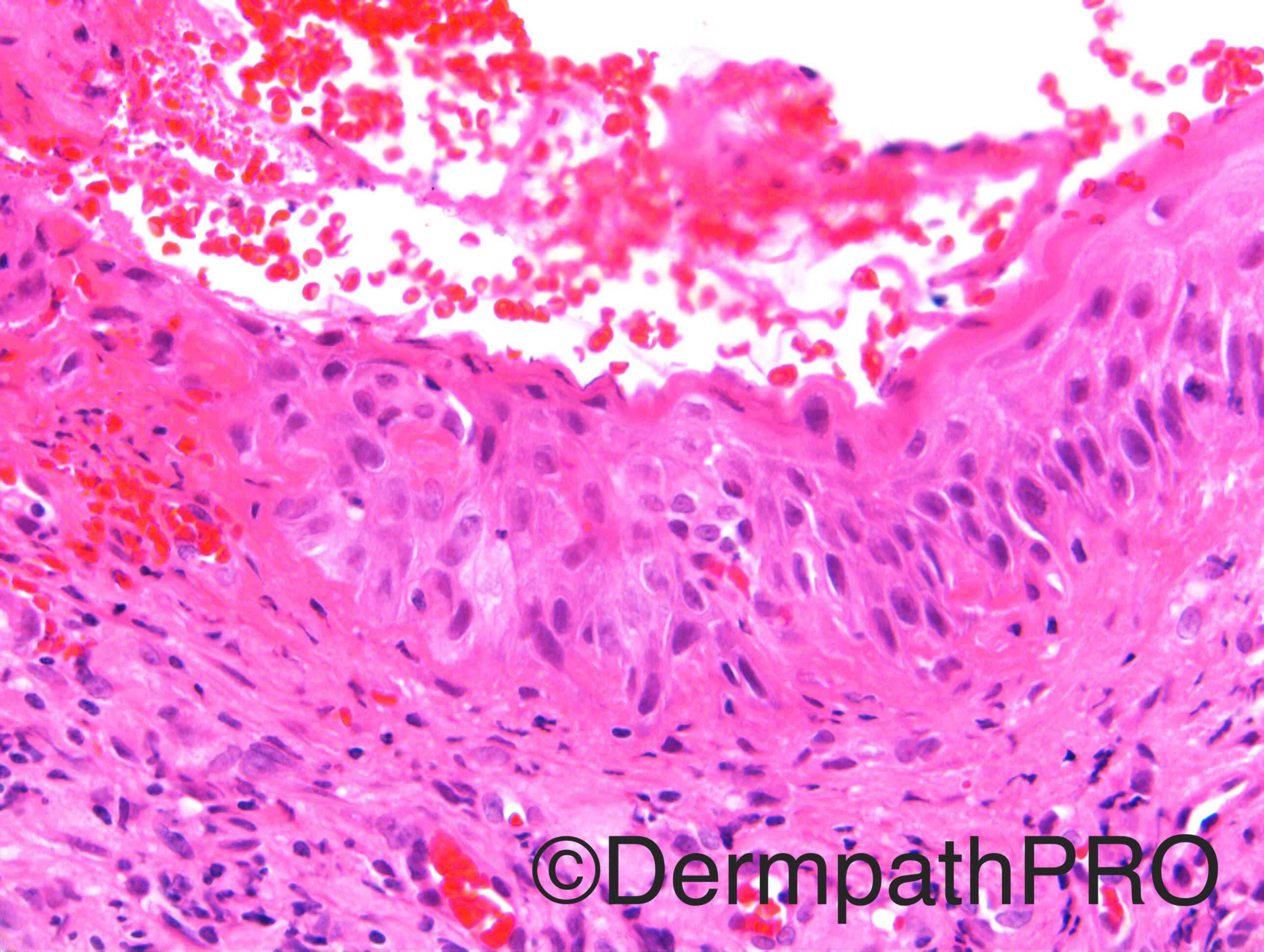
Nipple fibrino-emorragic ulceration, with reactive atypical epithelia cells ( do not look like pagetoid...). Radiation induced ulcer of the nipple?

Share this comment


Link to comment
share_externally

vincenzo polizzi

Posted

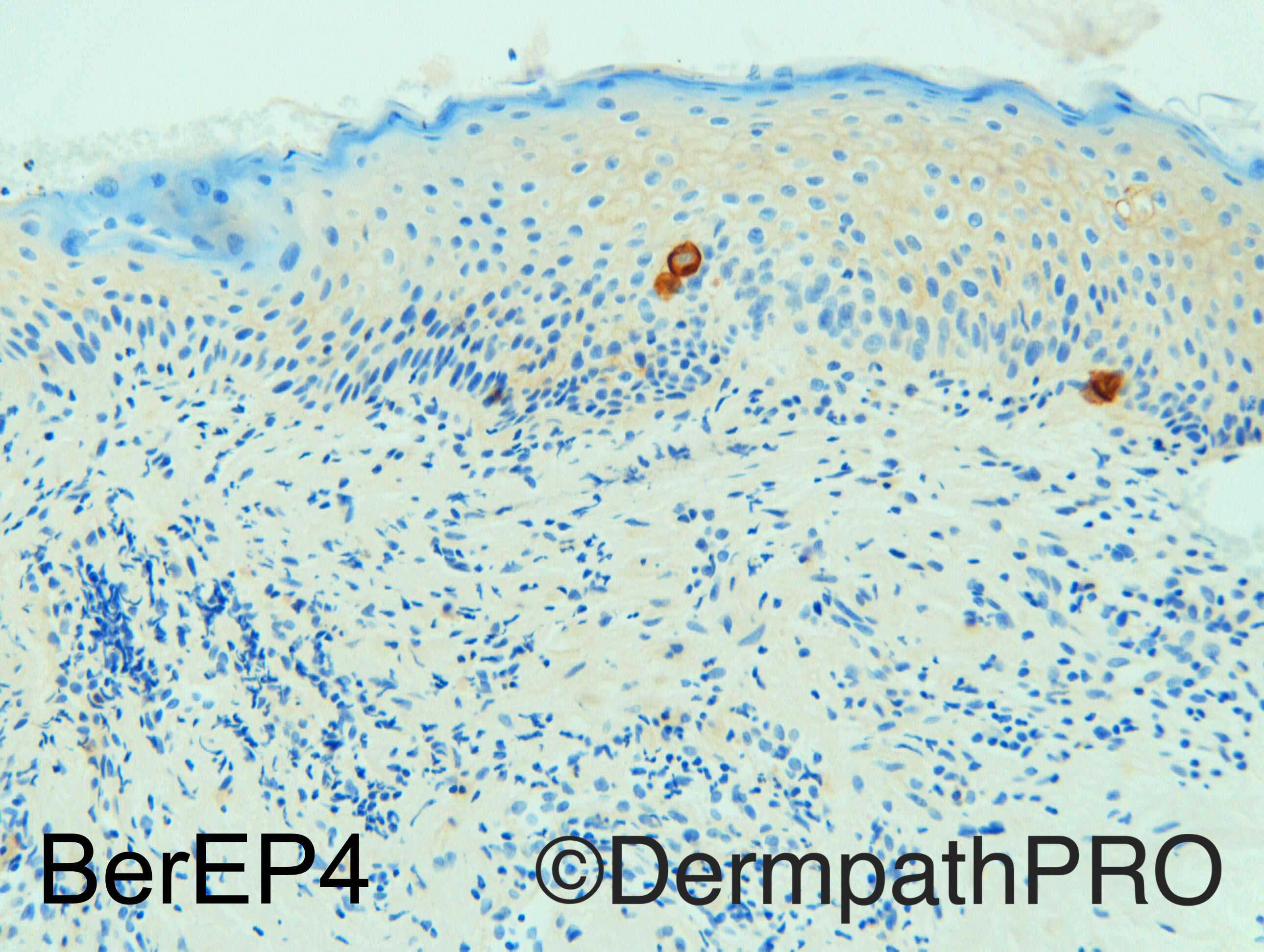
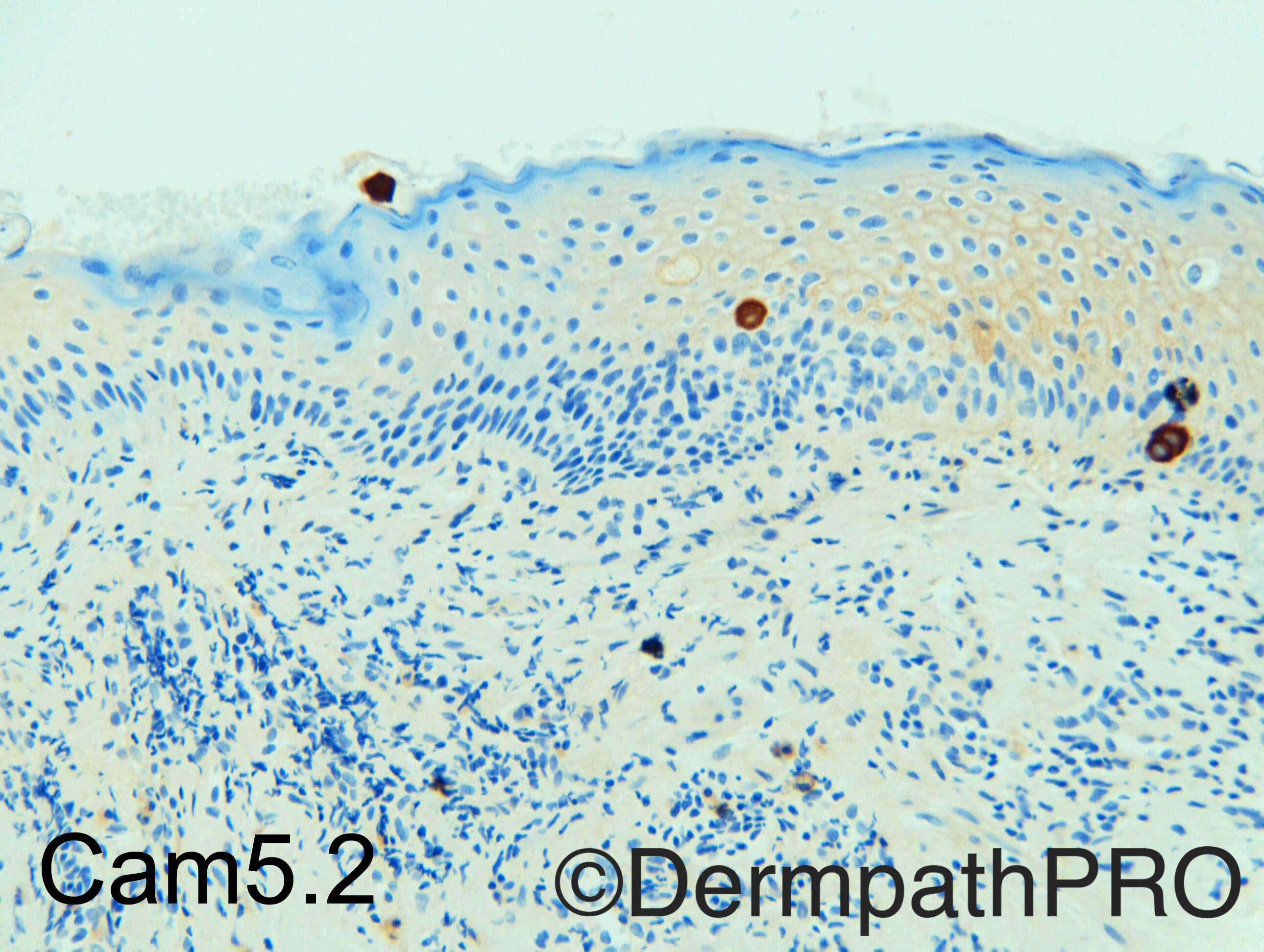
Luminal expression of Cam5.2 and CK7, isn't it?

Share this comment


Link to comment
share_externally

Raul Perret

Posted

Those are Toker cells highlighted by the IHQ, it would be interesting to include HER2neu and or polyclonal CEA as those markers are positive in Paget's (I dont think there is need of doing them in this case).

Share this comment


Link to comment
share_externally

Raul Perret

Posted

Yes it could be radiation induced ulcer/dermatits if the clinical fits, that is a good suggestion

Share this comment


Link to comment
share_externally

Dr. Mona Abdel-Halim

Posted

I think Vincenzo's suggestion is good.

Share this comment


Link to comment
share_externally

Nitin Khirwadkar

Posted

I agree with Vincenzo's suggestion of radiation induced ulceration. However, it is a tricky case. Both Toker cells and Paget's show a similar immunoprofile (CK7+). Rarely Toker cells, can be Her2 neu+ (and Paget's can be negative), and can also express ER, and can show atypia!  CD138 can be helpful, along with p53 (positive in Paget's), and negative in Toker cells. I haven't personally used CD138. The age group is right for Paget's disease. Need correlation with radiology. Did she have history of radiation? I would be cautious, and recommend rebiopsy, after the ulcer heals. 

Share this comment


Link to comment
share_externally

Arti Bakshi

Posted

Yes, Toker cells and Paget's show similar immunoprolfie, but morphologically this is not Paget's. Also CAM 5.2 doe not show any positivity in the area of ulceration (1st image), only scattered cells in the adjacent epidermis; so would go with Toker cells.

The cause of the ulcer is uncertain in these images…radiation induced ulcer may fit, provided there is a history. Cant seen any radiation fibroblasts or typical vascular changes though!

Share this comment


Link to comment
share_externally

Dr. Richard Carr

Posted

This is a very difficult case.  I gave you the exact information that came with the request but subsequent enquiry revealed there was a previous history of contralateral mastectomy for extensive DCIS and ipsilateral WLE for DCIS with radiotherapy that caused "significant radiation burns". These two punch biopsies were initially reported as showing ulceration with abundant foreign material within the exudate. No evidence of neoplasia.


At MDM review discussion mild spongiosis was noted. She was treated with potent topical steroids. On review at 3 months the area was larger and remained suspicious given the past medical history.  Patient was very reluctant to undergo another biopsy but finally acquiesced and I received the specimen which showed florid Paget's disease.  I then reviewed the prior biopsies which in my opinion showed unexplained ulceration, quite striking and suspicious cellular atypia leading me to suspect subtle Paget's disease.  The immunostains showed a few clusters and individual cells but the majority of atypical cells appeared to be compatible with reactive keratinocytes.  In my opinion the initial biopsies should have been reported as showing ulceration of undetermined cause i.e. non-diagnostic and additional levels & immunostains performed. I suspect this is Paget's and not just Toker cell's but it is hard to be definitive.


Learning Points:

As a general rule be very suspicious of nipple biopsies and have a low threshold for deeper levels and immunostains, try to be fully cognizant of the complete clinical history.

Share this comment


Link to comment
share_externally


Join the conversation

You can post now and register later. If you have an account, sign in now to post with your account.

Guest
Add a comment...

×   Pasted as rich text.   Paste as plain text instead

  Only 75 emoji are allowed.

×   Your link has been automatically embedded.   Display as a link instead

×   Your previous content has been restored.   Clear editor

×   You cannot paste images directly. Upload or insert images from URL.

Loading...

×
×
  • Create New...