Jump to content
In this section we have spot diagnoses posted on a daily basis since June 2010, now over 4000! You can review the archived cases and read the suggested diagnoses by users and the final comment by the contributors.
Case are uploaded each week day by 10 am UK time with the correct diagnosis will generally be posted at 8 pm UK time. Why not view the most recent spot diagnosis and proffer a diagnosis?

Case Number : Case 1446- 8 January Posted By: Guest

Please read the clinical history and view the images by clicking on them before you proffer your diagnosis.
Submitted Date :
   (0 reviews)

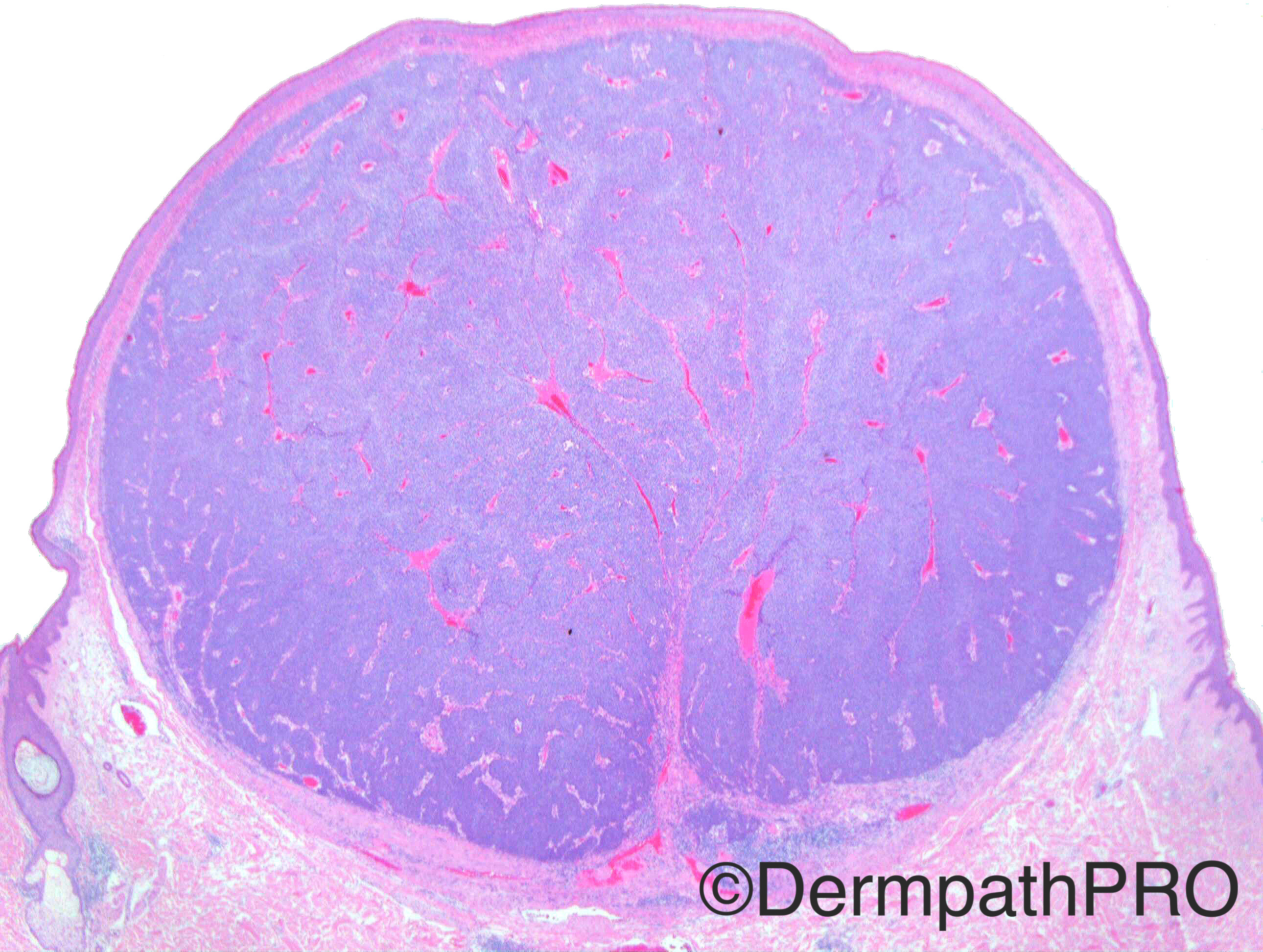
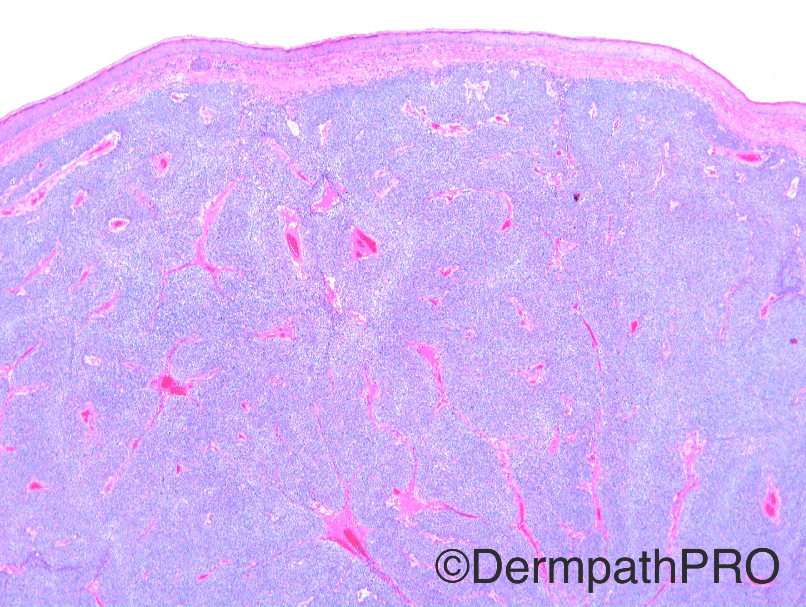
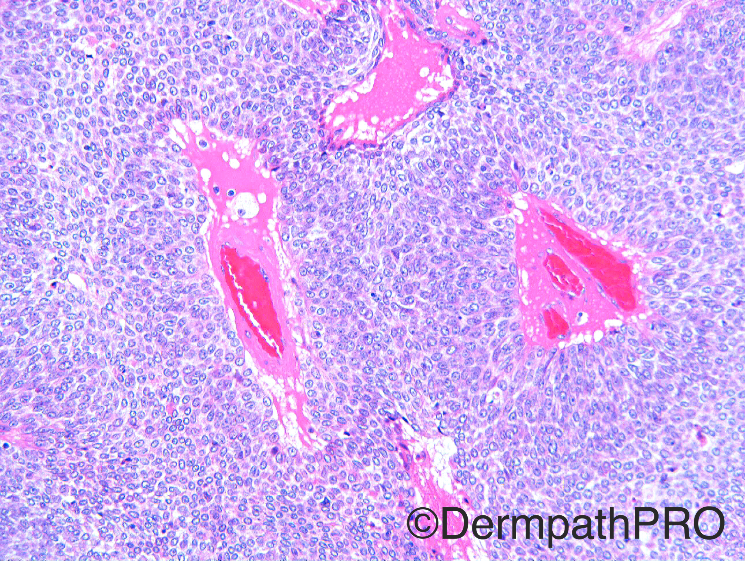
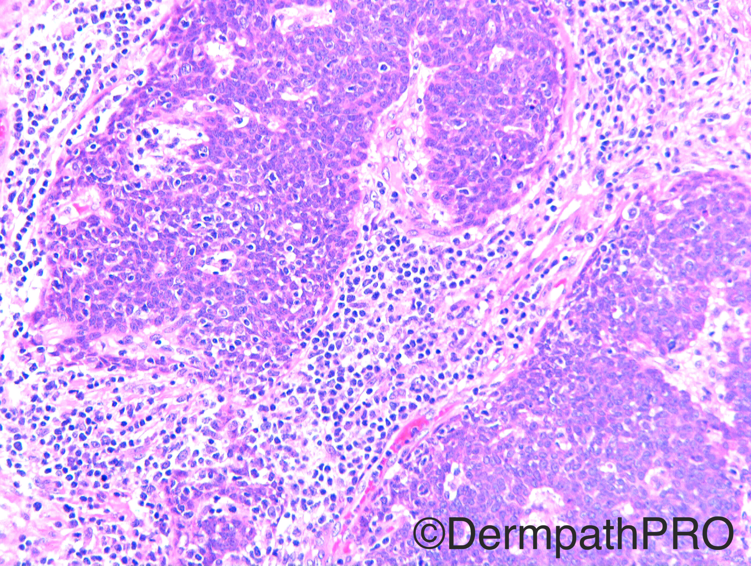
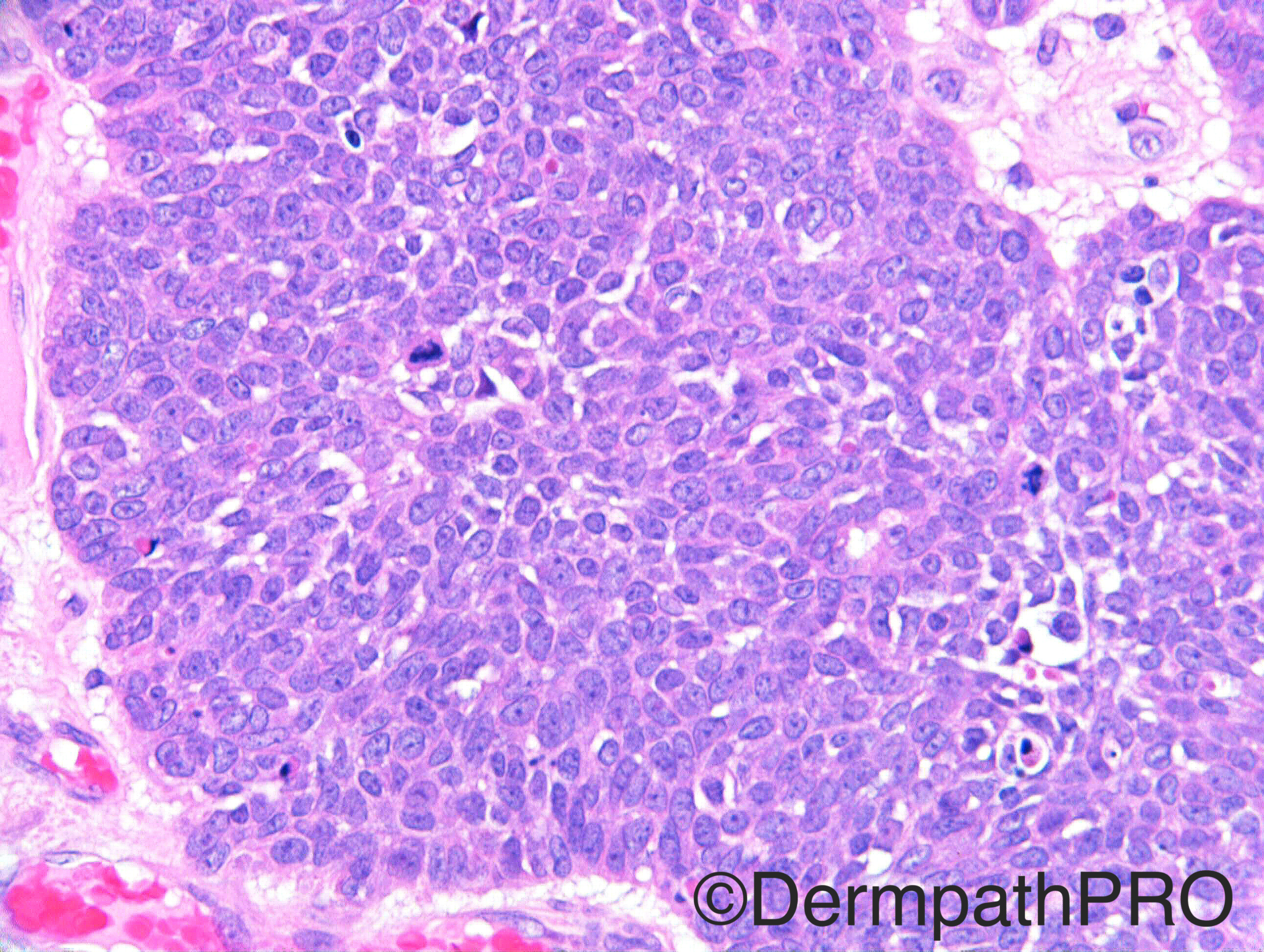
Case History: Nodular lesion anterior chest. ?BCC Case c/o Dr Nitin Khirwadkar

Case posted by Dr Richard Carr

  Report Record

User Feedback


Dr. Mona Abdel-Halim

Posted

I think it is sebaceoma with scarce mature sebocytes. The lesion is very well circumscribed, no significant cytological atypia. Muir Torre syndrome should be verified.

Share this comment


Link to comment
share_externally

Raul Perret

Posted

I think this is one of those borderline tumors where differential between sebaceoma and low grade sebaceous carcinoma is extremely difficult/subjective, I dont know still if there is a difference in prognosis, the lesion seen here is superficial, small (less than one cm?), well circunscribed and the margins are free; metastatic potential should be around 0%. I think I would render a diagnosis of low grade sebaceous carcinoma because  there is a grade of atypia and mitotic figures that are over my ''tolerance'' and would discard Muir Torre as was remarked by the collegues. I read a paper about a group that used a panel that included ki67, p53 and bcl-2 among other markers, I would like to know what is your experience regarding them Dr. Carr.

Cabral ES et al. Distinction of benign sebaceous proliferations from sebaceous carcinoma by immunohistochemistry. Am J of dermatopathology 2006 Dec;28(6):465-71.

Have a good week everyone

Share this comment


Link to comment
share_externally

Dr. Richard Carr

Posted

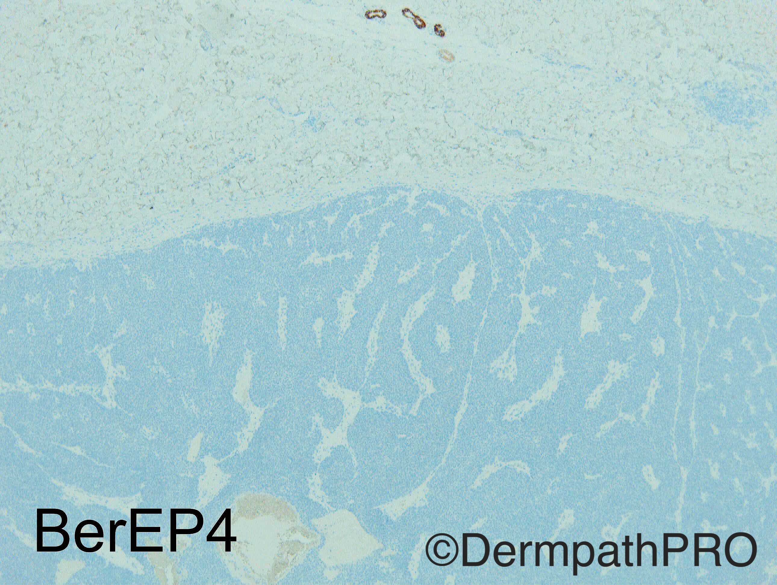
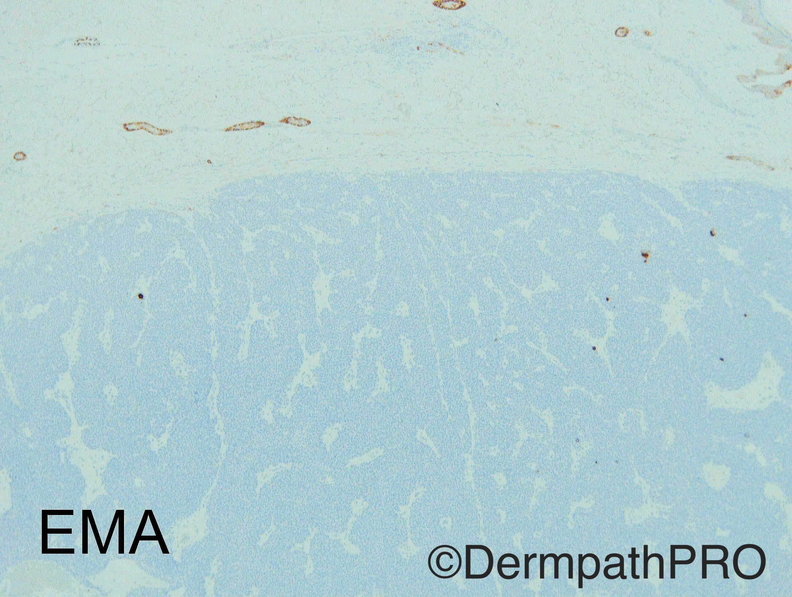
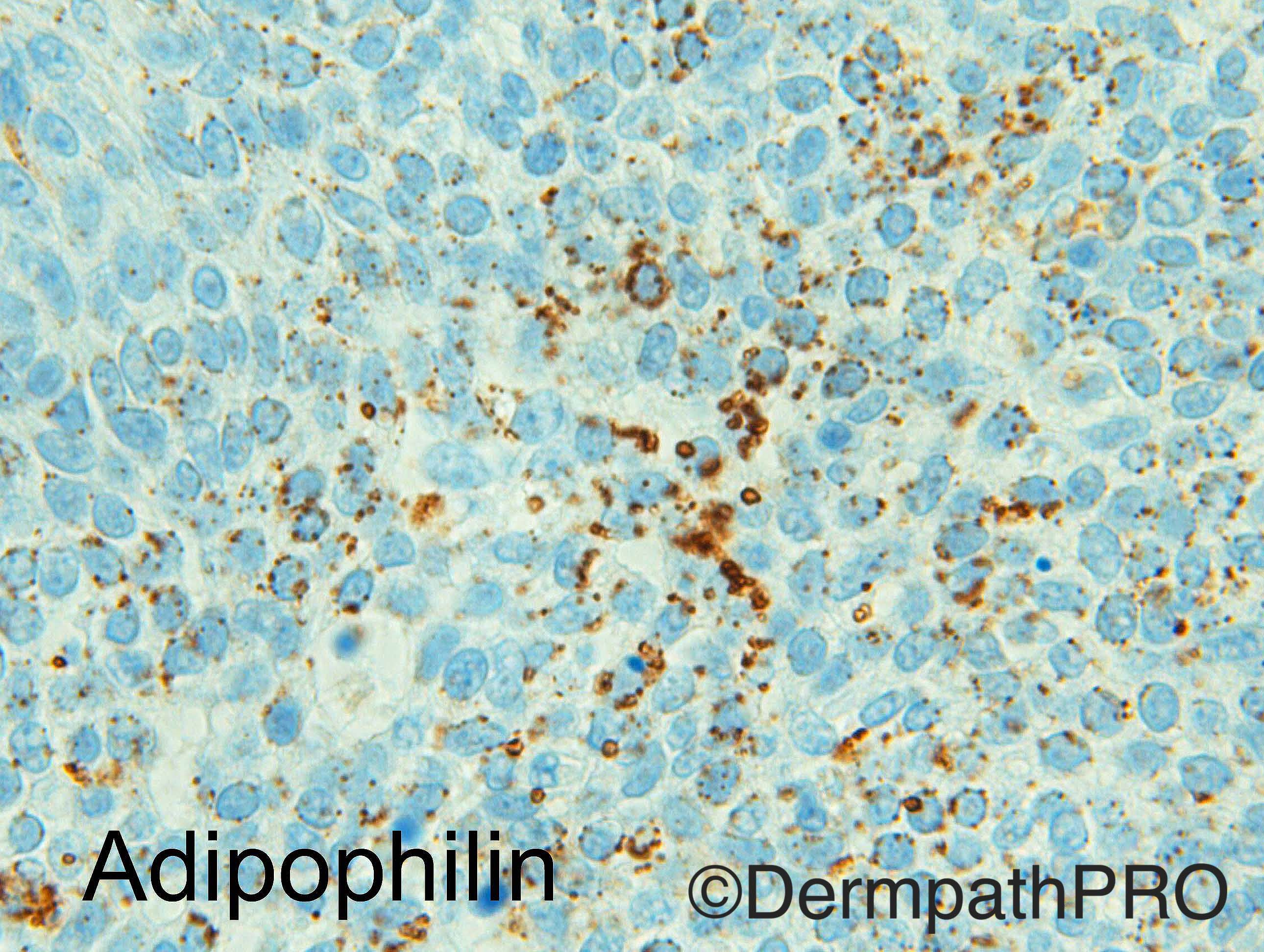
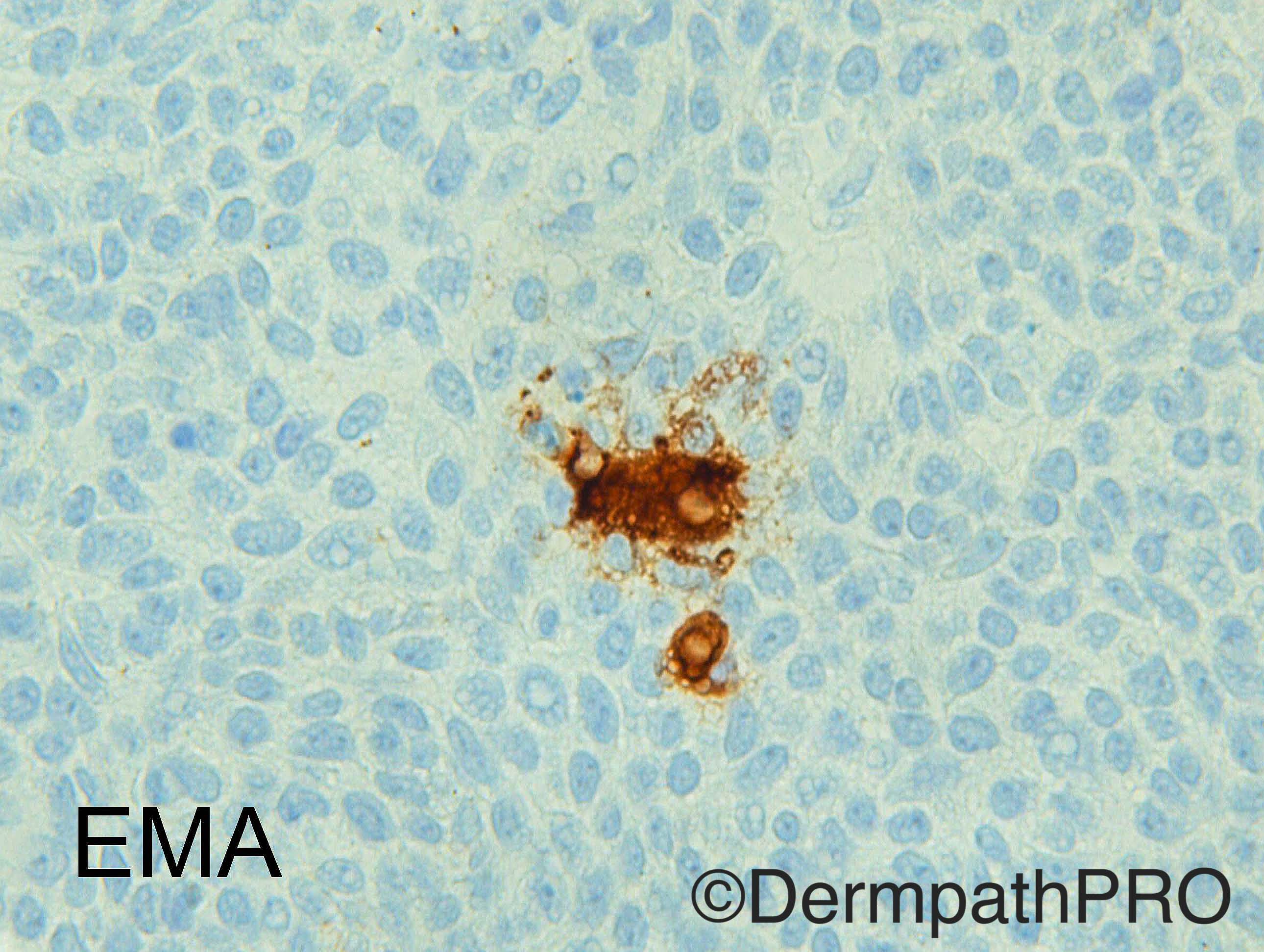
I favoured sebaceoma (typical relatively uniform amphophilic rounded/oval germinative cells, lack of amorphous stroma for hidradenoma or glandular differentiation, and negative BerEP4 and EMA in the germinative and intermediate cells) with no "obvious" mature sebocyctes on H&E but focal EMA and adipophilin distinct microvesiclar (as opposed to tiny non-specific granular) staining in support of the diagnosis.  In my experience they can be quite mitotic especially if superficial and ulcerated.  Anecdotally we have found more widepread EMA and BerEP4 in a majority of sebaceous carcinomas.


I have not systematically studied p53, Ki67 and Bcl2 but thanks for the reference.  I do have a low threshold for doing p53 in mitoitcally active adnexal neoplasms and a null or diffuse strong nuclear pattern might certainly support a lesion that has disturbing features for a diagnosis of malignancy. 


I would still mention the possibility of Muir Torre in my report even if it is in a negative sense e.g. "solitary sebaceoma appears to have a low association with Muir Torre."

Share this comment


Link to comment
share_externally


Join the conversation

You can post now and register later. If you have an account, sign in now to post with your account.

Guest
Add a comment...

×   Pasted as rich text.   Paste as plain text instead

  Only 75 emoji are allowed.

×   Your link has been automatically embedded.   Display as a link instead

×   Your previous content has been restored.   Clear editor

×   You cannot paste images directly. Upload or insert images from URL.

Loading...

×
×
  • Create New...