Jump to content
In this section we have spot diagnoses posted on a daily basis since June 2010, now over 4000! You can review the archived cases and read the suggested diagnoses by users and the final comment by the contributors.
Case are uploaded each week day by 10 am UK time with the correct diagnosis will generally be posted at 8 pm UK time. Why not view the most recent spot diagnosis and proffer a diagnosis?

Case Number : Case 1451- 15 January Posted By: Guest

Please read the clinical history and view the images by clicking on them before you proffer your diagnosis.
Submitted Date :
   (0 reviews)

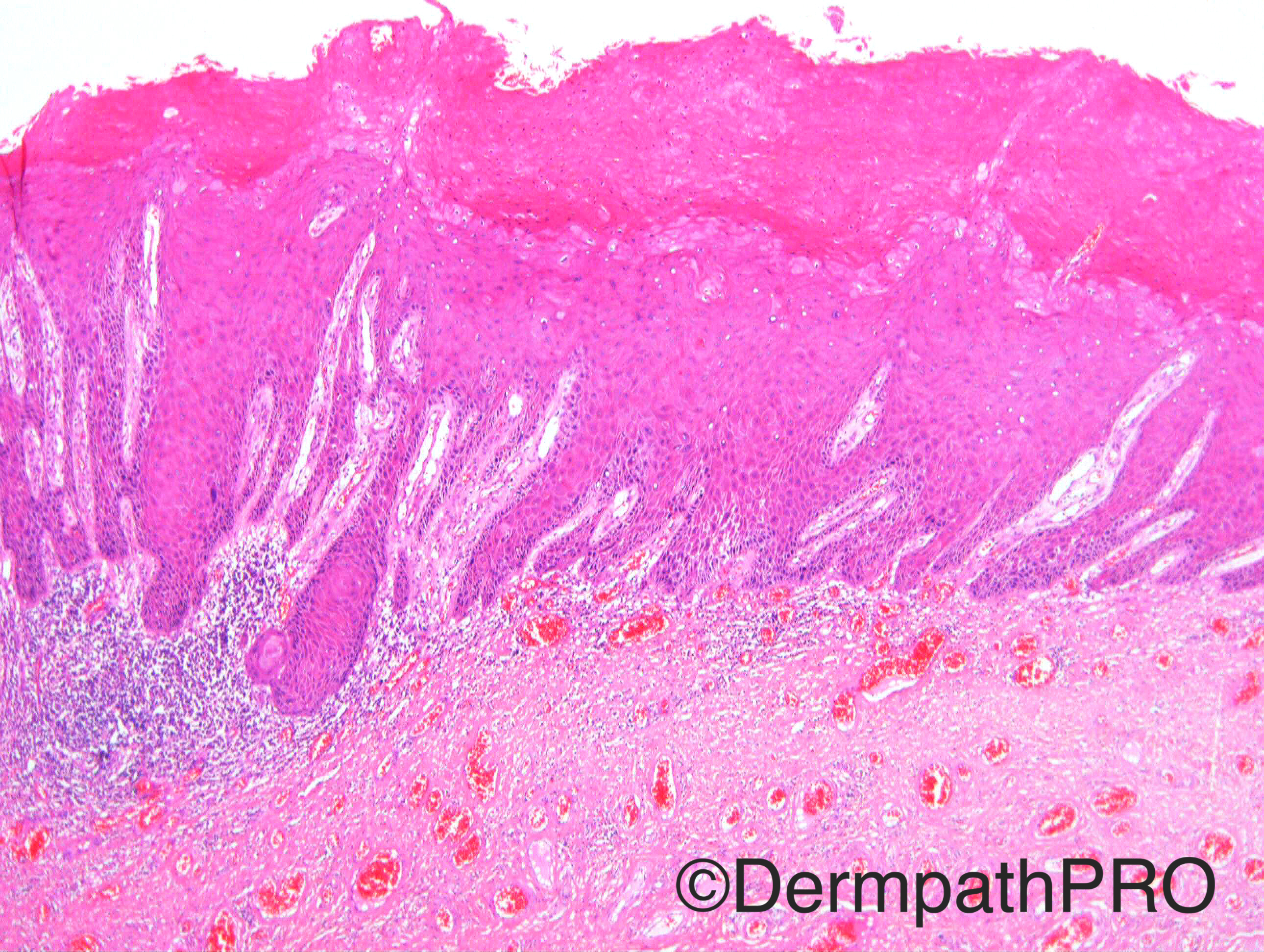
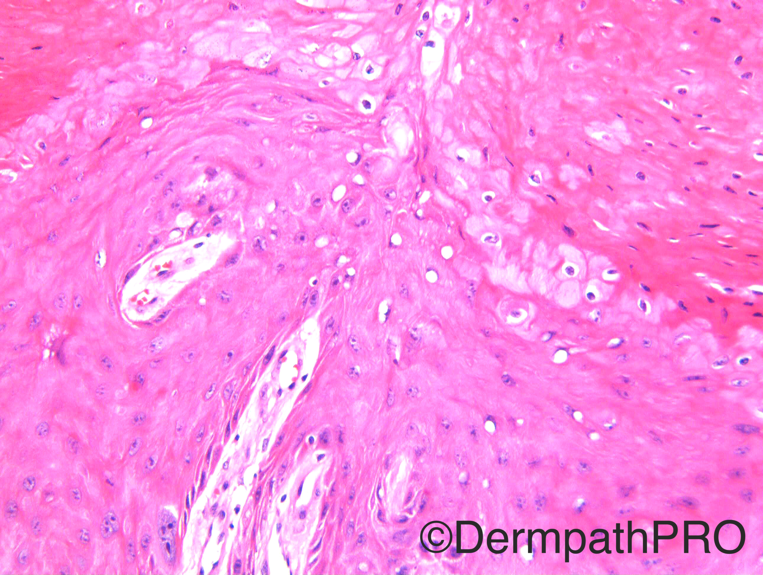
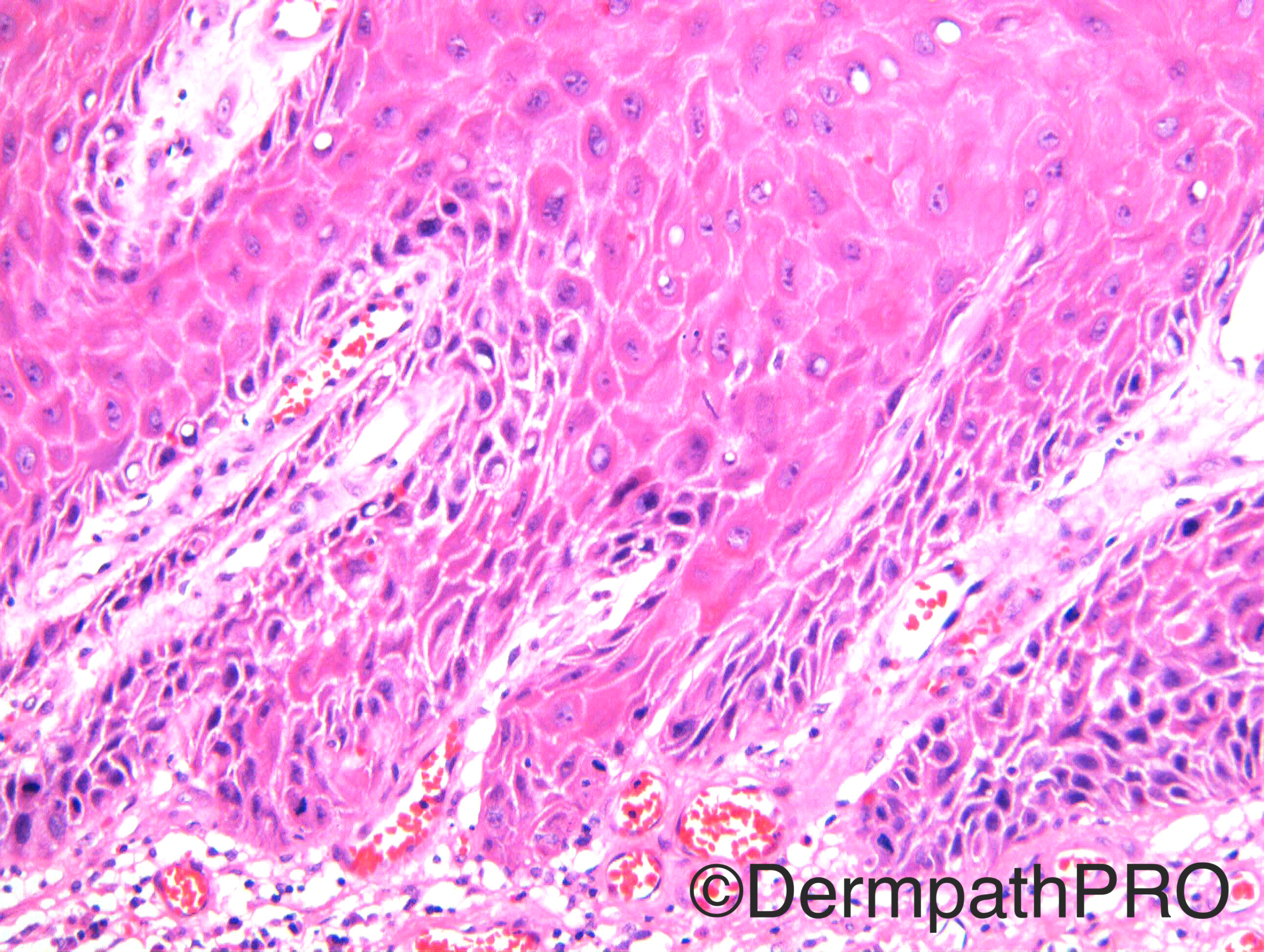
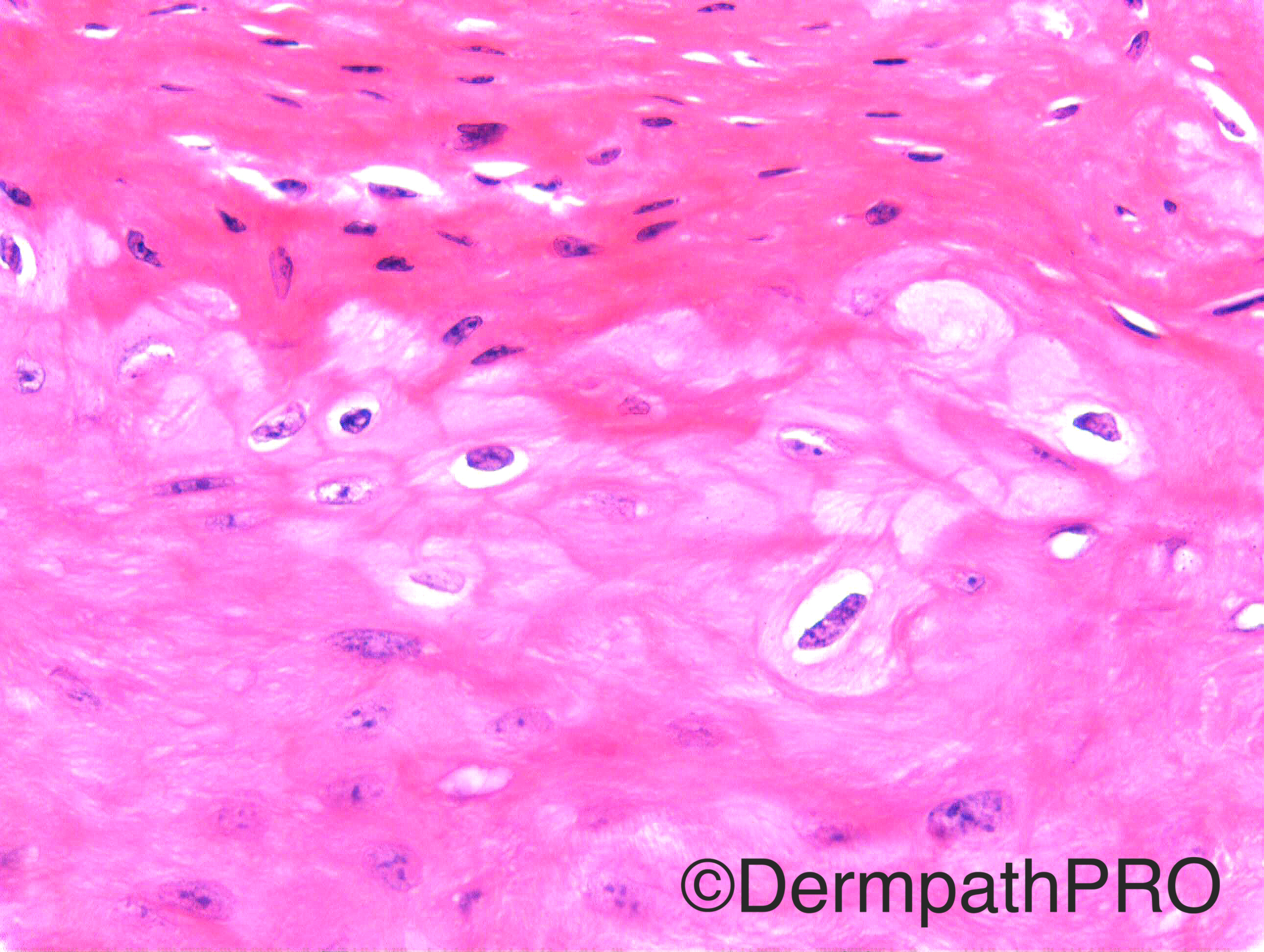
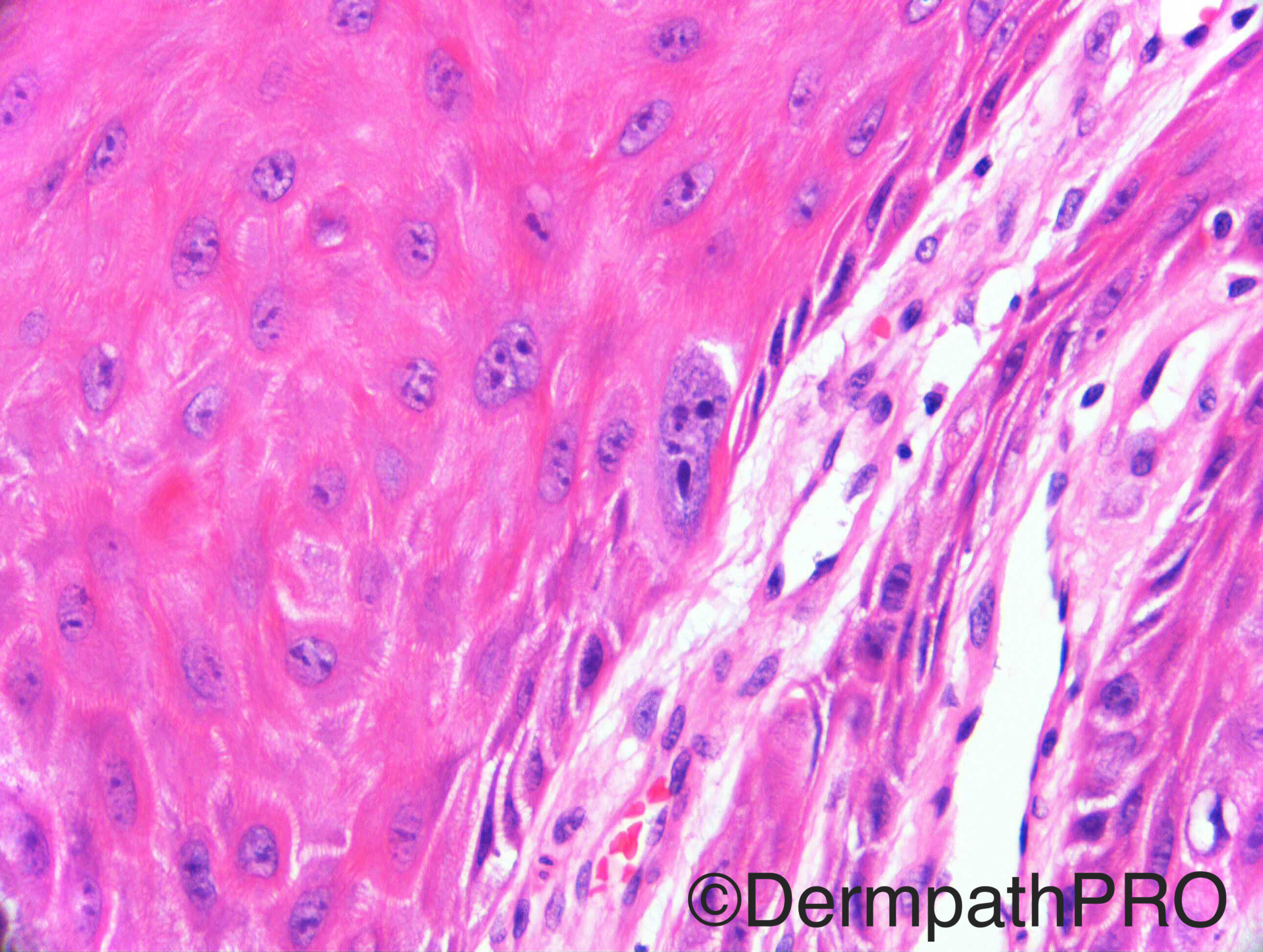
Case History: M70. Growth on glans penis. Foreskin showed BXO/male genital lichen sclerosus. (Case not for The Scream-ish!).

Case posted by Dr Richard Carr


  Report Record

User Feedback


Dr. Mona Abdel-Halim

Posted

Differentiated squamous intraepithelial lesion for further work up with P53,P16 and Ki67

Share this comment


Link to comment
share_externally

Guest Romualdo

Posted

Warty (condylomatous) squamous cell carcinoma. What is scream-ish?

Share this comment


Link to comment
share_externally

Raul Perret

Posted

Agree with warty squamous cell carcinoma. I think we can see microinvasion on the first picture. A differential could be the pseudohyperplastic variant of squamous cell carcinoma that like the warty and verrucous subtype are extremely well differentiated neoplasms. However, this variant (the pseudohyperplastic) tends to be multifocal, more frequent on the very elderly and lacks the clear viral effect seen here...

Share this comment


Link to comment
share_externally

Dr. Mona Abdel-Halim

Posted

Ok, this means that we r totally wrong... So thinking out of the box, I would propose another DDx: could this be just pseudoepitheliomatous hyperplasia with reactive atypia (hyperchromtic basal cells) and this lesion be: pseudoepitheliomatous keratotic and micaceous balanitis?? Anyways this condition has been associated with progression to verrucous carcinoma or SCC so we might still be on track !!!!

Share this comment


Link to comment
share_externally

Nitin Khirwadkar

Posted

Giant condylomata of Buschke and Löwenstein. The viral features are quite striking. PKMB, suggested by Mona is a possibility, and has malignant potential.

Share this comment


Link to comment
share_externally

Arti Bakshi

Posted

Like Mona's original suggestion of differentiated PeIN. Not convinced of invasive tumour and the nuclei in the last 2images are too 'ghastly' for pseudoepitheliomatous hyperplasia.

Share this comment


Link to comment
share_externally

Raul Perret

Posted

Ahhhhhhhhhh okkkkkkk the nuclei in the last picture looks like the face in the painting. Hahahaha it took a while....

Share this comment


Link to comment
share_externally

Sasi Attili

Posted

I agree with Mona.......look forward to the answer

Share this comment


Link to comment
share_externally

Dr. Richard Carr

Posted

I thought this would do for differentiated PeIN or differentiated squamous cell carcinoma in situ if you prefer.  Could not resist the little riddle!  In the next adjacent x2 field there was a larger exophytic well differentiated squamous cell carcinoma with pushing invasion.  I thought there may be some viral changes here but I have to admit to uncertainty but I do think the nuclear features in the parakeratosis are atypical and commensurate with a neoplastic rather than hyperplastic process.  Not sure the IHC will be overly useful in this case as we would probably just expect some supra-basal Ki67, perhaps slight up regulation of basal third wild pattern p53 and p16 only (I did it anyway for interest and it was as stated).  Bottom line is that these cases can be very difficult on biopsy and if not sure about PeIN to make sure you express uncertainty and keep patient under follow-up.

Share this comment


Link to comment
share_externally


Join the conversation

You can post now and register later. If you have an account, sign in now to post with your account.

Guest
Add a comment...

×   Pasted as rich text.   Paste as plain text instead

  Only 75 emoji are allowed.

×   Your link has been automatically embedded.   Display as a link instead

×   Your previous content has been restored.   Clear editor

×   You cannot paste images directly. Upload or insert images from URL.

Loading...

×
×
  • Create New...