Jump to content
In this section we have spot diagnoses posted on a daily basis since June 2010, now over 4000! You can review the archived cases and read the suggested diagnoses by users and the final comment by the contributors.
Case are uploaded each week day by 10 am UK time with the correct diagnosis will generally be posted at 8 pm UK time. Why not view the most recent spot diagnosis and proffer a diagnosis?

Case Number : Case 1453- 19 January Posted By: Guest

Please read the clinical history and view the images by clicking on them before you proffer your diagnosis.
Submitted Date :
   (0 reviews)

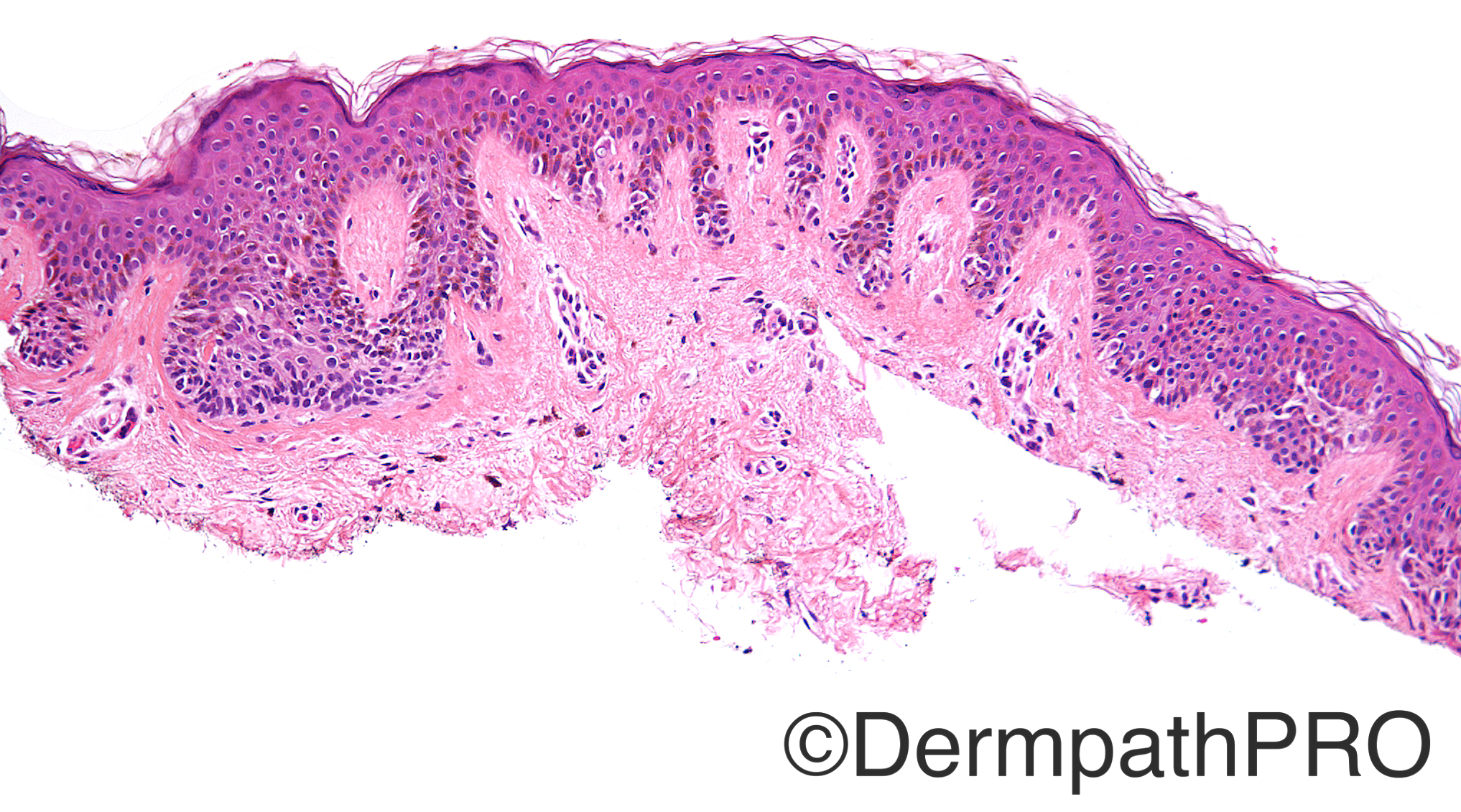
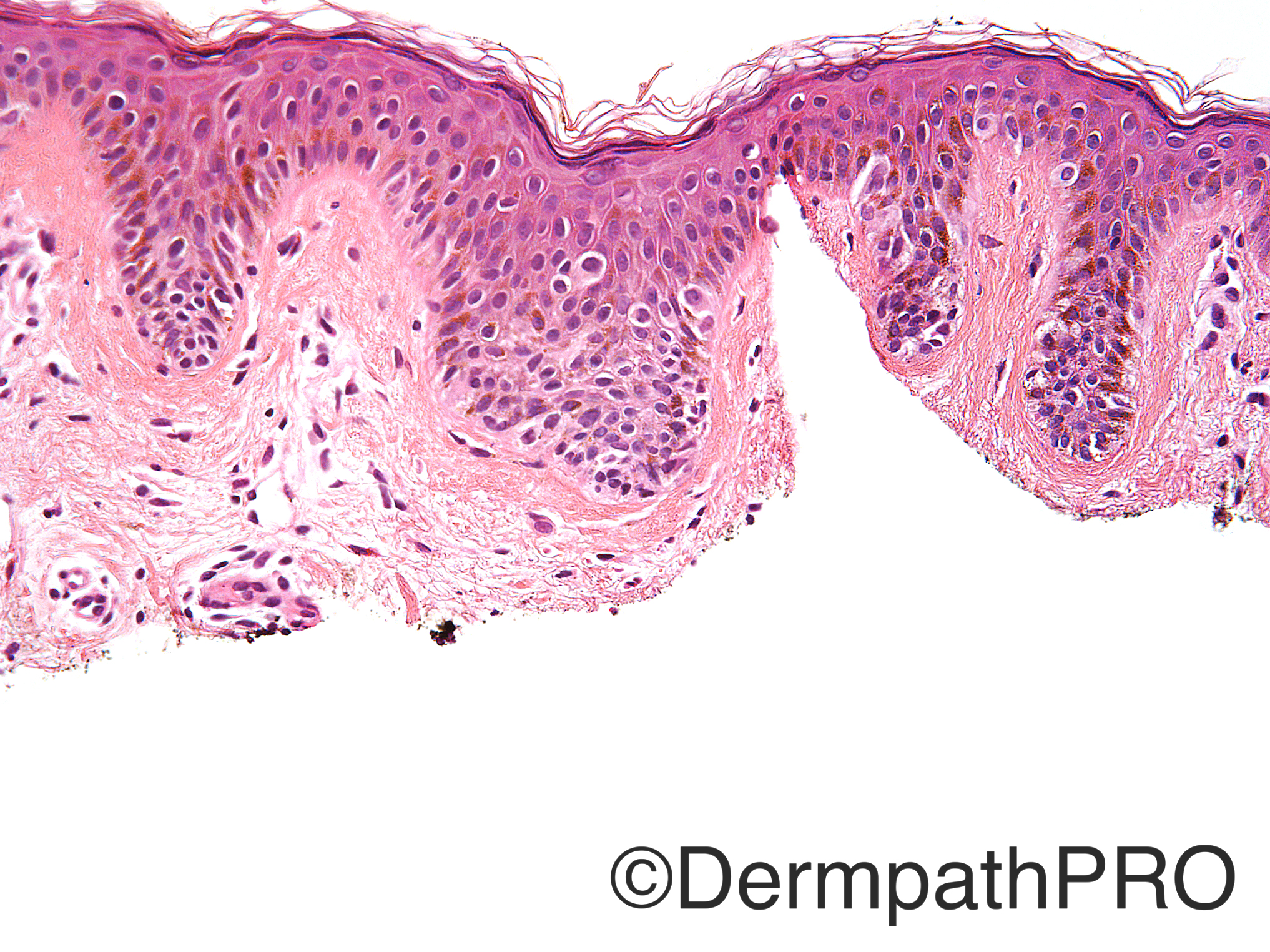
Case History: 47 year old woman with atypical nevus near the umbilicus.

Case posted by Dr Uma Sundram


  Report Record

User Feedback


Raul Perret

Posted

Junctional melanocytic nevus of special site (umbilicus)

Share this comment


Link to comment
share_externally

Nitin Khirwadkar

Posted

Agree with Raul. Junctional melanocytic proliferation with 'site related atypia'. Was this an excision or incisional biopsy?

Share this comment


Link to comment
share_externally

Dr. Mona Abdel-Halim

Posted

I can see that many rete ridges are elongated, there is fibroplasia of the papillary dermis and bridging between nests, so I think this is junctional dysplastic nevus (mildly dysplastic). I don't feel worried about melanoma in situ but definitely Melan A will be a good idea.

Share this comment


Link to comment
share_externally

Raul Perret

Posted

Dr. Hurt just for the purpose of learning, in your experience what is the usefulness of melanocytic markers in this kind of lesions? do you use to have a better assesment of melanocytic population (architecture)? Thanks in advance.

Share this comment


Link to comment
share_externally

vincenzo polizzi

Posted

Fibroplasia, no ascent cells, little epidermal infiltration of melanocytes push toward diagnosis of junctional nevus; but the irregular nesting pattern of atypical cells (more in fig4), although small nests, looks like expansive. Difficult case. I agree with Melan-A.

Share this comment


Link to comment
share_externally

Dr. Richard Carr

Posted

It was clinically atypical and yet not excised. Is this normal practice? Certainly risky. I have been trying to advise our clinical colleagues for years either leave it alone (if you are not worried) or if you are at all worried then excise with a clear margin of at least 2 to 3mm of normal skin (beyond any faint halo of pigmentation) if you want a confident histopathological opinion (this is more true than ever for small, recently grown, often impossible to classify lesions). Having said all that I favour a mildly atypical junctional melanocytic proliferation with no overtly worrying features, commensurate with the anatomic location, but "if there is any clinical concern complete excision with clear margins would be recommended".

Share this comment


Link to comment
share_externally

Uma Sundram

Posted

I think these types of cases are so challenging! This was an attempted excision but the lesional cells did extend to the margins. We did Melan A and saw that the cells did not show full thickness upward scatter although as you can see from the H+E stained sections, there is multifocal limited scatter of melanocytes. I shared this with my group and we were in agreement that there is atypia, probably commensurate with the site (umbilicus). Recommended either close clinical follow up (if the patient was comfortable with this) or a narrow re excision to ensure that the lesion was completely removed. Over the years I have often done just Melan A on cases like this. This is a clean and relatively unambiguous stain. It allows one to see the expanse and architecture of the lesion and better assess lateral circumscription. As you can see, in H+E stained sections relatively small melanocytes often blend in with keratinocytes, making assessment difficult.

Share this comment


Link to comment
share_externally


Join the conversation

You can post now and register later. If you have an account, sign in now to post with your account.

Guest
Add a comment...

×   Pasted as rich text.   Paste as plain text instead

  Only 75 emoji are allowed.

×   Your link has been automatically embedded.   Display as a link instead

×   Your previous content has been restored.   Clear editor

×   You cannot paste images directly. Upload or insert images from URL.

Loading...

×
×
  • Create New...