Jump to content
In this section we have spot diagnoses posted on a daily basis since June 2010, now over 4000! You can review the archived cases and read the suggested diagnoses by users and the final comment by the contributors.
Case are uploaded each week day by 10 am UK time with the correct diagnosis will generally be posted at 8 pm UK time. Why not view the most recent spot diagnosis and proffer a diagnosis?

Case Number : Case 1456- 22 January Posted By: Guest

Please read the clinical history and view the images by clicking on them before you proffer your diagnosis.
Submitted Date :
   (0 reviews)

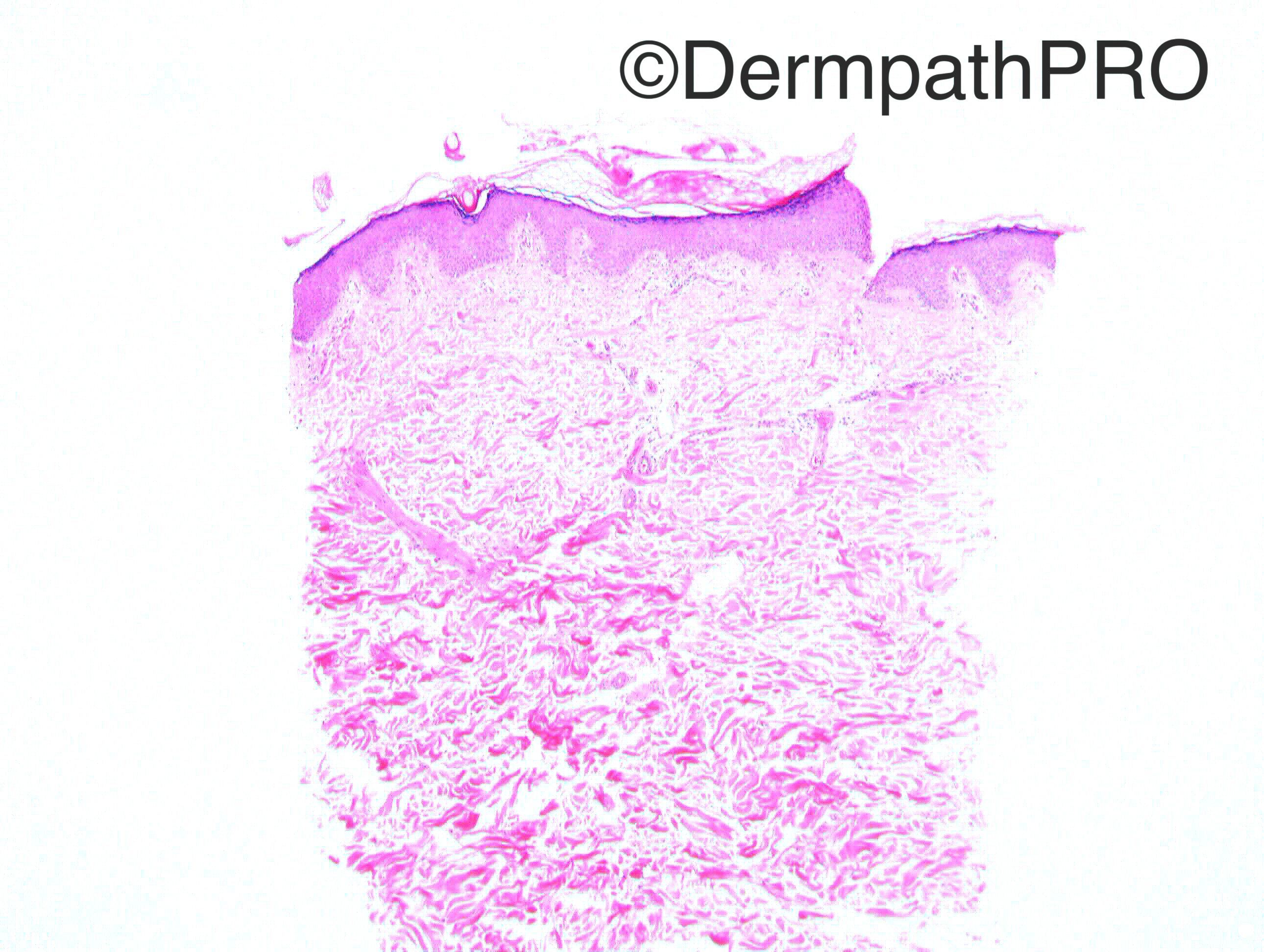
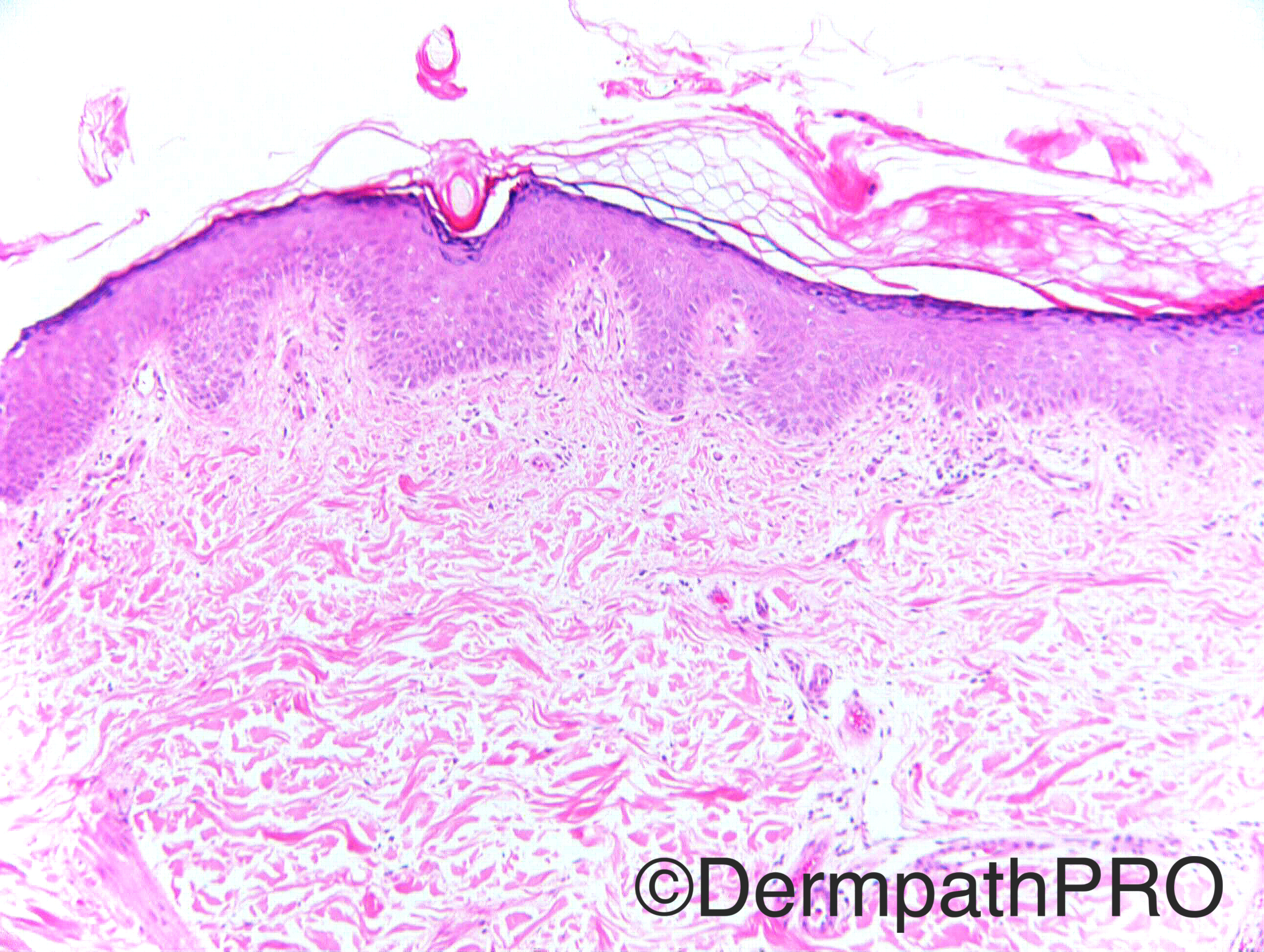
Case History: F60. Arm. Punch biopsy. Additional images to follow at 4pm GMT

Case posted by Dr Richard Carr


  Report Record

User Feedback


Nitin Khirwadkar

Posted

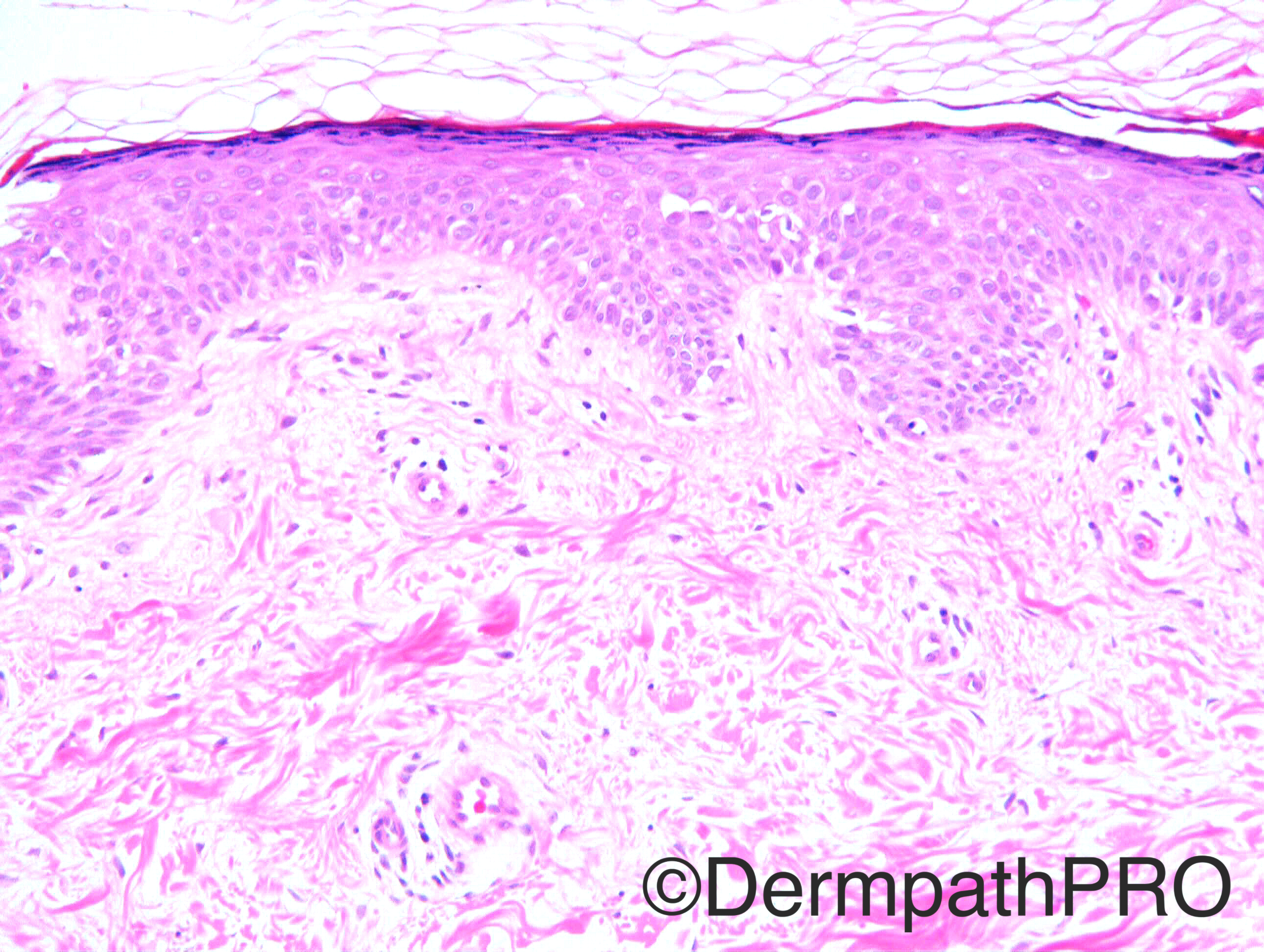
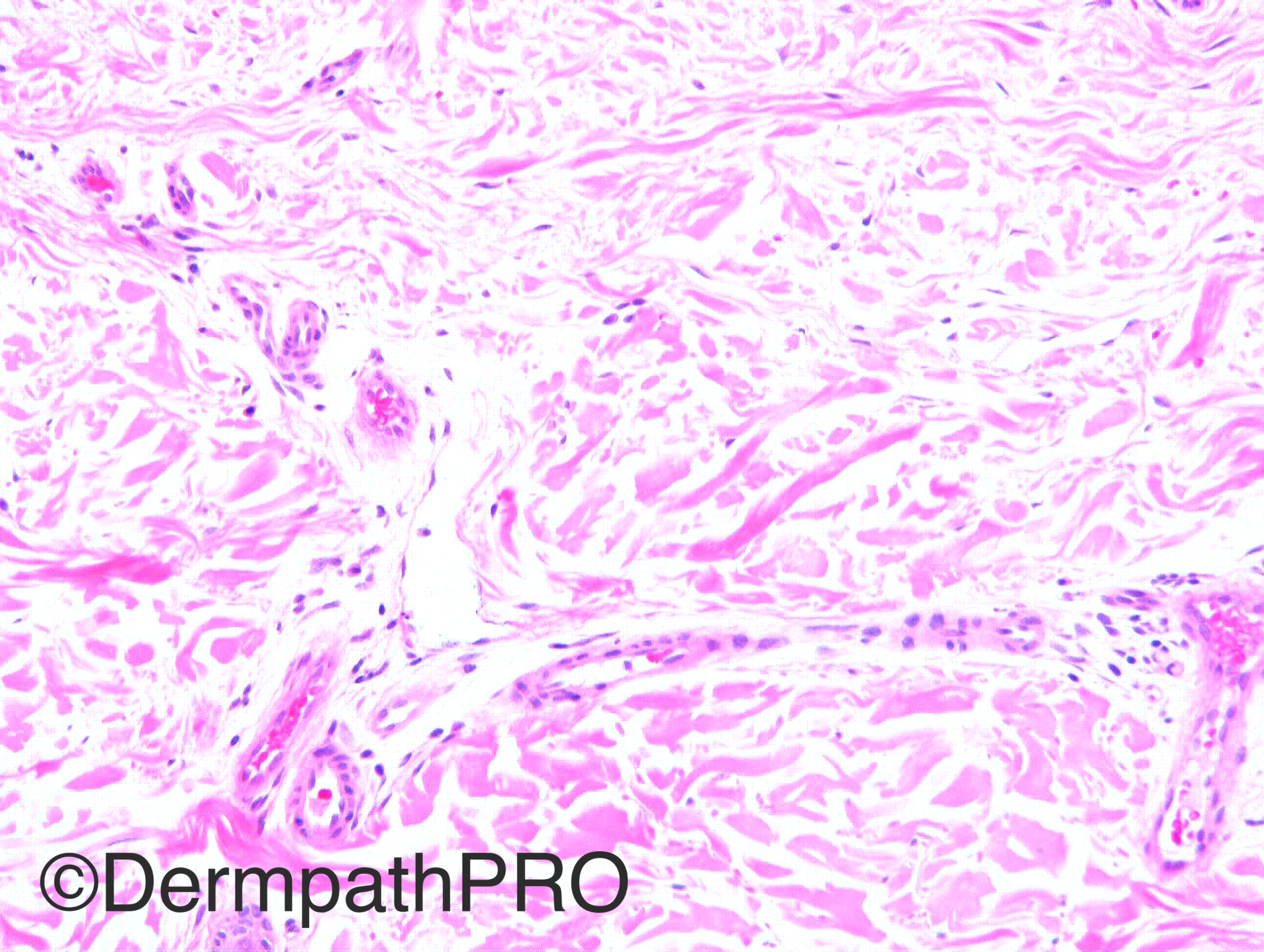
There is a focus of hyperparakeratosis. Small space between the stratum granulosum and the corneum, not sure if genuine or artefact. Focal, epidermal spongiosis. Separation of collagen fibers within the dermis, secondary to oedema/mucin. Some more images, special stains required.

Share this comment


Link to comment
share_externally

Raul Perret

Posted

I see abnormal keratinization with clefting, mild hypergranulosis, keratinocyte clefting on the stratum spinosum and mild acanthosis. On the superficial dermis minimal lymphocytic infiltrate surrounding vessels and dermal edema as Nitin suggested. I would like to see the next images but I was thinking more on a pattern similar to some type of ichthyosis... I would focus on the chapter of epidermal disorders of maturation and keratinization for now

Share this comment


Link to comment
share_externally

Dr. Mona Abdel-Halim

Posted

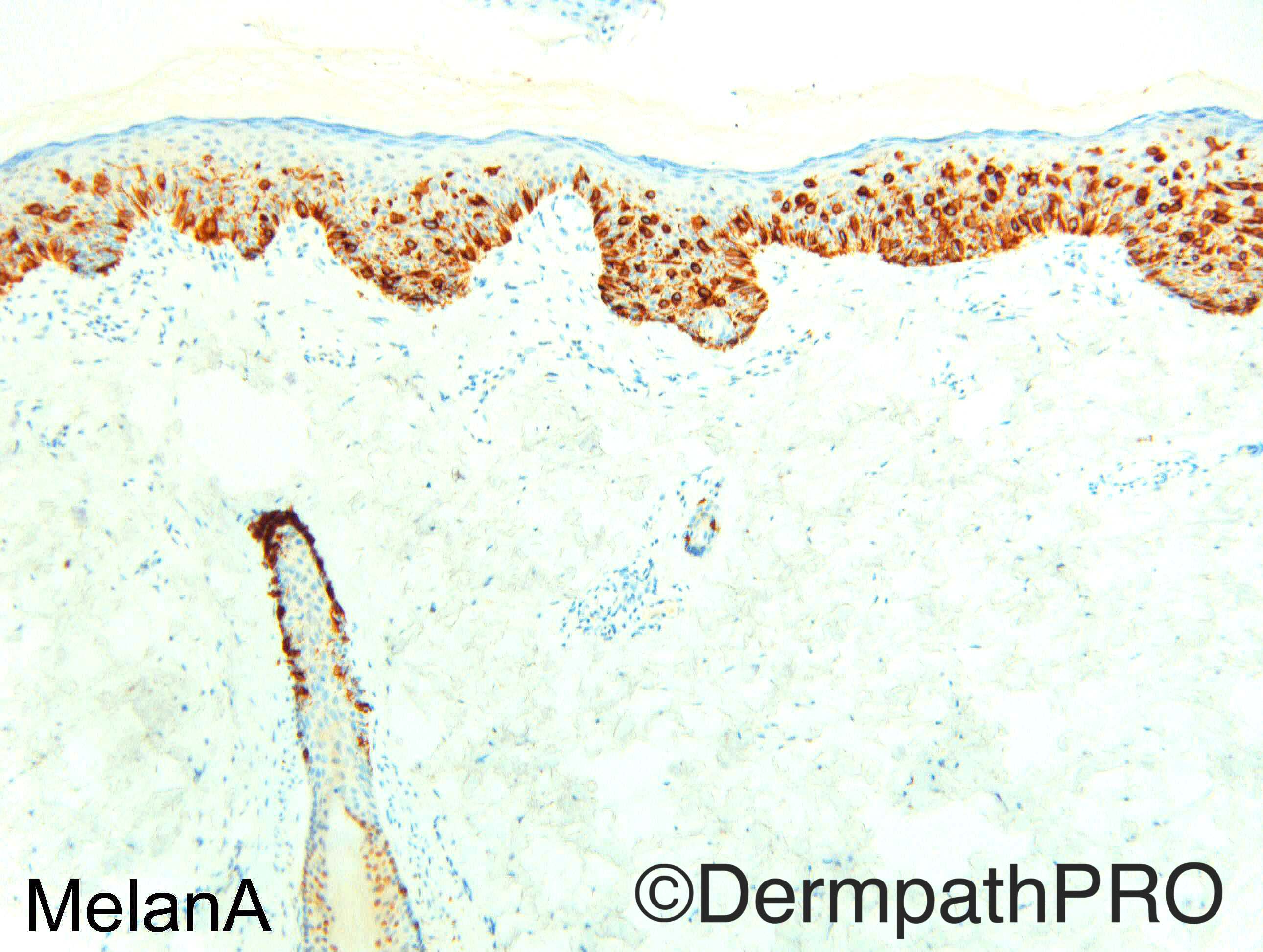
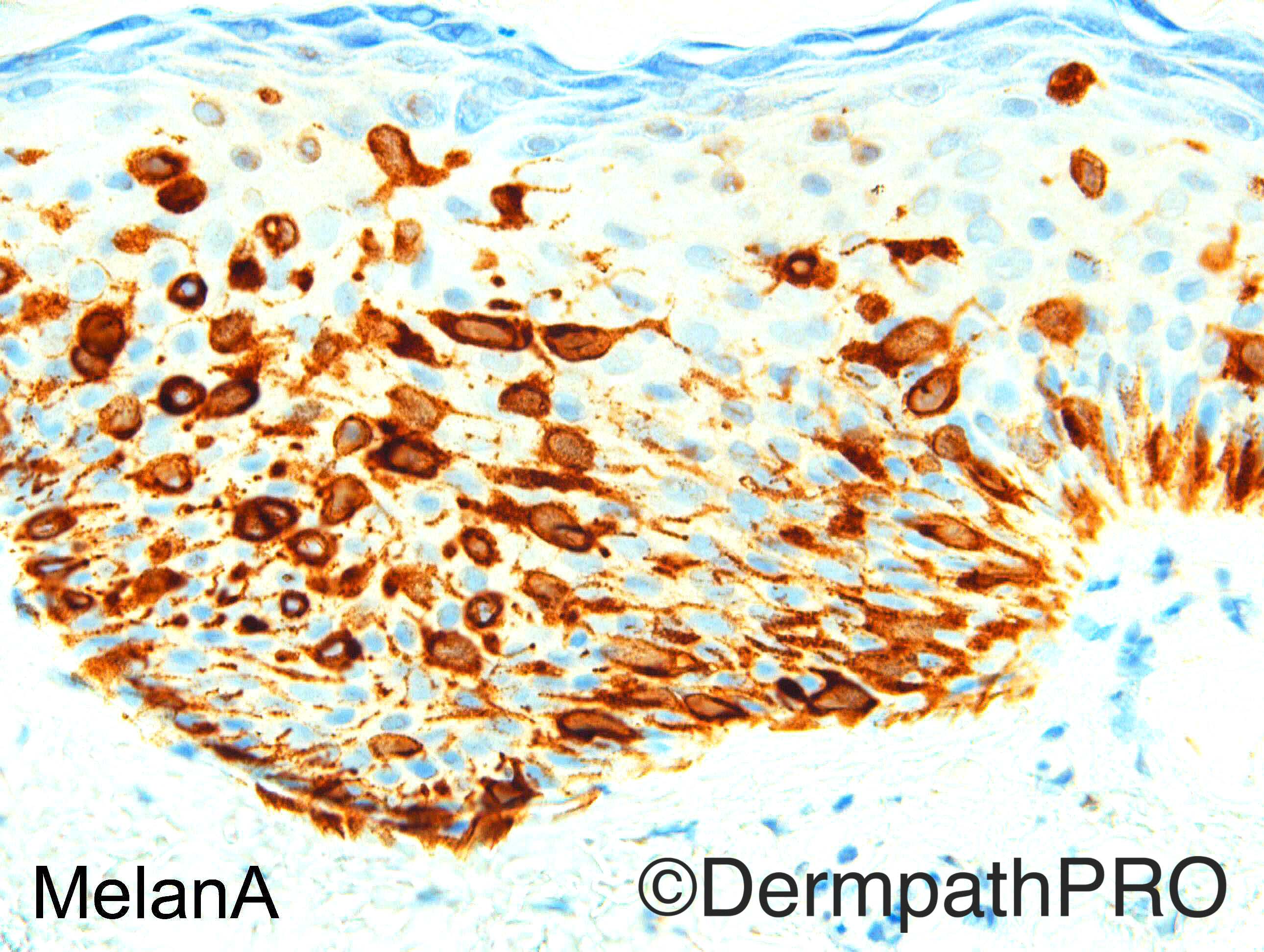
Wao, so subtle on H&E.. Without Melan A and clinical would have been very difficult to suspect MIS.

Share this comment


Link to comment
share_externally

Raul Perret

Posted

I know im a bit difficult today but there is something that I find suspicious here, I know that there is upward scatter of melanocytes (pagetoid) but they seem to be at a similar level (quite symmetric) and they dont seem to surpass the spinous layer. Altogether with some epidermal changes (mild hiperplasia, etc.) I was thinking of pagetoid spitz nevus as a differential to MIS, I would really like to have the slide to analize it thoroughly. Is it a small lesión Dr. Carr? like 0,5 cm or less?

Share this comment


Link to comment
share_externally

Nitin Khirwadkar

Posted

Wow!!! I was totally off track! Did not think about an intraepidermal melanocytic proliferation, until the Melan-A lighted up a proliferation of dendritic and non-dendritic melanocytes. There is prominent upward migration, along with pagetoid spread. Did this clinically come as a pigmented lesion, and was this a punch excision? Slight reservation to go for a MIS, although its nearly there. Why is there dermal oedema?

Share this comment


Link to comment
share_externally

Arti Bakshi

Posted

Melan A certainly dramatic! I think would favour lentigo maligna (depending on clinical)

Share this comment


Link to comment
share_externally

Guest Jim Davie MD

Posted

MelanA is showing downwards pagetoid spread along follicular and eccrine adnexa.   This supports MIS in the context of the prominent pagetoid scatter.  

Share this comment


Link to comment
share_externally

Dr. Richard Carr

Posted

My report:

Subtle pagetoid proliferation with mild cytological atypia.               
Positive:  Melan A, S100, p53 (apparently).                               
Negative:  Cam5.2.                                                       
Ki67:      Inconclusive, p16 non-contributory.                            
We favour melanoma in situ (amelanotic). 


Comment: I deliberately gave you up to x20 objective.  The melanocytic cells were subtle and could easily be missed even on a good day!  I guess cases like this are designed to catch us out.  If you plan to report on scanned images in the future (my guess is we will all be doing this in the very near future) make sure they are scanned at x40! and have a low threshold on "invisble cases" to put them aside, slow down and have a good think. Clinical correlation is obviously essential, in this case a lesion for 2 years ?BCC/AK.  I do not know the size.  I think there is enough here to warrent complete excision but the case will automatically be discussed at MDM in the UK.

Share this comment


Link to comment
share_externally

Dr. Mona Abdel-Halim

Posted

Thanks Dr Carr for sharing such an important case

Share this comment


Link to comment
share_externally


Join the conversation

You can post now and register later. If you have an account, sign in now to post with your account.

Guest
Add a comment...

×   Pasted as rich text.   Paste as plain text instead

  Only 75 emoji are allowed.

×   Your link has been automatically embedded.   Display as a link instead

×   Your previous content has been restored.   Clear editor

×   You cannot paste images directly. Upload or insert images from URL.

Loading...

×
×
  • Create New...