Jump to content
In this section we have spot diagnoses posted on a daily basis since June 2010, now over 4000! You can review the archived cases and read the suggested diagnoses by users and the final comment by the contributors.
Case are uploaded each week day by 10 am UK time with the correct diagnosis will generally be posted at 8 pm UK time. Why not view the most recent spot diagnosis and proffer a diagnosis?

Case Number : Case 1458- 26 January Posted By: Guest

Please read the clinical history and view the images by clicking on them before you proffer your diagnosis.
Submitted Date :
   (0 reviews)

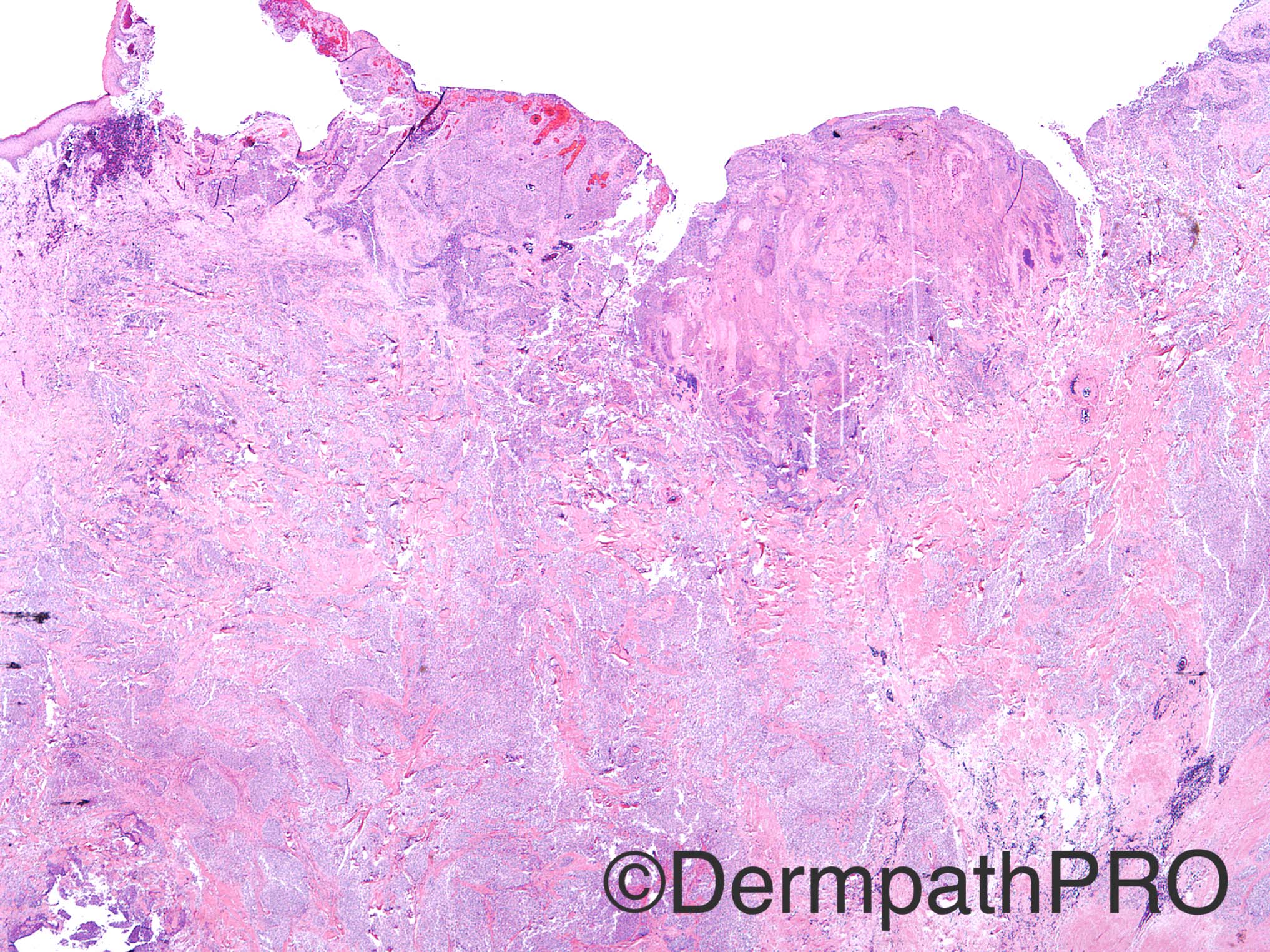
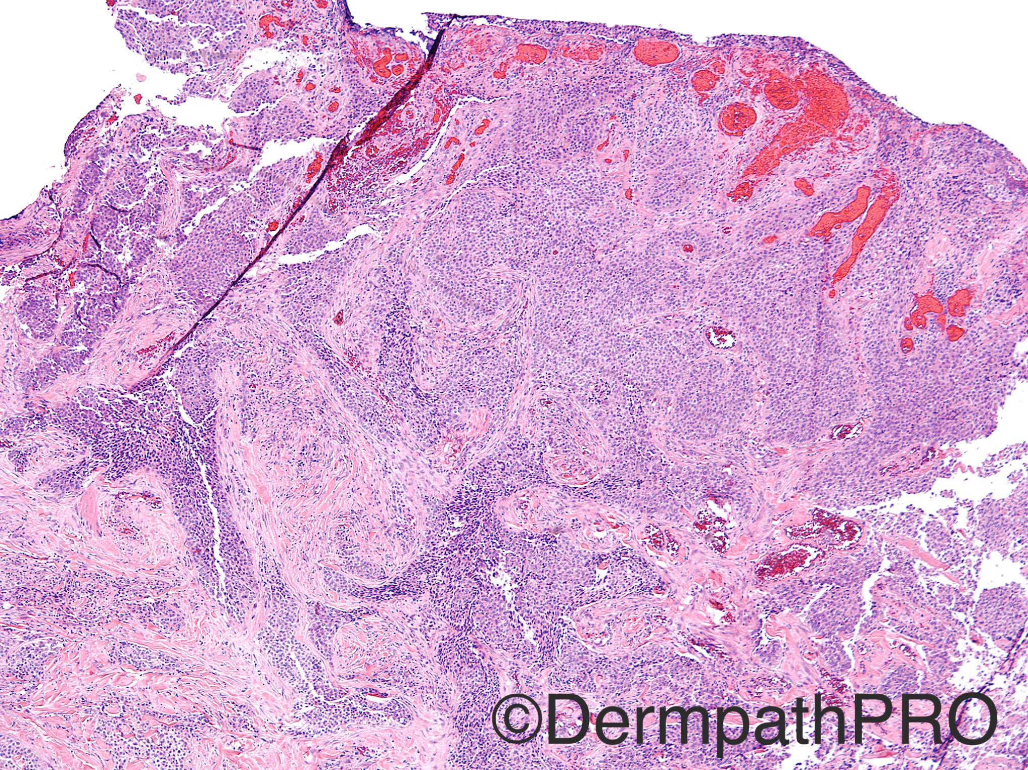
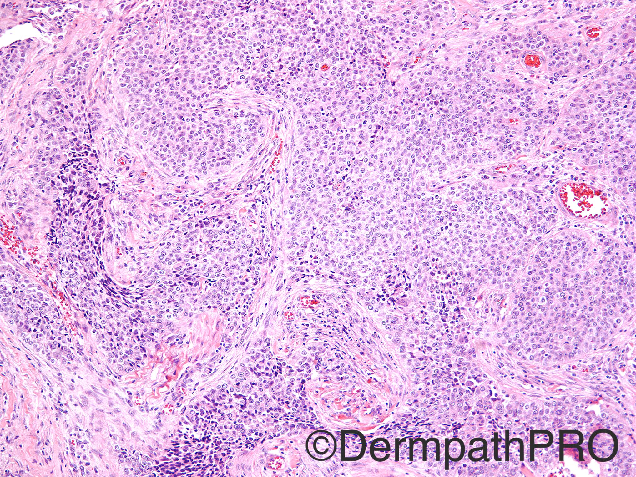
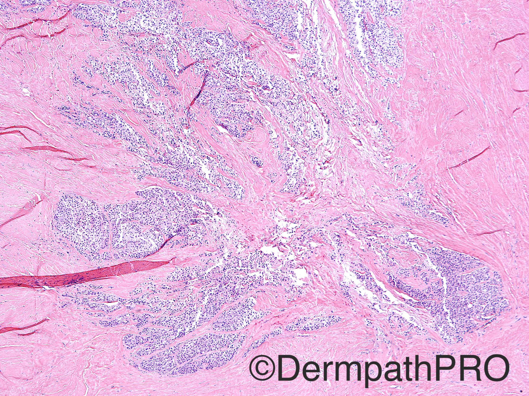
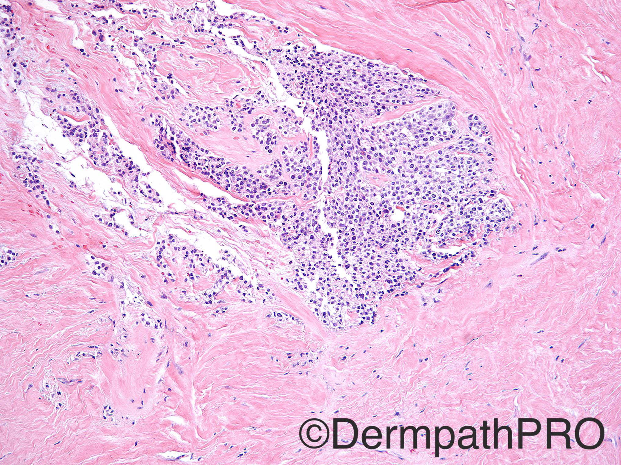
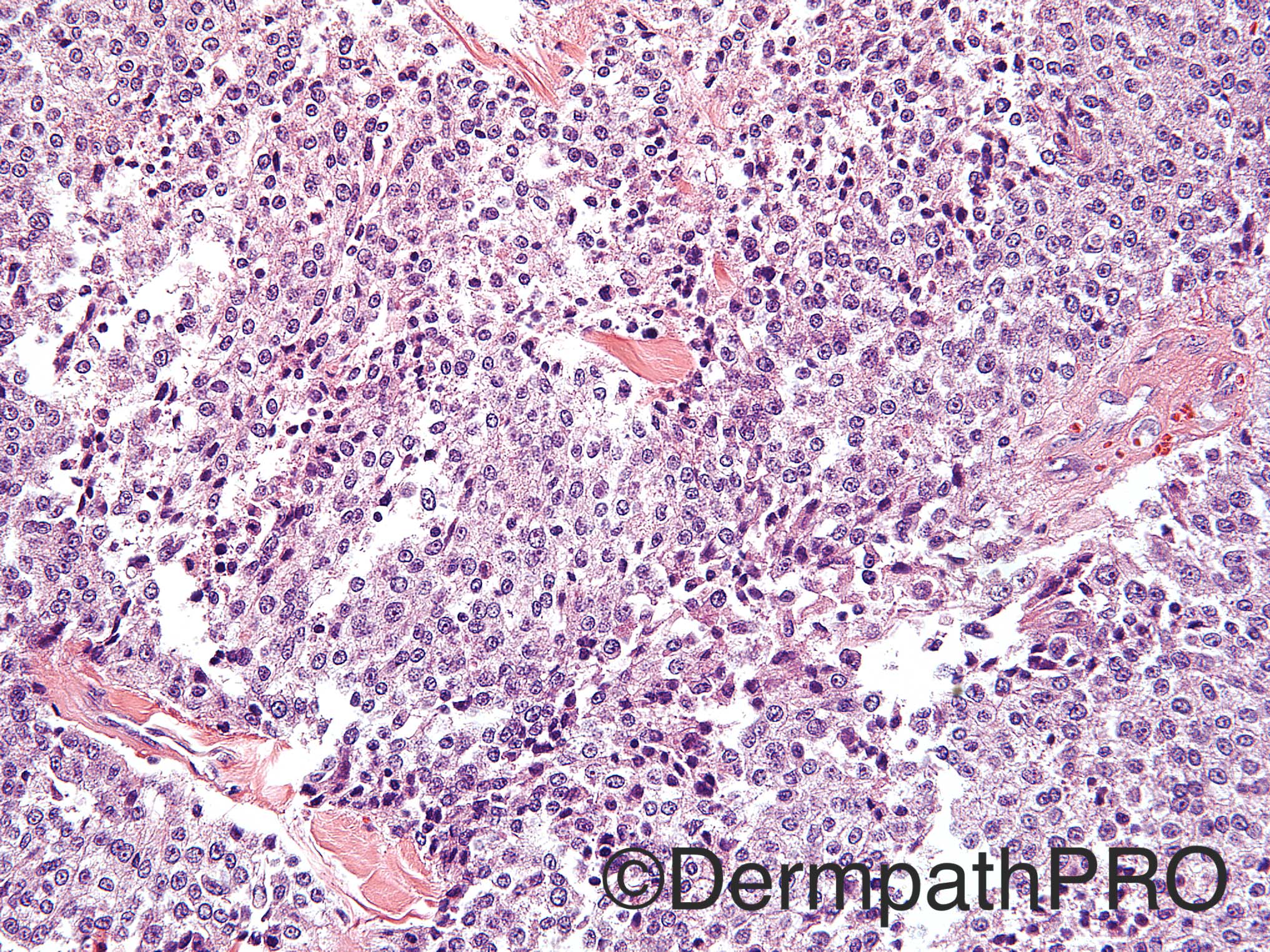
Case History: 77 year old male with lesion on right neck.

Case posted by Dr Uma Sundram


  Report Record

User Feedback


Arti Bakshi

Posted

An ulcerated cellular tumour composed of rather monomorphic cells with round central nuclei and moderate pale cytoplasm. The cytomorphology makes me think Glomus tumour (?atypical/malignant), although vascular pattern not particularly prominent. Other d/d- myoepithelioma

Will need wide panel of immunos. 

Share this comment


Link to comment
share_externally

vincenzo polizzi

Posted

Let me to argue better my diagnosis (TC): first i think there is a subtle surface connection of the tumor; then i see a clear appearance of the cells; last, a subtle peripheral palisade. Neck is a typical site for TC.

Share this comment


Link to comment
share_externally

Nitin Khirwadkar

Posted

Ulcerated tumour. Not sure if there is any intact residual epithelium. The first image shows a focus of what looks like tumour necrosis (unless an artefact). The tumour is widely infiltrative, and as Arti has mentioned, has a rather monomorphic appearance. The last image shows some fine granularity and clearing within the cytoplasm of the cells, along with mild and moderate cytonuclear atypia. This tumour needs a wide panel of immunos. Likely to be a primary tumour, epithelial or myoepithelial. Would also rule out a metastasis with IHC.

Share this comment


Link to comment
share_externally

Guest Romualdo

Posted

I also think this is a carcinoma and I liked Vincenzo's suggestion of trichilemmal carcinoma.

Share this comment


Link to comment
share_externally

Raul Perret

Posted

I agree that a wide panel of IHQ should be performed but morphology makes me think of clear cell hidradenocarcinoma/ malignant acrospiroma with clear cell change

Share this comment


Link to comment
share_externally

Dr. Richard Carr

Posted

A bit flumoxed (that means don't know).  Probably an undifferentiated carcinoma, might add some neuroendocrine markers, S100, CD99 (for PNET).

Share this comment


Link to comment
share_externally

Dr. Mona Abdel-Halim

Posted

Very difficult without IHC, favouring clear cell hidradenocarcinoma.

Share this comment


Link to comment
share_externally

Uma Sundram

Posted

Clear cell hidradenocarcinoma; melanoma markers, synaptophysin, chromogranin and CD99 were all negative.  Cytokeratins positive. Trichilemmal carcinoma would also fit; there were more papillary and cystic areas in other sections which led us to favor clear cell hidradenocarcinoma. A month later the patient had an involved regional lymph node which was diagnosed as malignant based on a cytology/FNA specimen. I was particularly impressed by the relative monotony of the lesional cells, but ulceration and infiltration led us to make the initial diagnosis of malignancy. The initial tumor was also relatively friable and very large, quite worrisome for malignancy clinically.

Share this comment


Link to comment
share_externally


Join the conversation

You can post now and register later. If you have an account, sign in now to post with your account.

Guest
Add a comment...

×   Pasted as rich text.   Paste as plain text instead

  Only 75 emoji are allowed.

×   Your link has been automatically embedded.   Display as a link instead

×   Your previous content has been restored.   Clear editor

×   You cannot paste images directly. Upload or insert images from URL.

Loading...

×
×
  • Create New...