Jump to content
In this section we have spot diagnoses posted on a daily basis since June 2010, now over 4000! You can review the archived cases and read the suggested diagnoses by users and the final comment by the contributors.
Case are uploaded each week day by 10 am UK time with the correct diagnosis will generally be posted at 8 pm UK time. Why not view the most recent spot diagnosis and proffer a diagnosis?

Case Number : Case 1461 - 29 January Posted By: Guest

Please read the clinical history and view the images by clicking on them before you proffer your diagnosis.
Submitted Date :
   (0 reviews)

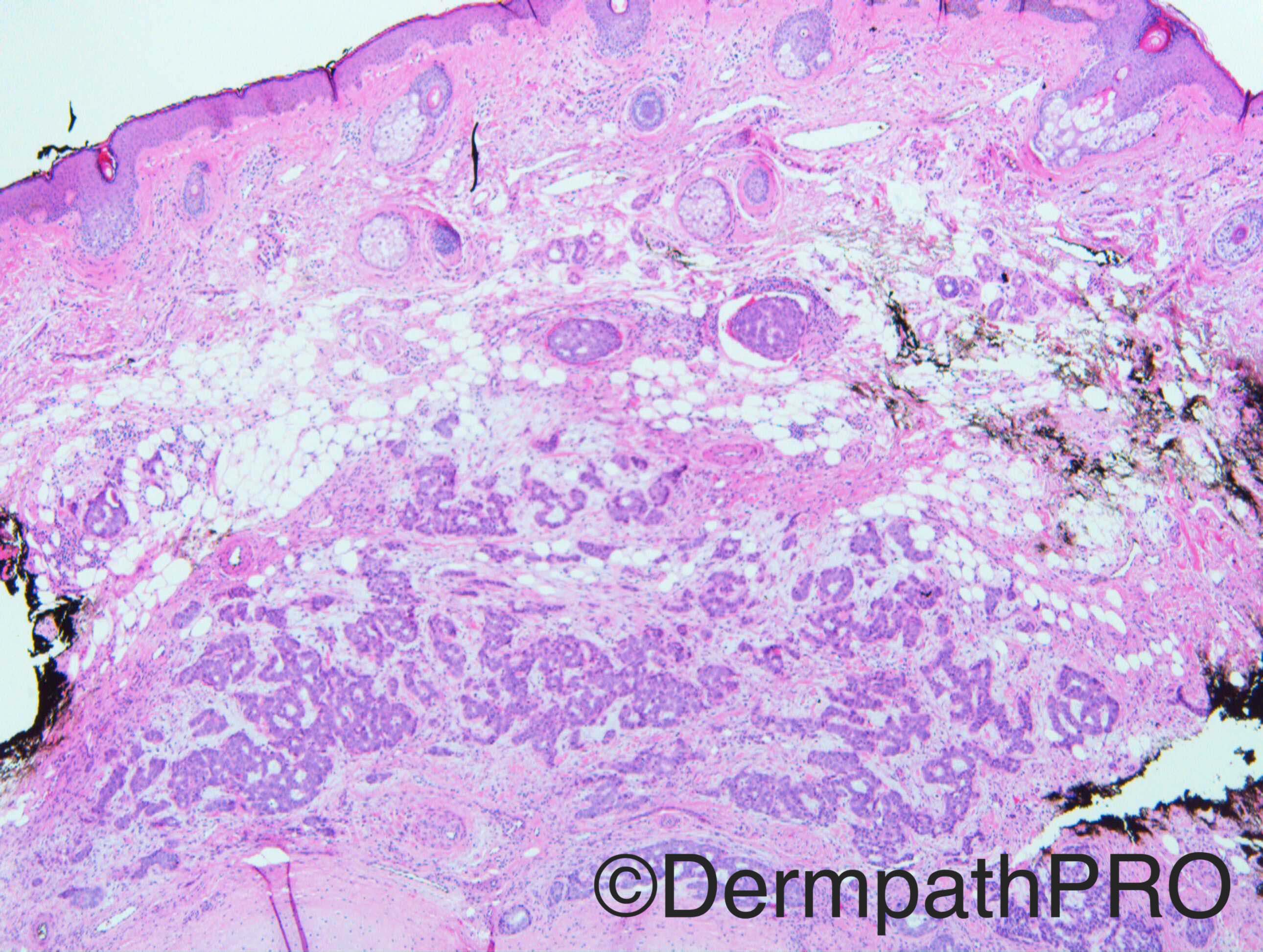
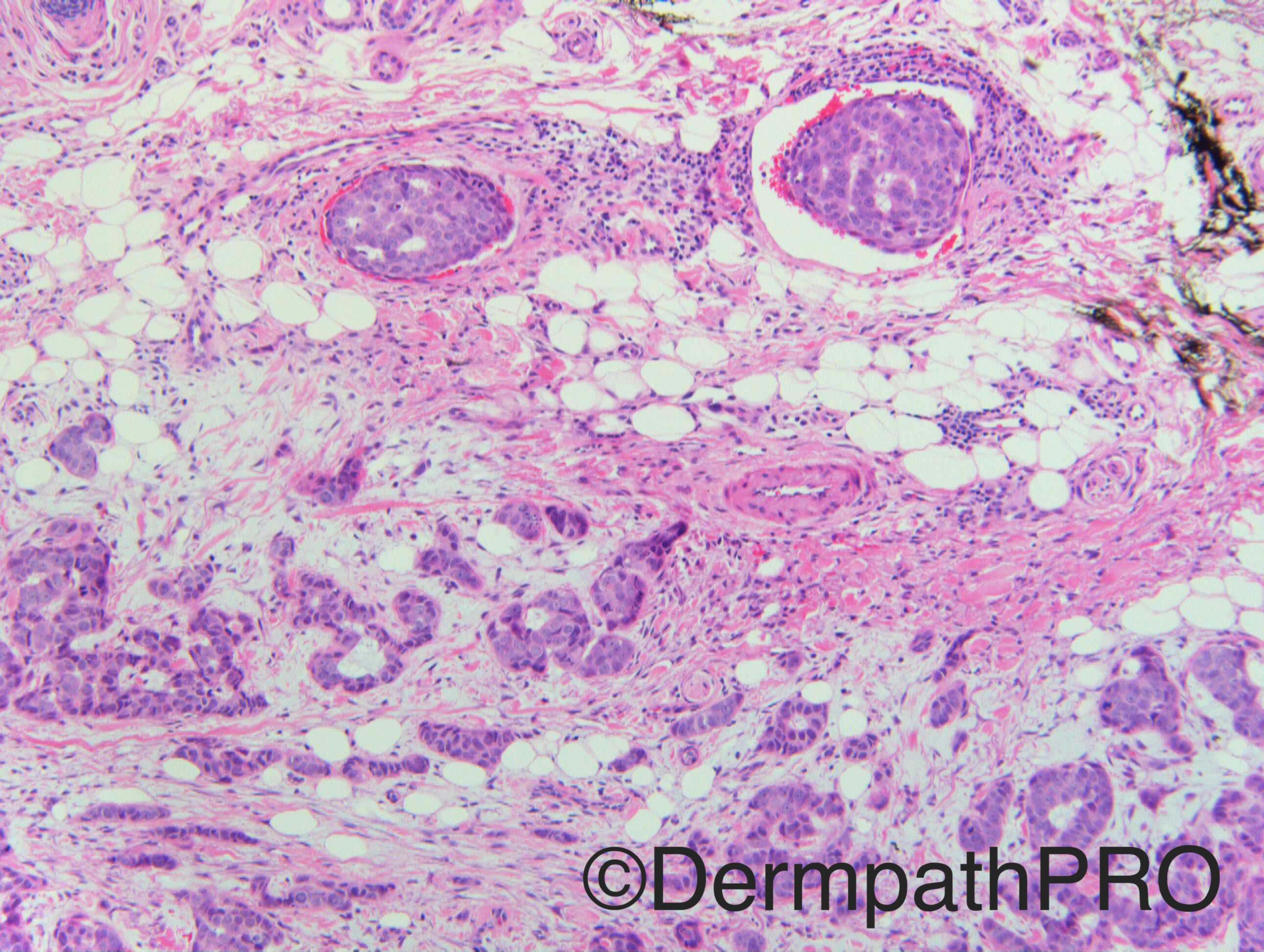
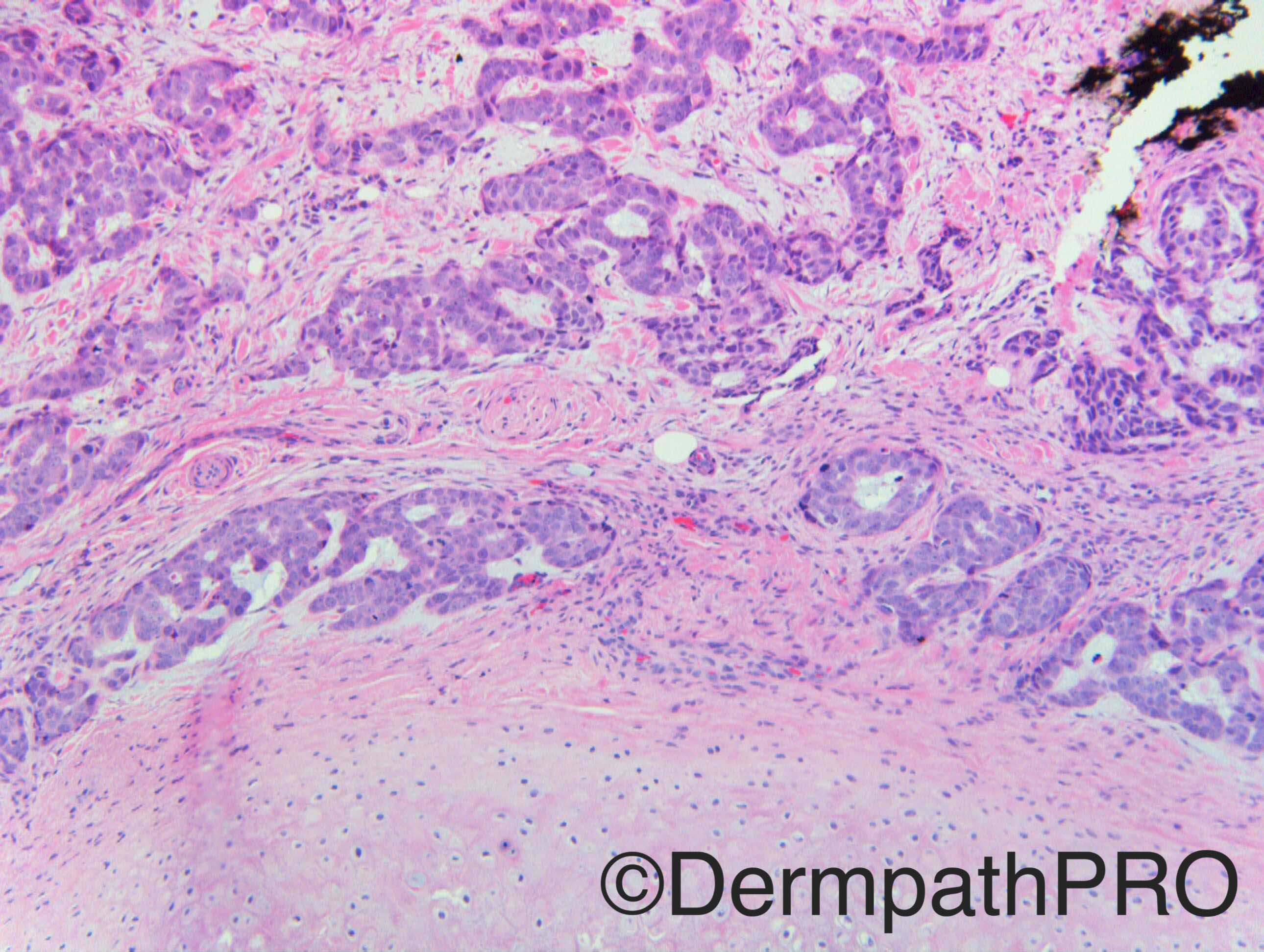
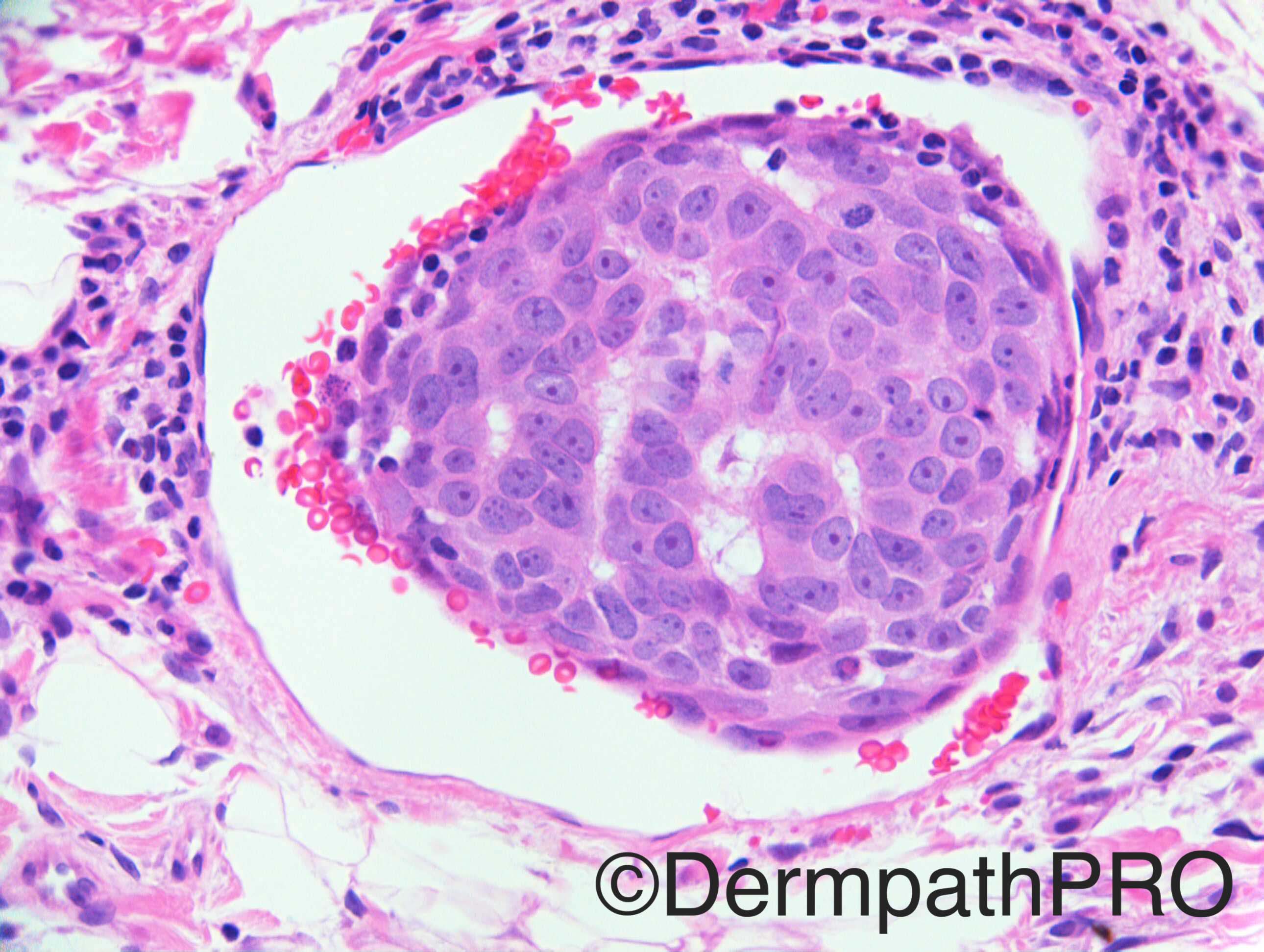
Case History: Ulcer on mid helical rim. This is from the 12 o’clock shave margin.

Additional images to follow

Case posted by Dr Richard Carr


  Report Record

User Feedback


Dr. Mona Abdel-Halim

Posted

Apocrine carcinoma with intravascular spread vs. Metastatic adenocarcinoma.

Share this comment


Link to comment
share_externally

Nitin Khirwadkar

Posted

Apocrine adenocarcinoma. DD, metastasis from a breast primary.

Share this comment


Link to comment
share_externally

Arti Bakshi

Posted

Agree with apocrine carcinoma vs mets.

Thought about ceruminous gland adenocarcinoma too, but this should be in realtion to EAC and 'helical rim' probably not the right site for this.

Share this comment


Link to comment
share_externally

Dr. Richard Carr

Posted

I have asked dermpathpro team to replace these images with my originals which show a subtle but important difference.  Hopefully this will happen before the IHC is posted.

Share this comment


Link to comment
share_externally

vincenzo polizzi

Posted

If metastatic, i would think to a Salivary Duct High Grade Carcinoma...but a primary cutaneous hydroadenocarcinoma is my first diagnosis. Angiolymphatic invasion is a typical findings.

Share this comment


Link to comment
share_externally

Guest Romualdo

Posted

I vote for metastatic carcinoma. If the patient is a man, from prostate, if a woman, from the breast.

Share this comment


Link to comment
share_externally

Raul Perret

Posted

Yeah makes me think of metastatic adenocarcinoma, I would consider lung primary too. I will wait to see the other images

Share this comment


Link to comment
share_externally

Dr. Richard Carr

Posted

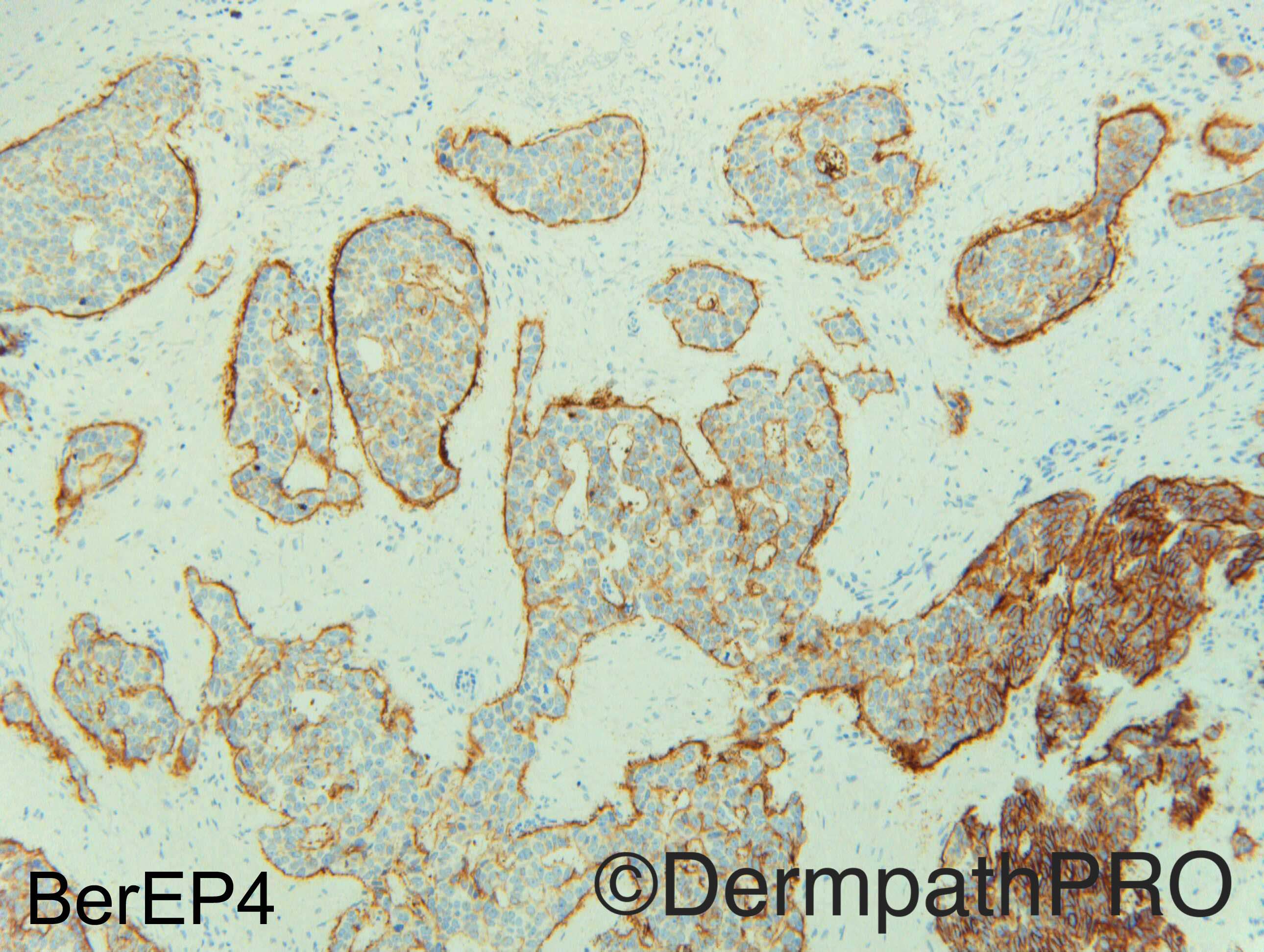
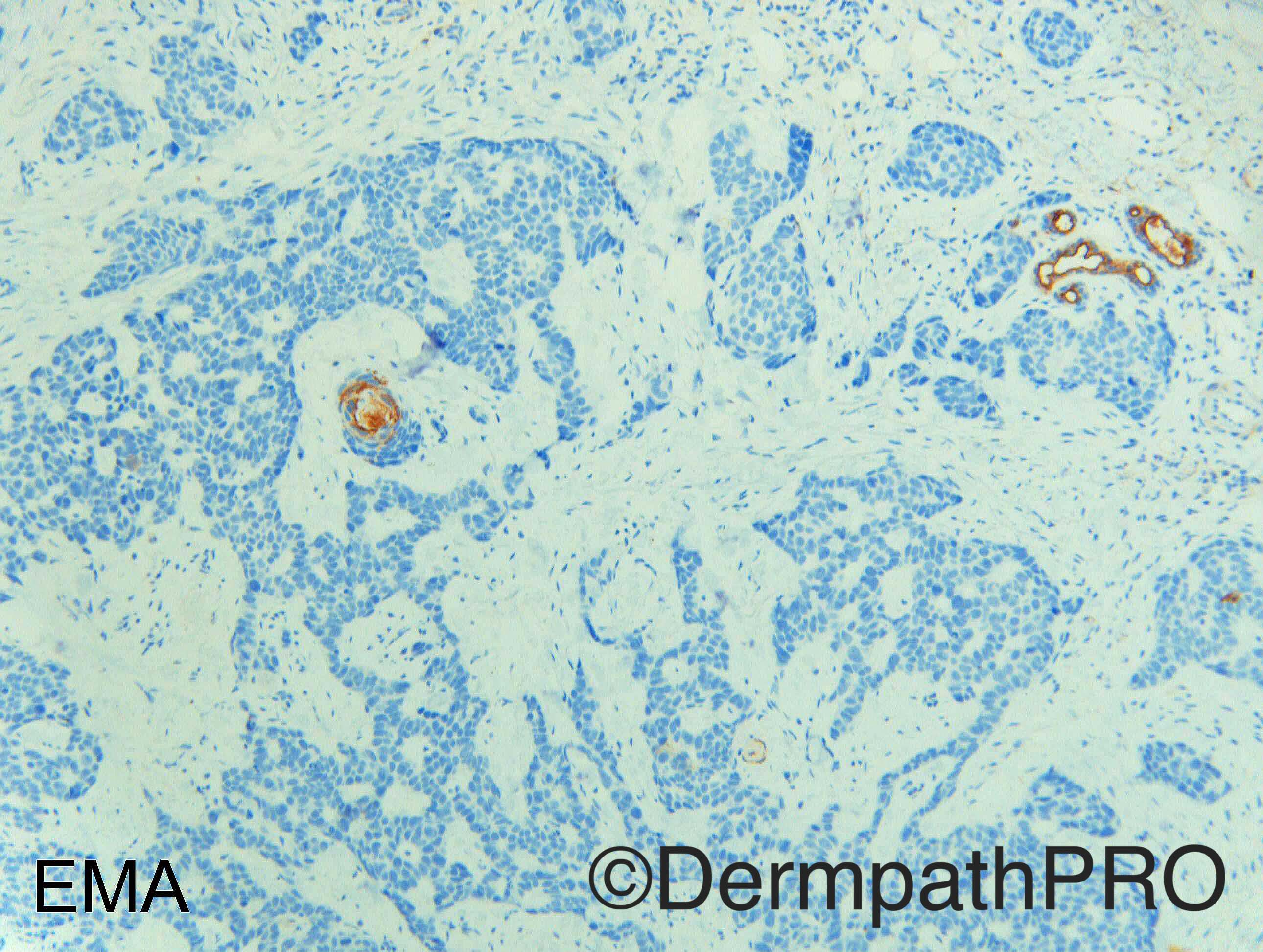
Okay looks like my original images have not made it on.  Basically they showed nice interstitial (blue-tinged) mucin around the tumour islands (these images have had the contrast upped and unfortunately the subtle clue is not really appreciable).  Now you have the IHC too.... time to re-think.  I'm still hoping the original images will get posted.

Share this comment


Link to comment
share_externally

Nitin Khirwadkar

Posted

Primary mucinous carcinoma. There is definite BerEP4 positivity. So, did think of an adenoid BCC. BerEP4 can be positive in primary cutaneous MC.

Share this comment


Link to comment
share_externally

Raul Perret

Posted

I agree that primary cutaneous mucinous carcinoma is a great option. Still we cannot rule out a metástasis as there is no in situ component in this images and we dont know the clinical. Also particular is the fact that the mucin is not abundant so this case should correspond to the mixed variant with and extensive invasive ductal component. On the other hand, even if there is an adenoid variant of BCC and even one with true glandular differentiation I dont see other features typical of BCC in this images so I would discard this dx.

 I would love to see the complete lesion...

Share this comment


Link to comment
share_externally

Arti Bakshi

Posted

Odd that EMA is completely negative (for primary mucinous Ca). Given that the mucin was surrounding tumour islands and  BerEP4 is strongly positive,  BCC with pseudo glandular pattern is a possibility.

But difficult to explain LV invasion and the deep location of tumour!  Looking forward to the answer!

Share this comment


Link to comment
share_externally

Dr. Richard Carr

Posted

Well done Arti (again!) I only showed you the shave margin.  It was a solitary, indolent lesion (clinically BCC), from the ear that looked like a fairly typical all be it infiltrative and slighly metatypical basal cell carcinoma.  Confirmed by moderate BerEP4 with peripheral accentuation (no luminal staining) and completely negative EMA in the basaloid cells (most unusual for any type of adenocarcinoma and almost a specific pattern for BCC).  I was surprised to see this very isolated lymphovascular invasion when I was cutting innumerable levels to see if tumour was clear of the shave margin.  I have only seen lymphovascular invasion a couple of times with BCC (thousands of cases reported).  I suspect we may miss it or it is just very rare.  Given the slightly metatypical look I guess there is a moderate risk for metastasis and patient is having radiotherapy (instead of a re-exision) and will be followed up as for an SCC.  There was no glandular differentiation (just pseudoglandular). Try to remember an old adage that "uncommon variants of common lesions are still more common than rarities" (although not necessarily on Dermpathpro!).

Share this comment


Link to comment
share_externally


Join the conversation

You can post now and register later. If you have an account, sign in now to post with your account.

Guest
Add a comment...

×   Pasted as rich text.   Paste as plain text instead

  Only 75 emoji are allowed.

×   Your link has been automatically embedded.   Display as a link instead

×   Your previous content has been restored.   Clear editor

×   You cannot paste images directly. Upload or insert images from URL.

Loading...

×
×
  • Create New...