Jump to content
In this section we have spot diagnoses posted on a daily basis since June 2010, now over 4000! You can review the archived cases and read the suggested diagnoses by users and the final comment by the contributors.
Case are uploaded each week day by 10 am UK time with the correct diagnosis will generally be posted at 8 pm UK time. Why not view the most recent spot diagnosis and proffer a diagnosis?

Case Number : Case 1570 - 01 July Posted By: Guest

Please read the clinical history and view the images by clicking on them before you proffer your diagnosis.
Submitted Date :
   (0 reviews)

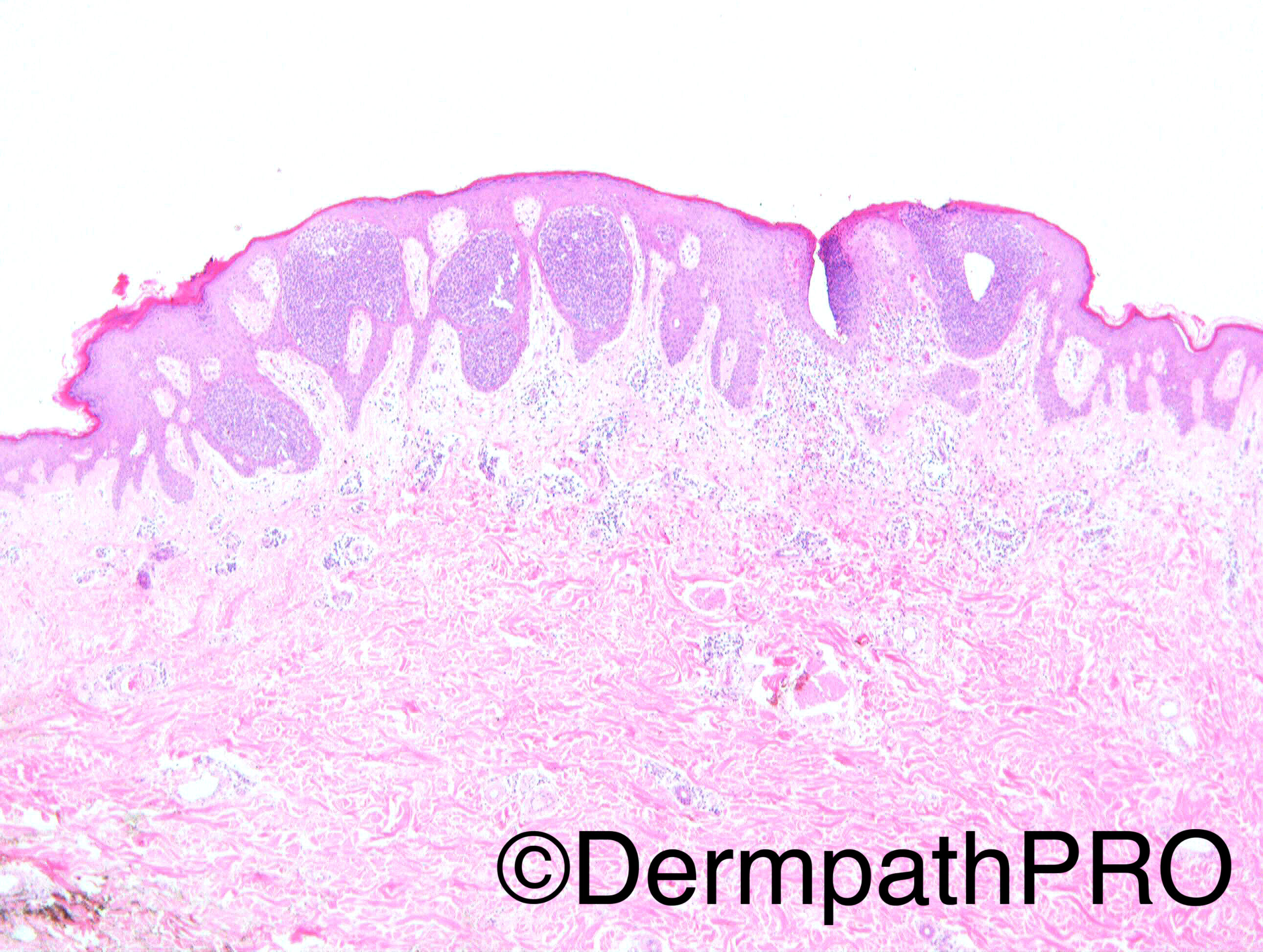
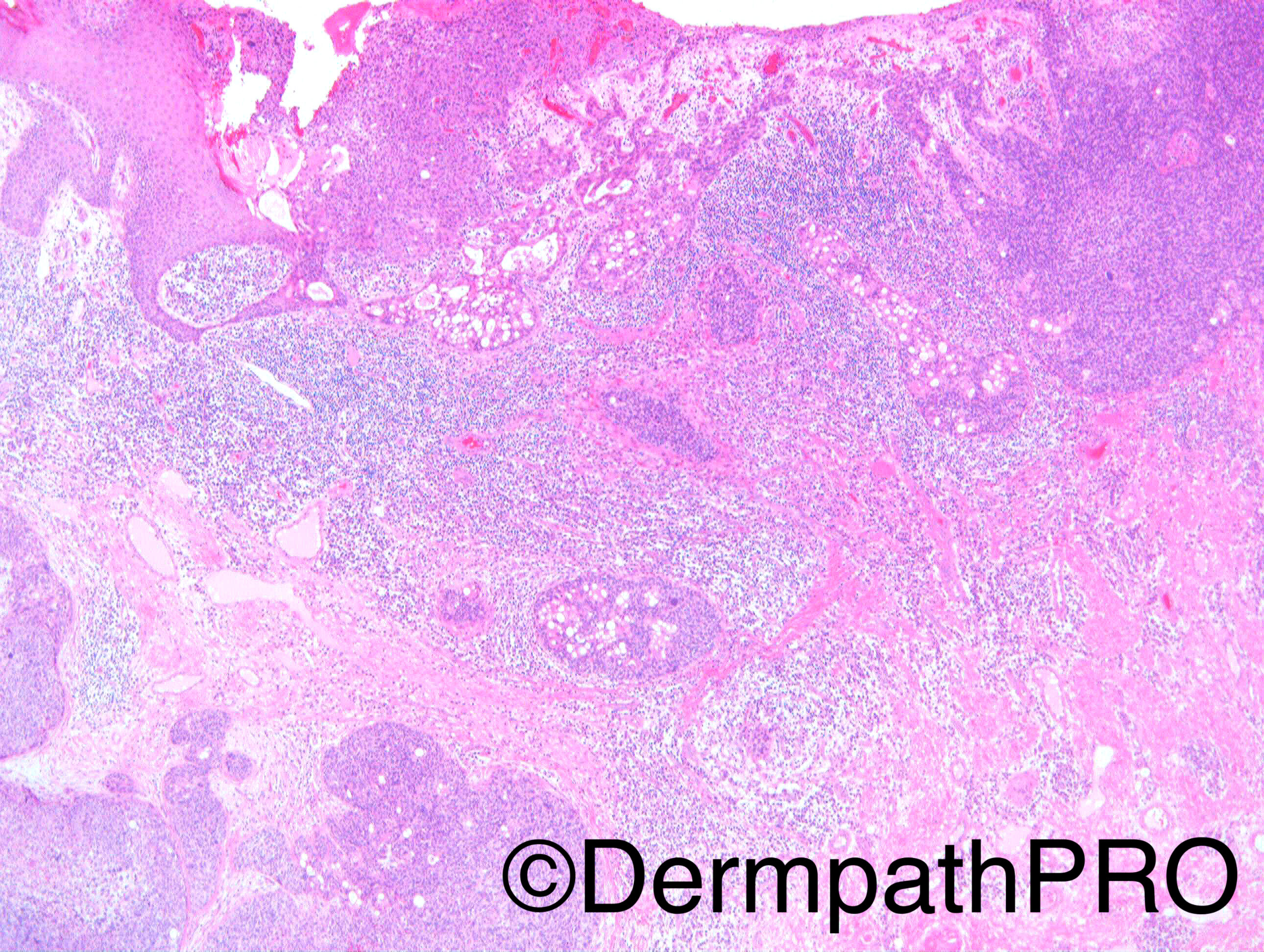
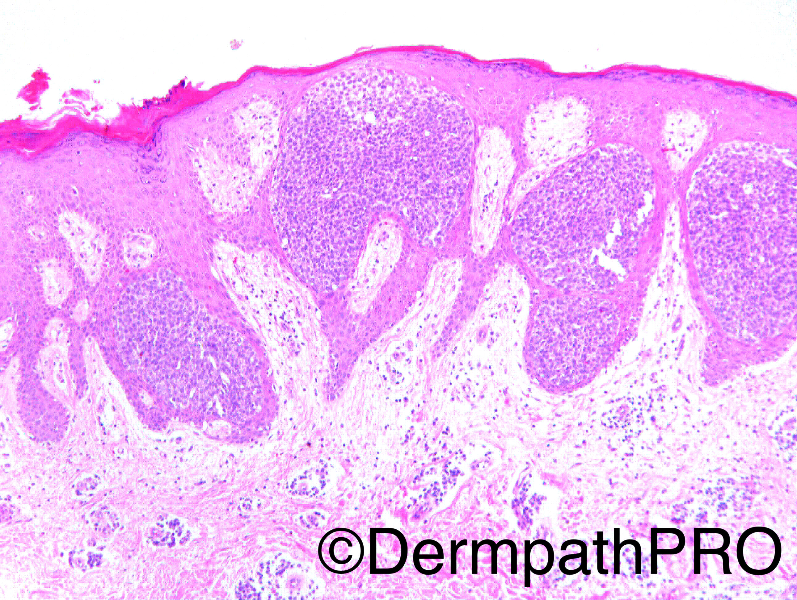
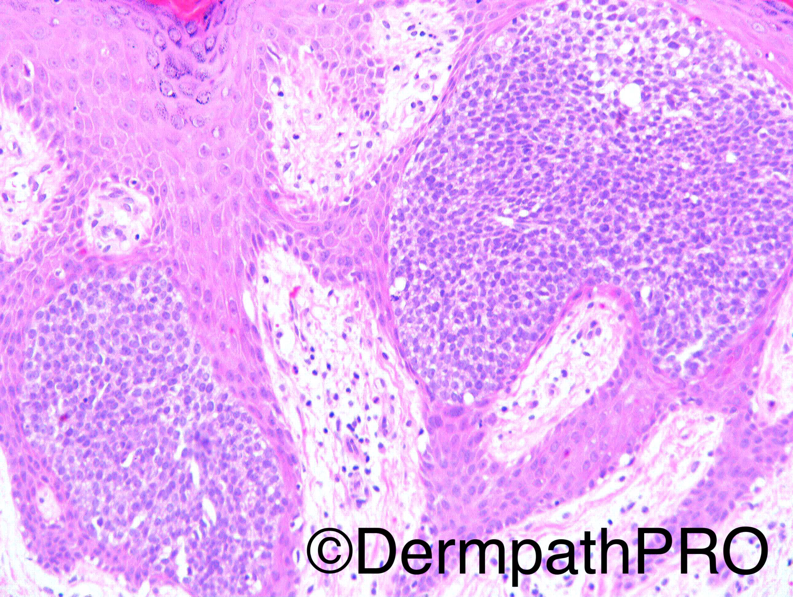
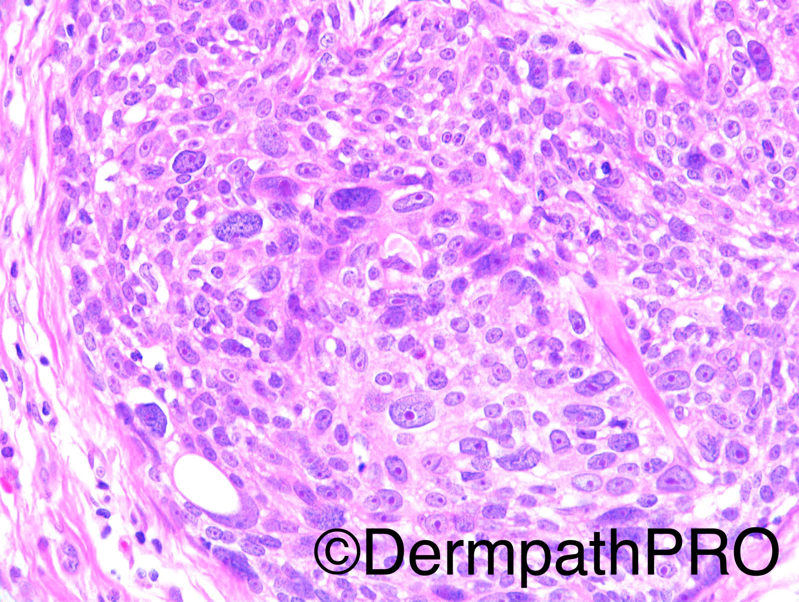
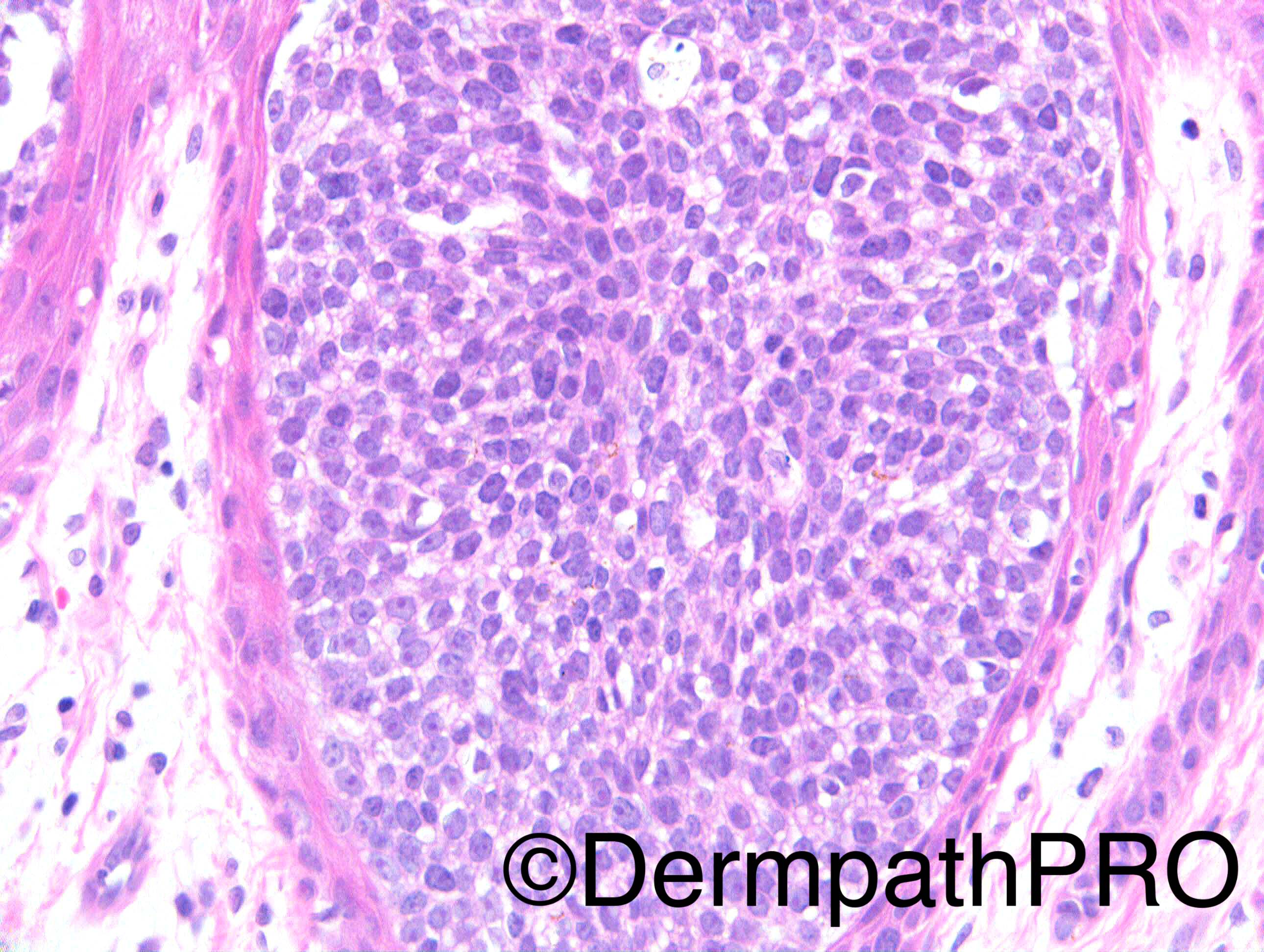
F85. Longstanding lesion, recently bled.


Dr Richard Carr


  Report Record

User Feedback


vincenzo polizzi

Posted

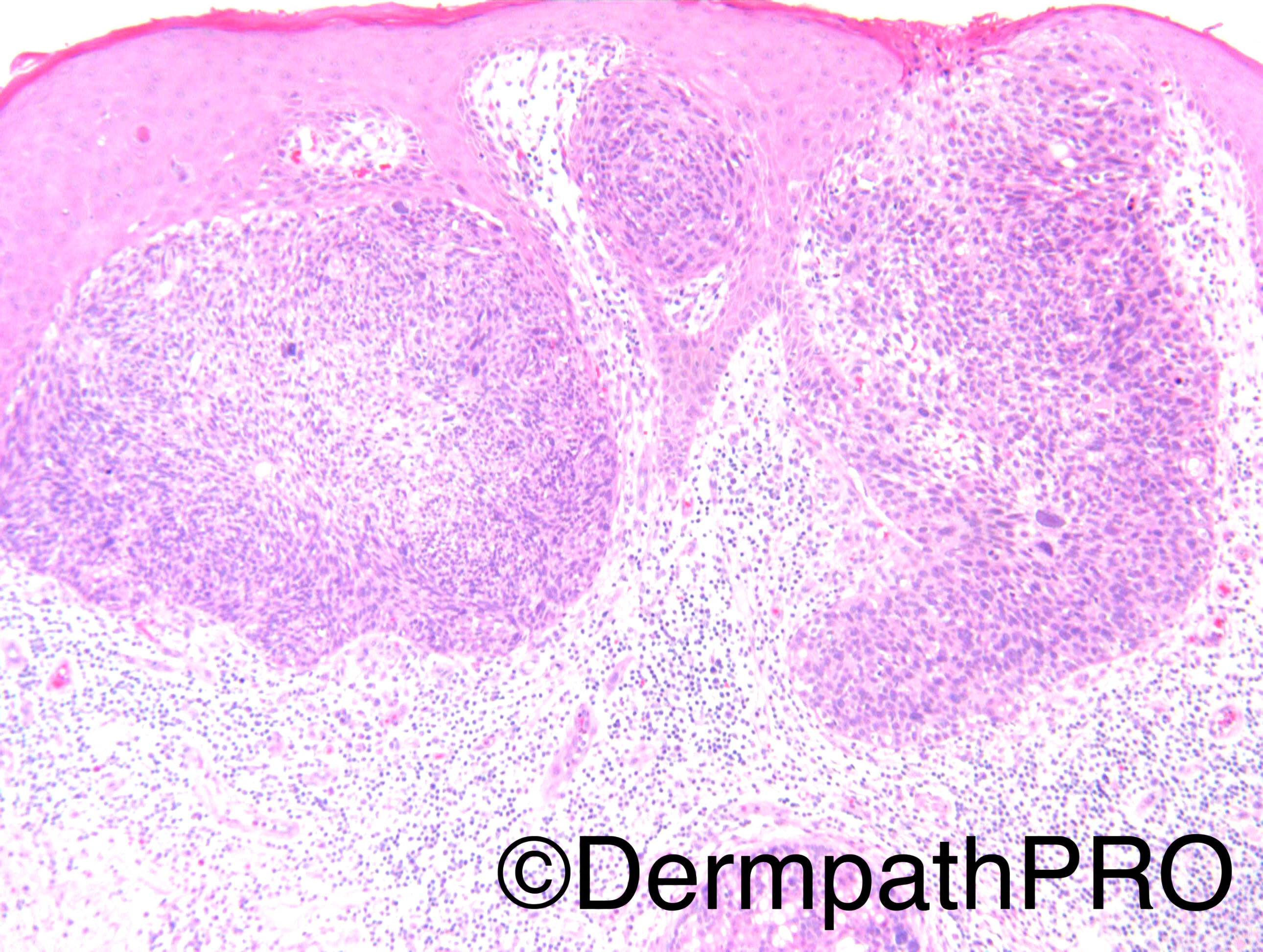
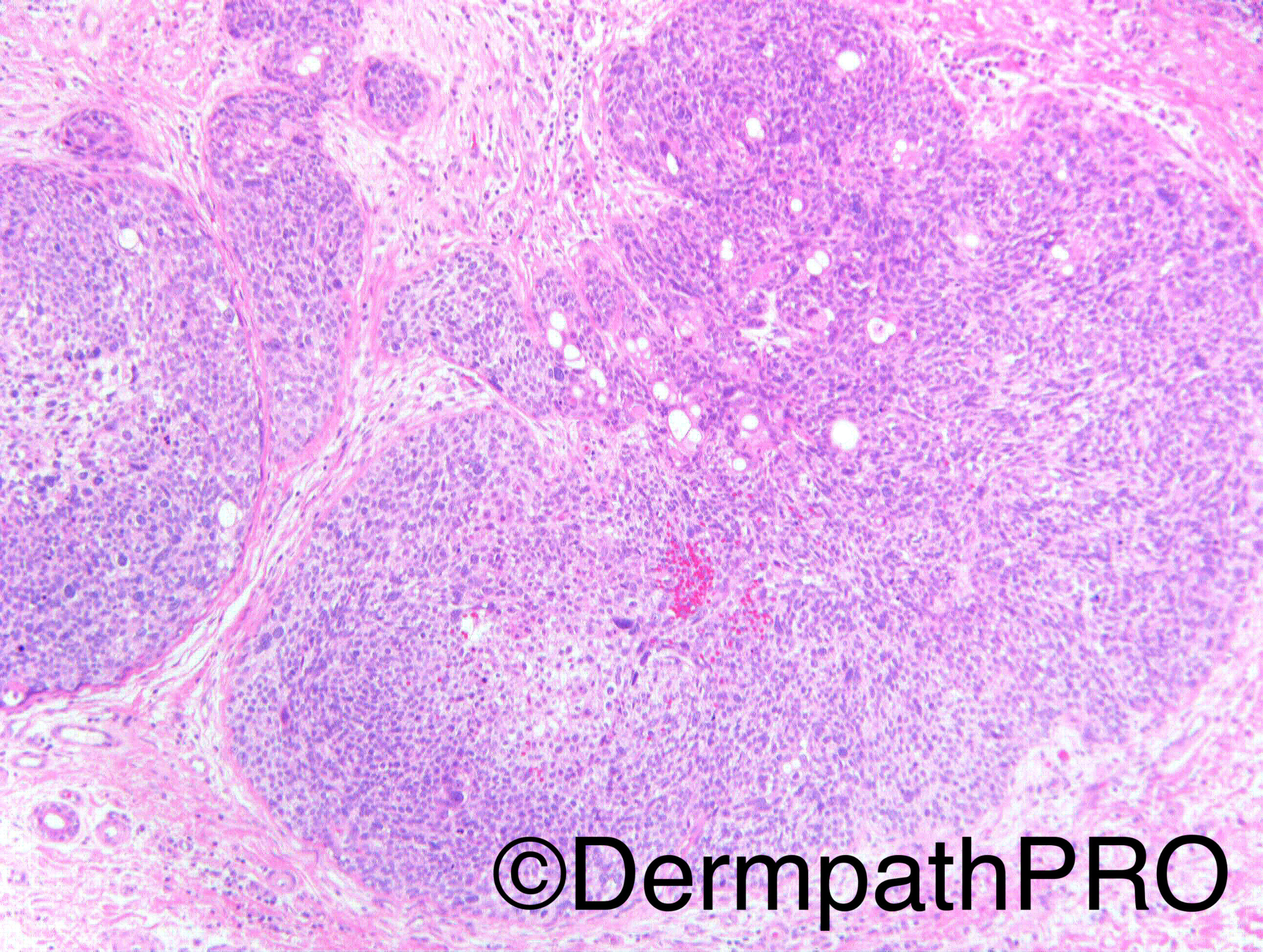
Eccrine Invasive Porocarcinoma with a residual in situ component

Share this comment


Link to comment
share_externally

Dr. Mona Abdel-Halim

Posted

Yes, porocarcinoma.

Share this comment


Link to comment
share_externally

vincenzo polizzi

Posted

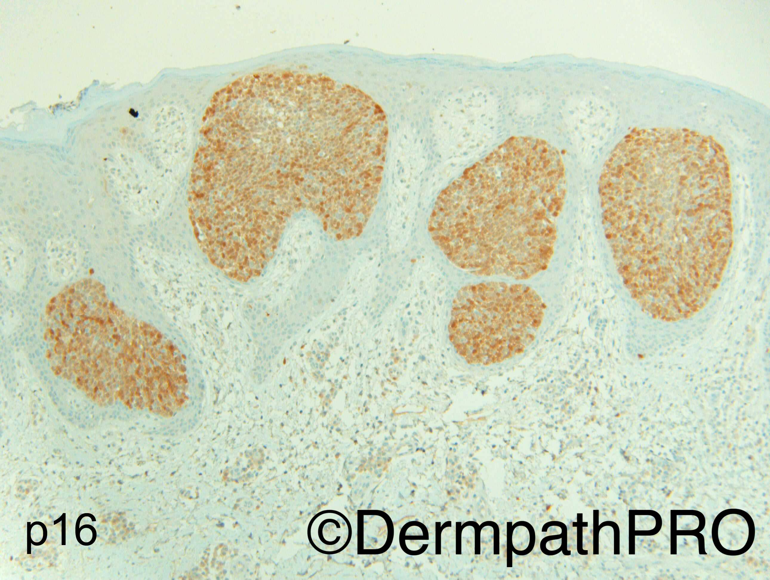
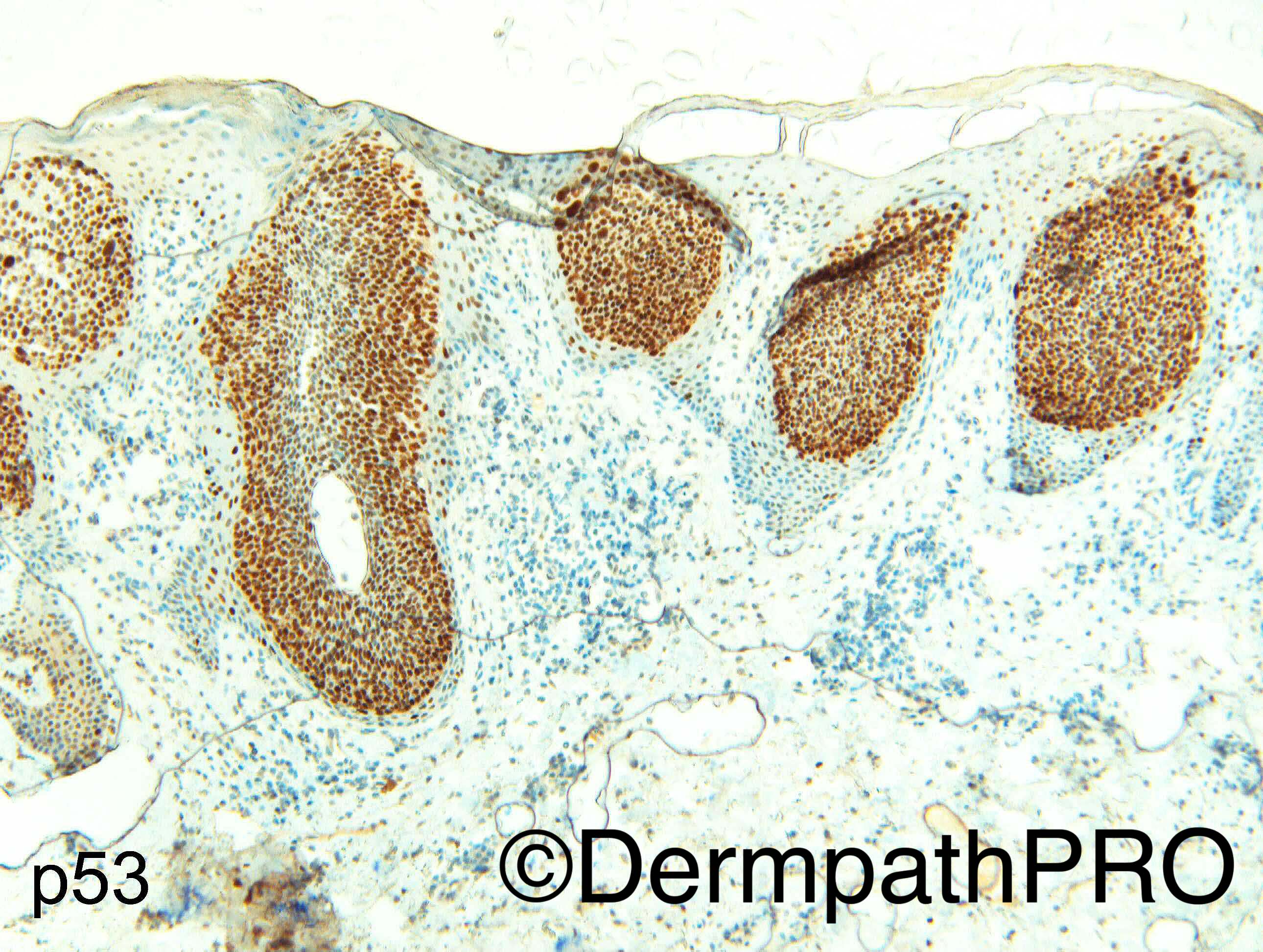
Nice p16 expression highlighting the malignant nature of the intraepidermal poromatous component...Dr Carr what do you think about the p53 usefulness in order to distinguish between benign and malignant lesions? I've studied something about this but i'm a bit confuse. Thanks for this very interesting case!

Share this comment


Link to comment
share_externally

Guest Arash Daryakarr

Posted

Agree with porocarcinoma

Share this comment


Link to comment
share_externally

Dr. Richard Carr

Posted

Case kindly received in referral by Dr Miroslav Radojkovic.


In view of the purely rounded borders I called this porocarcinoma / malignant hidroacanthoma in situ.  Porocarcinoma in situ can have rather deep pushing, but entirely circusmcript, borders as in this case.


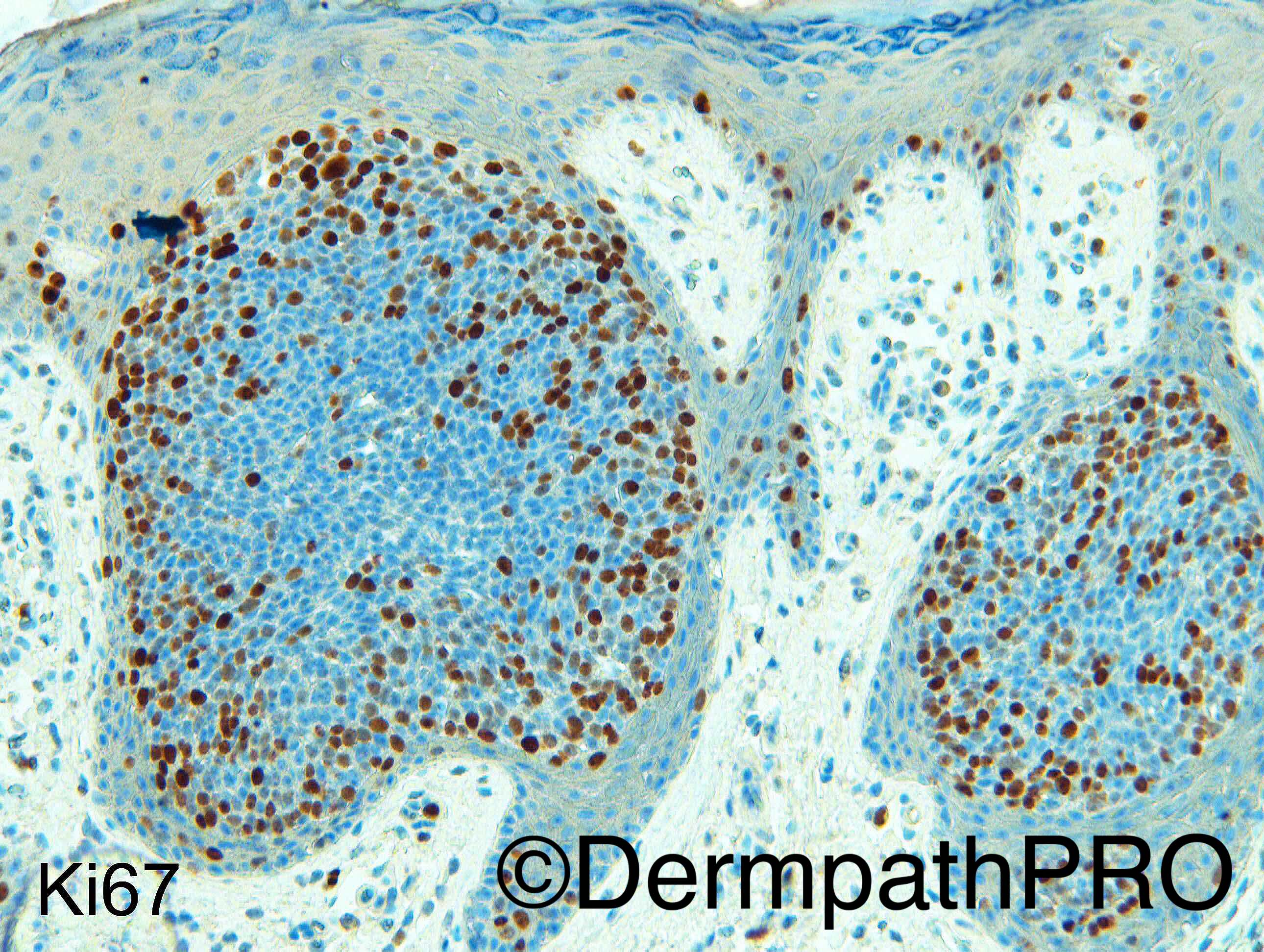
I find p53 (diffuse strong nuclear or completely negative, null phenotype, with good internal controls) quite useful being nearly 99% specific for malignancy (I've seen diffuse strong p53 once only in a lesion I called benign, an otherwise typical pilomatrixoma).  It is not so sensitive however (circa 50% for surface tumours - no doubt UV induced mutations mainly). Beware you can have quite prominent weak and moderate staining as wild-type up-regulation (but maturing central cells will be weak/negative) so my cut-off for diffuse strong is high e.g. ~80% nuclei with strong staining - personal experience. In clonal lesions I find the combination of p16, Ki67 and p53 to be highly useful in distinguishing benign from malignant.  You can see quite a variation of p16 and p53 in individual cases (p16+, p53 wild; p16-, p53 mutant/null, p16+/p53 mutant/null)


This case did have a benign hidroacanthomatous component - not illustrated.

Share this comment


Link to comment
share_externally


Join the conversation

You can post now and register later. If you have an account, sign in now to post with your account.

Guest
Add a comment...

×   Pasted as rich text.   Paste as plain text instead

  Only 75 emoji are allowed.

×   Your link has been automatically embedded.   Display as a link instead

×   Your previous content has been restored.   Clear editor

×   You cannot paste images directly. Upload or insert images from URL.

Loading...

×
×
  • Create New...