Jump to content
In this section we have spot diagnoses posted on a daily basis since June 2010, now over 4000! You can review the archived cases and read the suggested diagnoses by users and the final comment by the contributors.
Case are uploaded each week day by 10 am UK time with the correct diagnosis will generally be posted at 8 pm UK time. Why not view the most recent spot diagnosis and proffer a diagnosis?

Case Number : Case 1580 - 15 July Posted By: Guest

Please read the clinical history and view the images by clicking on them before you proffer your diagnosis.
Submitted Date :
   (0 reviews)

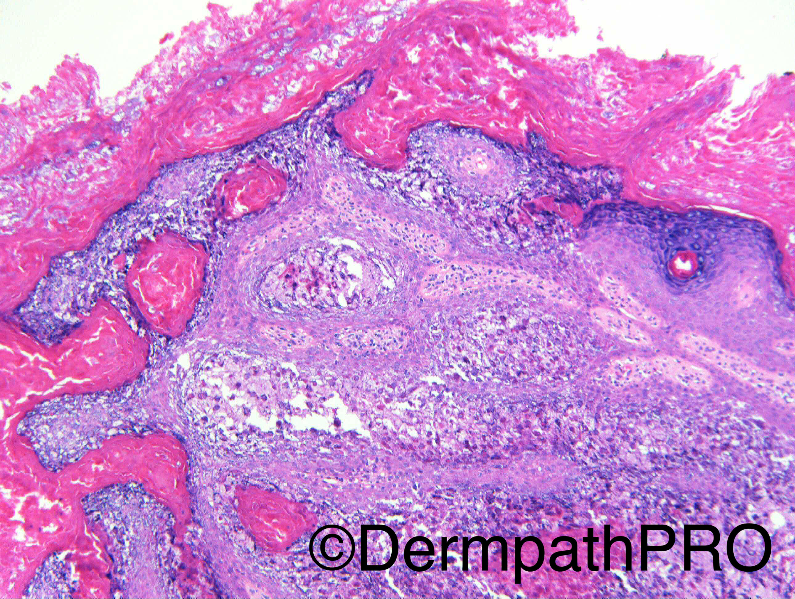
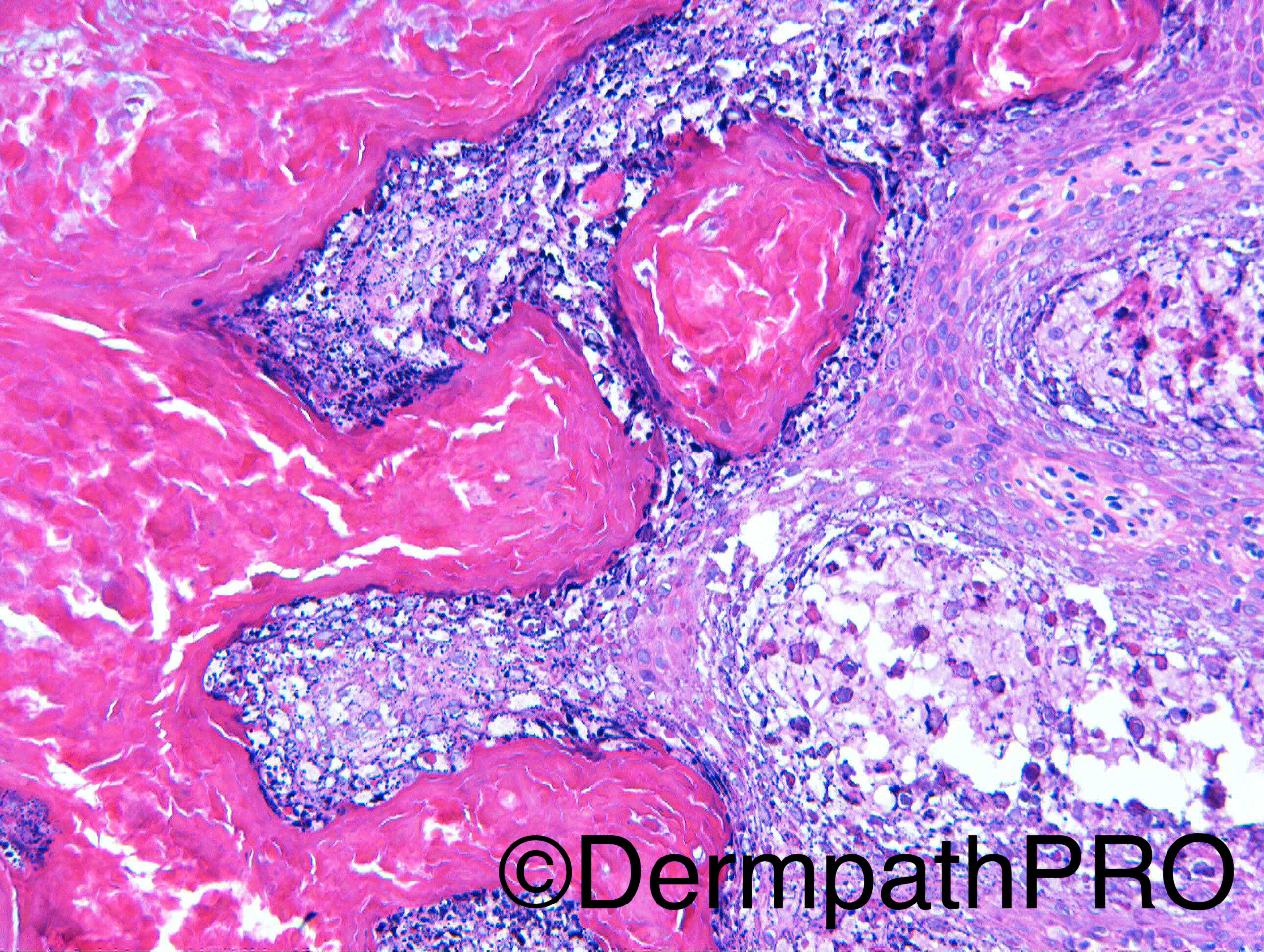
M60. Biopsy from GUM clinic. Site not stated! White irregular surfaced papule. ?Wart

Dr Richard Carr


  Report Record

User Feedback


Raul Perret

Posted

epidermolythic acanthoma (if unique)

Share this comment


Link to comment
share_externally

Agree with epidermolythic acanthoma

Share this comment


Link to comment
share_externally

Nitin Khirwadkar

Posted

Epidermilytic acanthoma. Multiple genital ones have been described.

Share this comment


Link to comment
share_externally

Raul Perret

Posted

Epidermilytic acanthoma. Multiple genital ones have been described.

Yes, you right I meant that I did not think it was part of a genodermatosis. Agree multiple genital and even disseminted cases have been reported

Share this comment


Link to comment
share_externally

Dr. Mona Abdel-Halim

Posted

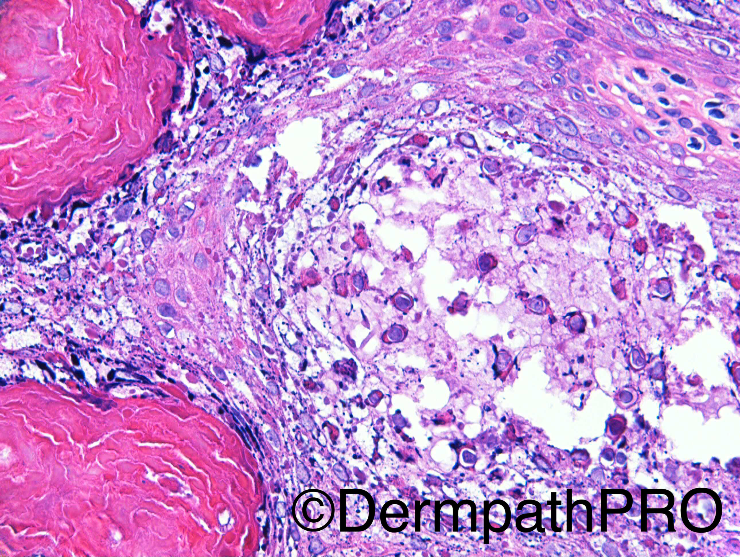
I just think that the last image is showing peri nuclear esinophilic condensations of WSN or am I wrong??
Also WSN can affect genital mucosa and according to McKee's Pathology of skin, foci of epidermolytic hyperkeratosis have been reported in a case of WSN.

Share this comment


Link to comment
share_externally

Dr. Richard Carr

Posted

Yes this is a rather wart-like example of epidermolytic acanthoma. To our knowledge it is not that rare on the genitalia but perhaps under-recognised and may be confused with viral warts although none of the reported cases that have been worked up has HPV genome been demonstrated. I don't think I have ever seen a white sponge naevus (genital, oral or otherwise) as a hot case but it should have typical clinical features and is usually present from early life.  Here the epidermolytic hyperkeratosis pattern is rather spectacular. There are now four genital cases of EA in my slide collection, all "routine" cases.

Share this comment


Link to comment
share_externally


Join the conversation

You can post now and register later. If you have an account, sign in now to post with your account.

Guest
Add a comment...

×   Pasted as rich text.   Paste as plain text instead

  Only 75 emoji are allowed.

×   Your link has been automatically embedded.   Display as a link instead

×   Your previous content has been restored.   Clear editor

×   You cannot paste images directly. Upload or insert images from URL.

Loading...

×
×
  • Create New...