Jump to content
In this section we have spot diagnoses posted on a daily basis since June 2010, now over 4000! You can review the archived cases and read the suggested diagnoses by users and the final comment by the contributors.
Case are uploaded each week day by 10 am UK time with the correct diagnosis will generally be posted at 8 pm UK time. Why not view the most recent spot diagnosis and proffer a diagnosis?

Case Number : Case 1555 - 10 June Posted By: Guest

Please read the clinical history and view the images by clicking on them before you proffer your diagnosis.
Submitted Date :
   (0 reviews)

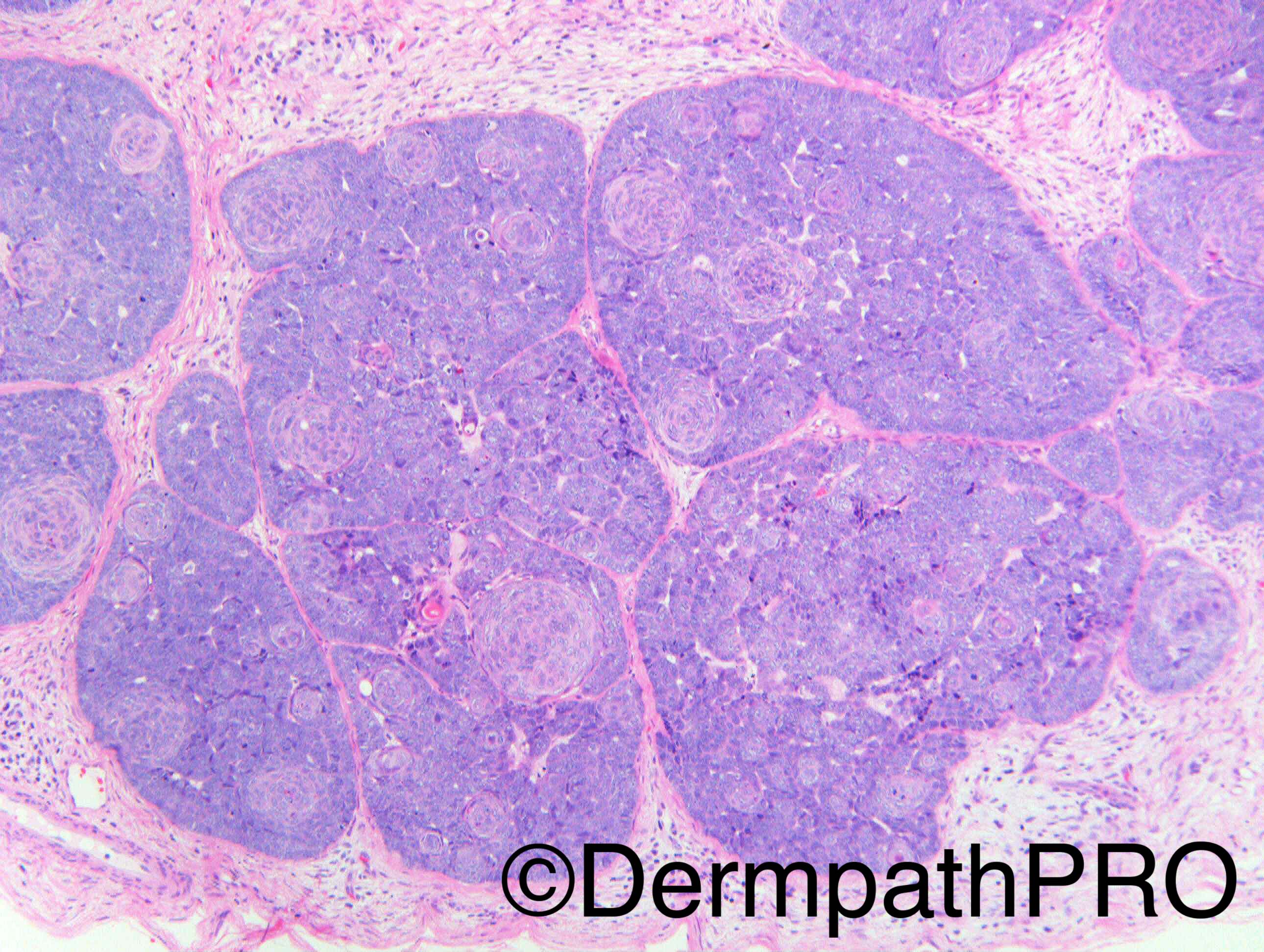
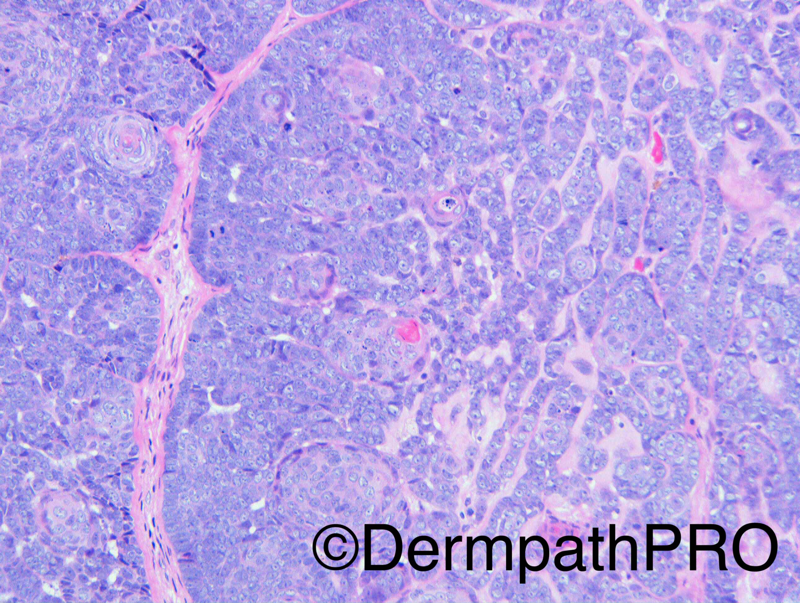
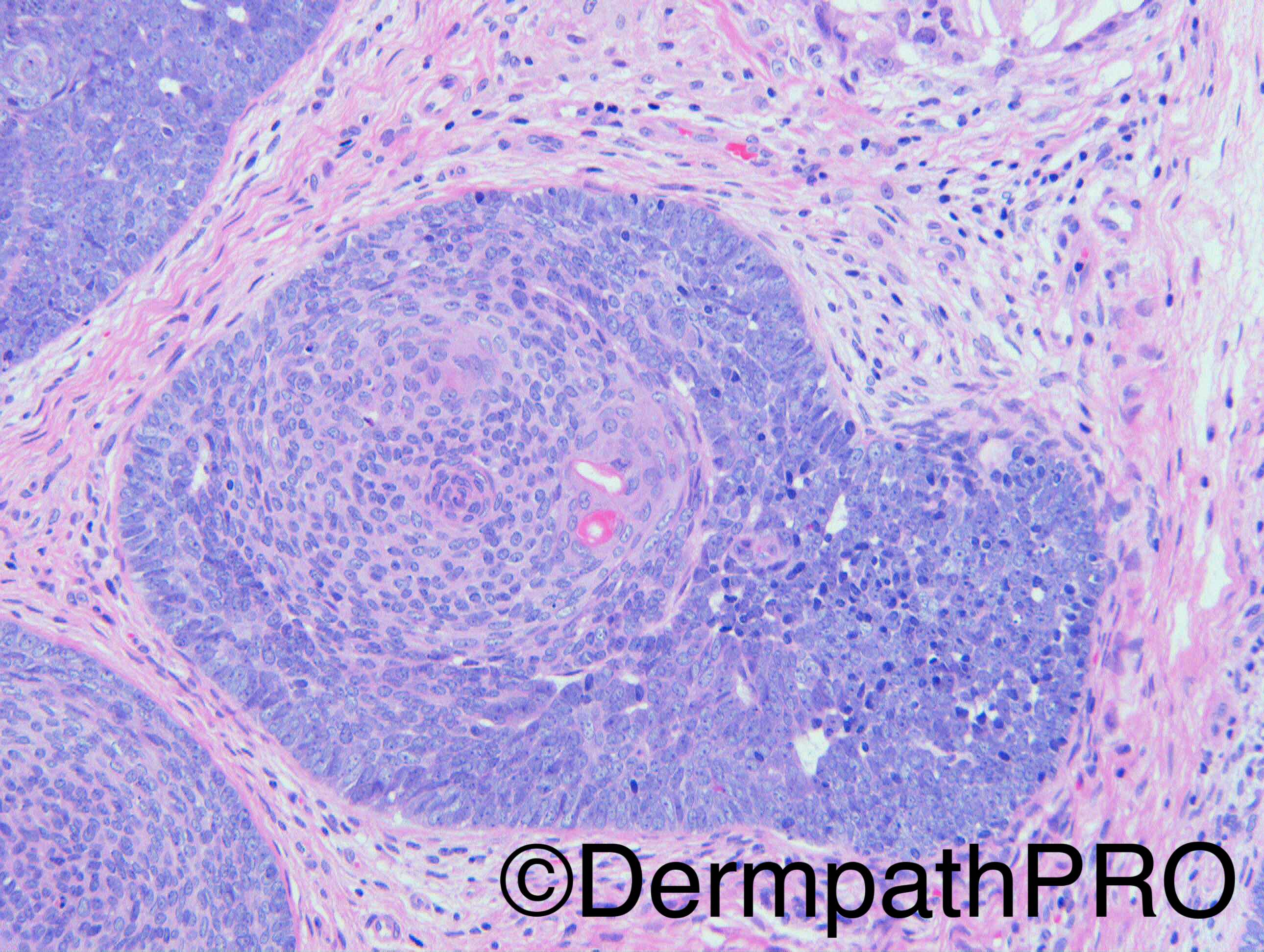
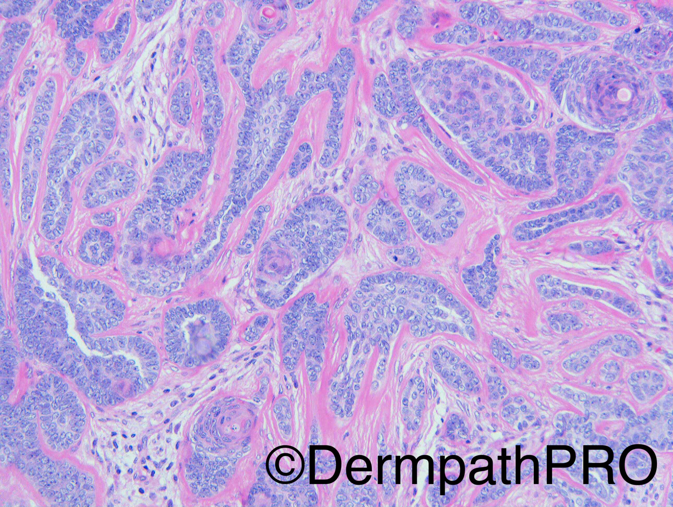
F60. Upper arm. Many years, itchy, firm, sub-dermal, mobile nodule, with small punctum.

Dr Richard Carr


  Report Record

User Feedback


vincenzo polizzi

Posted

A Trichoblastoma with distinctive cell balls of pale squamoid cells with peripheral condensation, reminiscent of Trichogerminoma...

Share this comment


Link to comment
share_externally

Raul Perret

Posted

I dont find this case easy. My impression is that we see a basaloid neoplasm with mainly a lobular architecture and cord like areas with extensive hyaline material surrounding them. I see some sebaceous ducts and some infundibulocystic areas (or maybe just cystically dilated sebaceous ducts?) there is atypia to my eyes (quite prominent) and mitosis. Some isolated palissading but no retraction artifact and no mucin. The Neoplasm seems mainly dermal but focally connected to epidermis? (lower right corner of first picture). I was thinking of sebaceous carcinoma, ddx with bcc with sebaceous differentiation so would do some immunos EMA, Adipophilin, Ber-ep4, CD10, CK20, P53 would probably throw some neuroendocrine markers too. Trichoblastoma should be a differential but sincerely It was not my first impression, anyways I may be wrong. Really funny perforating phenomenon of the neoplasm, nice case

Share this comment


Link to comment
share_externally

Nitin Khirwadkar

Posted

Basaloid tumour with focal epidermal attachment. Focal, labyrinthine and jigsaw puzzle arrangement along with a predominant multi nodular growth pattern. Not sure if there is definite follicular mesenchyme type stroma. Prominent sebaceous ducts and infundibulocystic structures. No retraction artefact or stream mucin. I haven't seen individual sebocytes (mature/immature). Some of the pictures show overt atypia and brisk mitoses in the busload cells. Agree that we need some IHC (highlighted by Raul).


Could be a sebaceoma/sebaceous carcinoma. Did ponder about a trichoblastic carcinoma. However, would be difficult to diagnose TC in absence of an existing trichoblastoma.


Great, but a difficult case. Waiting for the answer :)

Share this comment


Link to comment
share_externally

Dr. Mona Abdel-Halim

Posted

I am finding it difficult, first thought trichoblastoma (trichogerminomatous variant) with ductal differentiation. But the 7th image is showing hyaline basement membranes reminiscent of ORS differentiation so I am also thinking of panfolliculoma but still can't see areas of matrical differentiation. Waiting to learn :-))

Share this comment


Link to comment
share_externally

Arti Bakshi

Posted

Odd feature for trichoblastome is that biphasic nature is not very obvious in these images. The specific follicular stroma (if present) is rather focal, maybe in image 6. Agree that there is some evidence of pan follicular differentiation in the form of infundibular and isthmic differentiation (sebaceous ducts with cell contains abundant cytoplasm). Not seen any sebocytes though to think sebaceous carcinoma. There is epidermal connection and areas of prominent atypia/ mitoses...odd features for both TB and panfolliculoma. So I am wondering if there is a component of BCC, in a background of trichoblastoma/panfolliculoma.

Share this comment


Link to comment
share_externally

Dr. Richard Carr

Posted

Case c/o Dr George Powell.  My report:

Many thanks for sending this rather wonderful case of a widely trichogerminomatous variant of a large nodular trichoblastoma (benign hair follicle tumour). The diagnosis is supported by condensation of papillary mesenchymal cells (highlighted by CD10 and CD34) in addition to widespread, moderately numerous, Merkel cells (CK20, EMA). In the sections submitted the lesion appears shelled out completely with a narrow soft tissue margin.


Appears I did not photograph the IHC.  Note the lobulated border of the stroma at low power - a typical feature of trichoblastomas. The cell balls here are rather typical of the entity. These lesions often are dominated by epithelium compared with stroma unlike most other TE/TB. In this case the papillary meschymal component is relatively inconspicuous but seen in image 6. There was focal inner root sheath differentiation (not illustrated). Foci of sebaceous differentiation were inconspicuous. The basement membrane thickening is prominent and I think that is exceedingly rare in BCC (I am not entirely convinced by all cases illustrated in the literature - i.e. some of them are trichoblastomas although I don't doubt it can also be seen rarely in BCC).

Share this comment


Link to comment
share_externally


Join the conversation

You can post now and register later. If you have an account, sign in now to post with your account.

Guest
Add a comment...

×   Pasted as rich text.   Paste as plain text instead

  Only 75 emoji are allowed.

×   Your link has been automatically embedded.   Display as a link instead

×   Your previous content has been restored.   Clear editor

×   You cannot paste images directly. Upload or insert images from URL.

Loading...

×
×
  • Create New...