Jump to content
In this section we have spot diagnoses posted on a daily basis since June 2010, now over 4000! You can review the archived cases and read the suggested diagnoses by users and the final comment by the contributors.
Case are uploaded each week day by 10 am UK time with the correct diagnosis will generally be posted at 8 pm UK time. Why not view the most recent spot diagnosis and proffer a diagnosis?

Case Number : Case 1486- 04 March Posted By: Guest

Please read the clinical history and view the images by clicking on them before you proffer your diagnosis.
Submitted Date :
   (0 reviews)

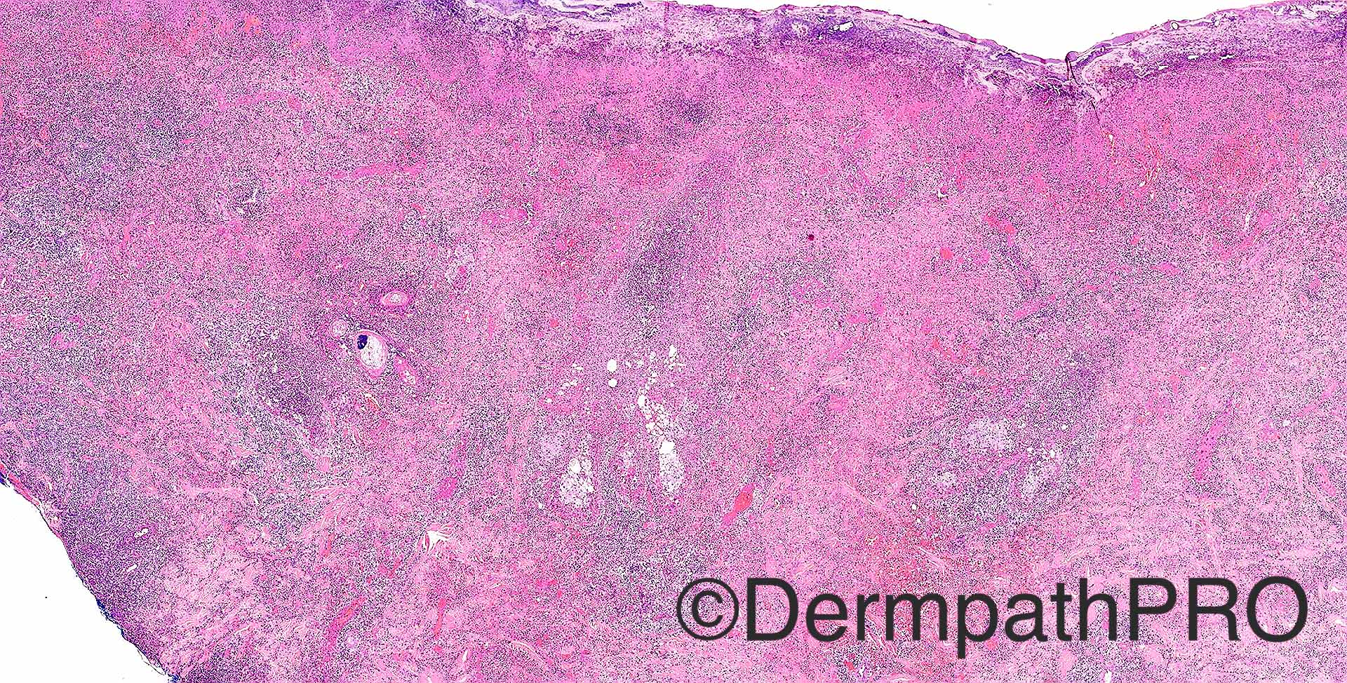
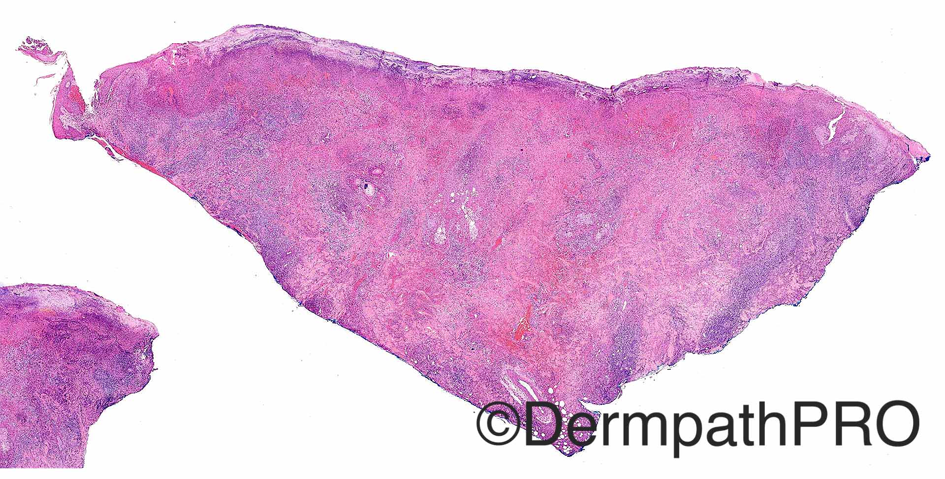
Case History: F30. Lesion chest wall, 2/52 tender & swollen. 12 to 14 weeks pregnant. Case c/o Dr Rand Hawari

Case posted by Dr Richard Carr


  • Like 1
  Report Record

User Feedback


Dr. Richard Carr

Posted

If you attended the ISDP session in New Dehli, please try to withhold the temptation to give the answer!

Share this comment


Link to comment
share_externally

vincenzo polizzi

Posted

  1. ???Herpetic changes...?necrosis caused by vascular trombosis

Share this comment


Link to comment
share_externally

Raul Perret

Posted

I will give it a go. It is clearly an acute ulcer, rich in neutrophils and lymphocytes

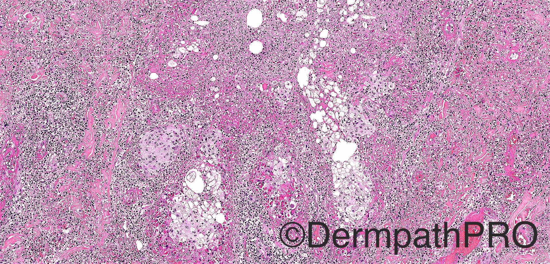
(mainly in the periphery). The dermal infiltrate seems to surpass the edges of the ulcer and looks quite Deep. There is also this particular infiltrate that to my eyes adopts the shape of follicular units with some foamy histiocytes present (necrotizing folliculitis with abscess formation?). I think we should first rule out an infectious cause but this morphology and the clinical presentation makes me think of pyoderma gangrenosum.

Share this comment


Link to comment
share_externally

vincenzo polizzi

Posted

Also was thinking of pyoderma gangrenosum. but there are particular nuclear and cellular changes in sebocytes...Very difficult!

Share this comment


Link to comment
share_externally

Raul Perret

Posted

I think that CPC is extremely important in this case. Infectious causes of ulcers due to Herpes family of virus (including zoster) have specific clinical settings that help differentiate from pyoderma.

Share this comment


Link to comment
share_externally

Arti Bakshi

Posted

Agree that the infiltrate does appear to be centered on folliculosebaceous units (although quite extensive elsewhere too)....like the suggestion of herpes folliculitis

Share this comment


Link to comment
share_externally

Very difficult case. I also though about herpes infection because of the sebaceous inflammation. There´s a disease called neutrophilic sebaceous adenitis, but the histology is different as is the clinical description.

Share this comment


Link to comment
share_externally

Dr. Mona Abdel-Halim

Posted

I think also that this is herpetic folliculitis.

Share this comment


Link to comment
share_externally

Guest Romualdo

Posted

I am not able to see typical nuclear alterations suggestive of herpetic infections. I liked Raul's suggestion of pyoderma gangrenosum.

Share this comment


Link to comment
share_externally

vincenzo polizzi

Posted

The eosinophilic cytoplasm degeneration is reminiscent of some viral change, herpes or pox ( farmyard pox- like ), but also am not able to see typical nuclear changes... 

Share this comment


Link to comment
share_externally

Nitin Khirwadkar

Posted

Largely ulcerated surface, pan-dermal mixed, predominantly acute inflammation with a secondary vasculitis. These are likely to be necrotic sebaceous glands, along with ballooning and acantholysis. The last image shows occasional nuclei with changes suspicious of Herpes. I would certainly do a HSV IHC. Infection high on the list!

Share this comment


Link to comment
share_externally

I cannot see cytopathic changes.  I think this is pyoderma gangrenosum.

Share this comment


Link to comment
share_externally

Dr. Richard Carr

Posted

This is a very rare case of Cowpox. Histological features in this case are typical and include intense, pan-dermal, folliculocentric (a clue), inflammatory process with extensive epidermal necrosis of the whole folliculo-sebaceous unit accompanied by lymphocytes, CD30+ enlarged cells, eosinophils and plasma cells. Striking, large, eosinophilic,  intra-cytoplastmic inclusion (Guarnieri) bodies. Sparse eosinophilic homogenous intra-nuclear inclusions and sparse multi-nucleated forms were also noted but lacked the typical slate grey nuclei seen in herpes.  Before the penny dropped (after I reviewed a lecture given to me by Dr Heinz Kutzner that included an identical case) I also considered herpes. Coxpox is an orthopox virus (same group as Smallpox and Vaccinia - the subsequent vaccine developed for Smallpox). Edward Jenner, in 1796, using fluid obtained from scratches on the hand of the dairymaid Sarah Nelmes injected a boy, James Phipps to protect him from smallpox having made the observation that Dairy maids seemed to be protected from the dreaded disease. See Spot Diagnosis 1196 for a reaction post vaccination posted by Dr Hafeez Diwan.

In the current case the typical histological features and PCR for orthopox virus was kindly confirmed by Dr Heinz Kutzner. The disease is usually passed to humans who have been scratched or bitten from Cats or rats (sometimes bought as domestic pets) that are carrying the disease.  The time-line is relatively characteristic as follows: Day 1-6: inflamed macule; Day 7-12 popular and blister; Day 13-20 blood filled blister, pus, ulceration (+/- secondary infection); Weeks 3-6 deep-seated, hard, crusted sore with surrounding erythema and swelling; Weeks 6-12 eschar sloughs leaving a scar.  There may be accompanying systemic symptoms: malaise, fever, vomiting, sore throat, conjunctivitis, lymphadenopathy. The disease may disseminate and be life threatening in the immunocompromised or in patients with other skin conditions. Treatment is symptomatic with antibiotics for secondary infection. The differential diagnosis includes Orf and milker's nodules (pseudocowpox) which is a parapox virus and histologically similar to Orf being characterised by less conspicuous eosinophilic inclusions in epidermal keratinocytes.  I am pleased to report in the current case the lesion healed spontaneously and the pregnancy progressed without incident. There have been rare outbreaks of this disease from time to time in recent decades in Europe usually linked to the sale and importation of infected rats as domestic pets.

Share this comment


Link to comment
share_externally

Raul Perret

Posted

Great case. Dr was there any other clinical information when you received the case, like clinical suspicion of infectious entity? did the patient have mentioned being bitten in the region by an animal or was in contact with cats, rats, etc?

Share this comment


Link to comment
share_externally

Dr. Richard Carr

Posted

No. The biopsy was done by a breast surgeon - hence the very generous biopsy - and the histological diagnosis was completely unexpected. Patient had several pets and visited other homes in their job but there was no clear history we could ascertain (after making the diagnosis) although often lesions are on exposed areas like the arms, face or neck area and this may be a clue too. Hopefully this is a case of once seen never forgotten!

Share this comment


Link to comment
share_externally


Join the conversation

You can post now and register later. If you have an account, sign in now to post with your account.

Guest
Add a comment...

×   Pasted as rich text.   Paste as plain text instead

  Only 75 emoji are allowed.

×   Your link has been automatically embedded.   Display as a link instead

×   Your previous content has been restored.   Clear editor

×   You cannot paste images directly. Upload or insert images from URL.

Loading...

×
×
  • Create New...