Jump to content
In this section we have spot diagnoses posted on a daily basis since June 2010, now over 4000! You can review the archived cases and read the suggested diagnoses by users and the final comment by the contributors.
Case are uploaded each week day by 10 am UK time with the correct diagnosis will generally be posted at 8 pm UK time. Why not view the most recent spot diagnosis and proffer a diagnosis?

Case Number : Case 1494- 16 March Posted By: Guest

Please read the clinical history and view the images by clicking on them before you proffer your diagnosis.
Submitted Date :
   (0 reviews)

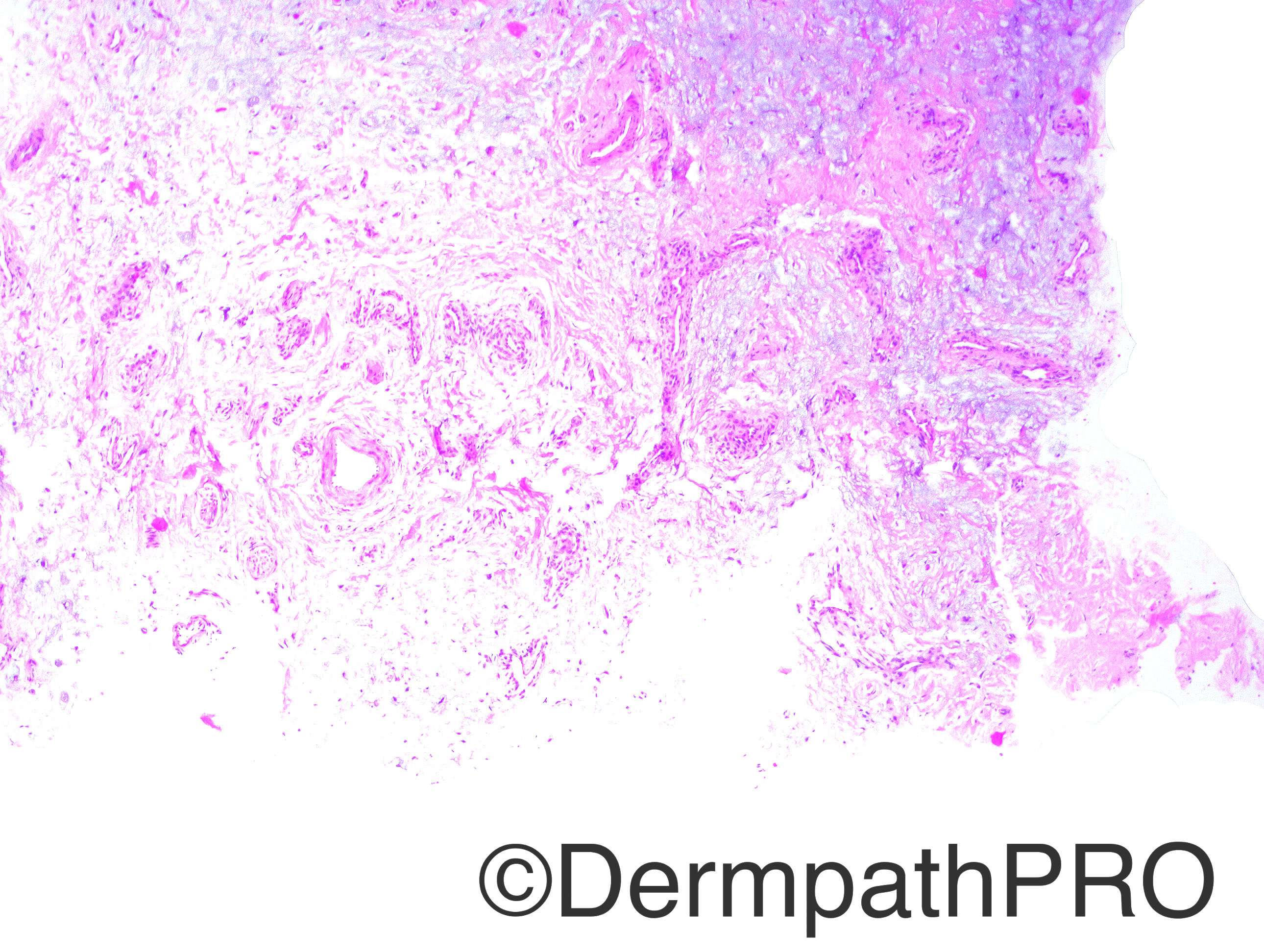
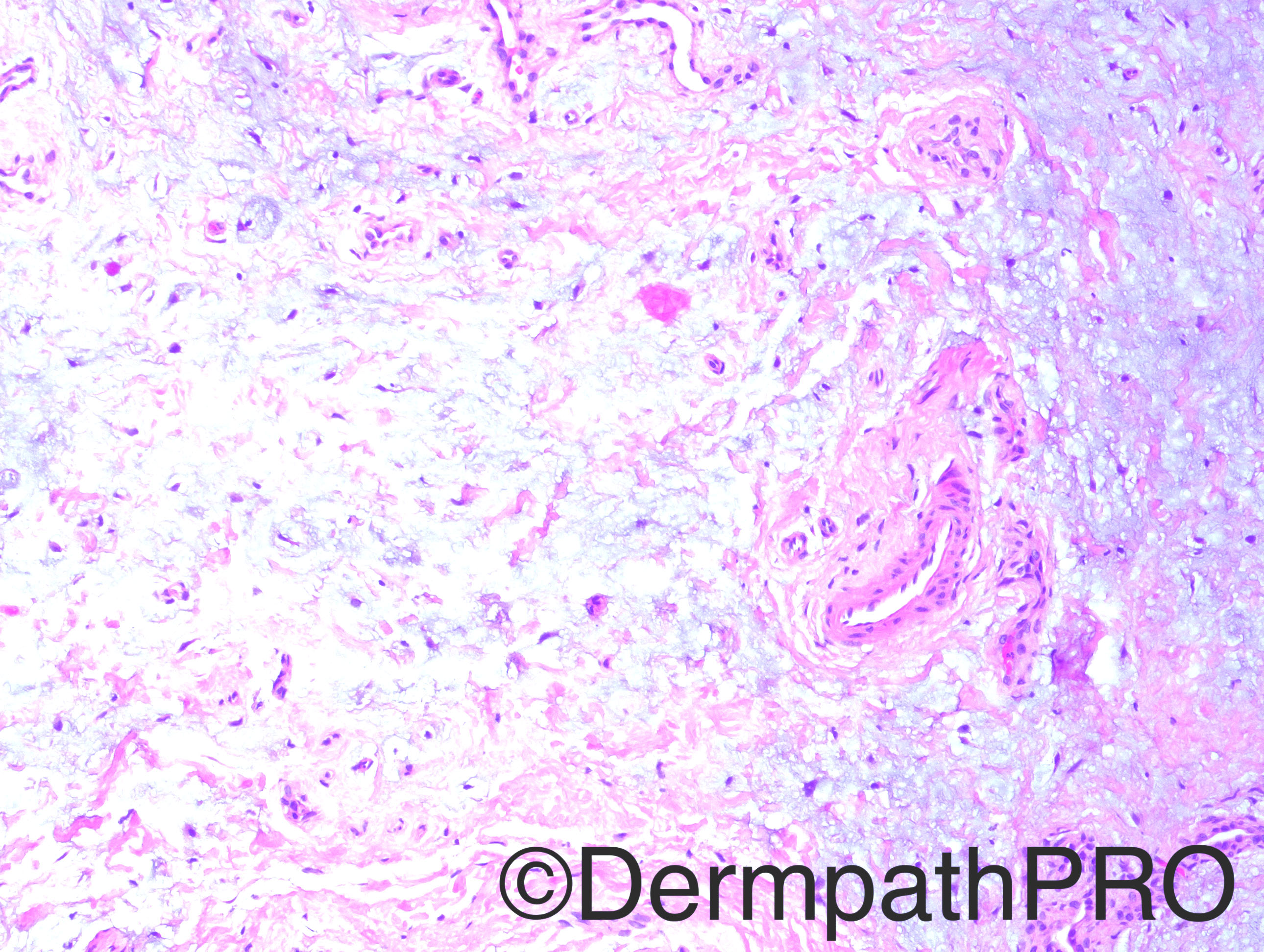
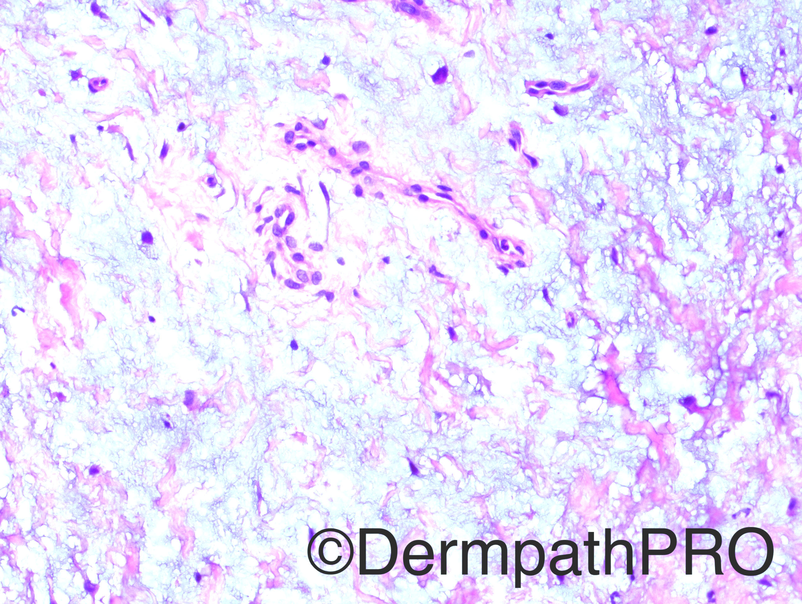
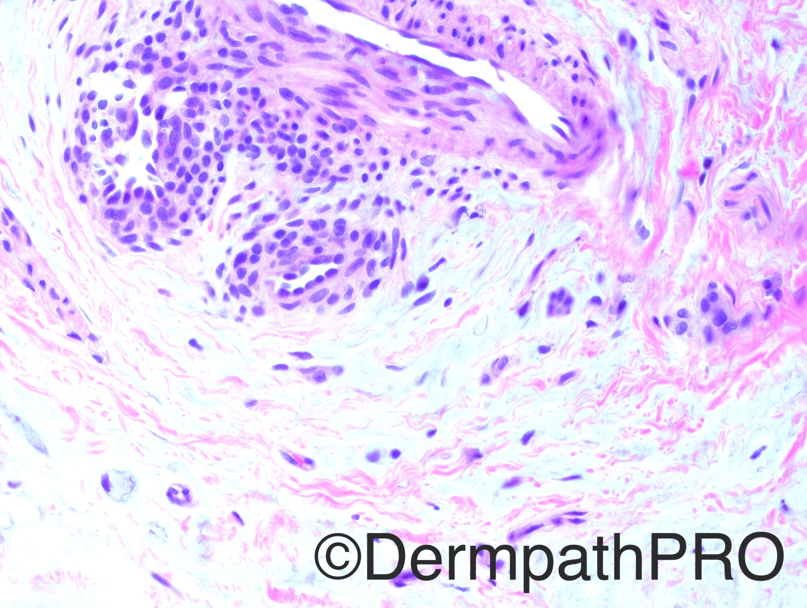
Case History: 10 year-old boy with a right little finger subungual lesion, with finger nail wound.

Case posted by Dr Hafeez Diwan


  Report Record

User Feedback


Dr. Mona Abdel-Halim

Posted

Superficial subungual angiomyxoma

Share this comment


Link to comment
share_externally

vincenzo polizzi

Posted

What about a CD34+ Glomus tumor with myxoid changes?
"CD34-positive glomus tumor: clinicopathologic and immunohistochemical analysis of six cases with myxoid stromal changes.
Mentzel T, et al. J Cutan Pathol. 2002."

Share this comment


Link to comment
share_externally

Raul Perret

Posted

My differential is between digital superficial acral fibromyxoma vs superficial angiomyxoma. This case lacks epithelial component and neutrophils (usually seen in superficial angiomyxoma) but it looks hypocellular for superficial acral fibromyxoma, some report EMA positivity in acral fibromyxoma that could help with the distinction. And I dont know if im right but superficial angiomyxoma reported in fingers is quite rare (only found  2 articles that sadly i cannot read). I think is important to add after Reading a bit that the histological distinction of these entities does not have importance on treatment and prognosis: Both are treated with total excision and both can recur

Share this comment


Link to comment
share_externally

I favour digital fibromyxoma, largely based on location.  As Raul said, superficial angiomyxoma is a tumour of the torso and head. 

Share this comment


Link to comment
share_externally

Arti Bakshi

Posted

I was also struck by the lack of cellularity of the lesion. Also thought of a digital myxoid pseudocyst in addition to the d/d considered above.

Share this comment


Link to comment
share_externally

Dr. Hafeez Diwan

Posted

I thought the glomus cells were not enough to make it a glomus tumor.  The focus I have photographed was the only focus.  I went with myxoma (or angiomyxoma).

Share this comment


Link to comment
share_externally


Join the conversation

You can post now and register later. If you have an account, sign in now to post with your account.

Guest
Add a comment...

×   Pasted as rich text.   Paste as plain text instead

  Only 75 emoji are allowed.

×   Your link has been automatically embedded.   Display as a link instead

×   Your previous content has been restored.   Clear editor

×   You cannot paste images directly. Upload or insert images from URL.

Loading...

×
×
  • Create New...