Jump to content
In this section we have spot diagnoses posted on a daily basis since June 2010, now over 4000! You can review the archived cases and read the suggested diagnoses by users and the final comment by the contributors.
Case are uploaded each week day by 10 am UK time with the correct diagnosis will generally be posted at 8 pm UK time. Why not view the most recent spot diagnosis and proffer a diagnosis?

Case Number : Case 1495- 17 March Posted By: Guest

Please read the clinical history and view the images by clicking on them before you proffer your diagnosis.
Submitted Date :
   (0 reviews)

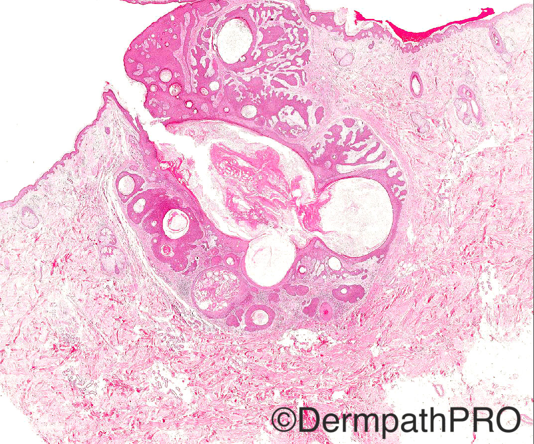
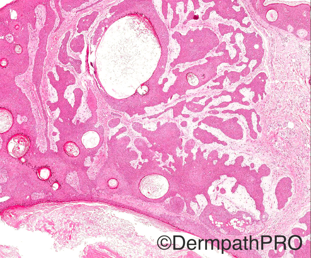
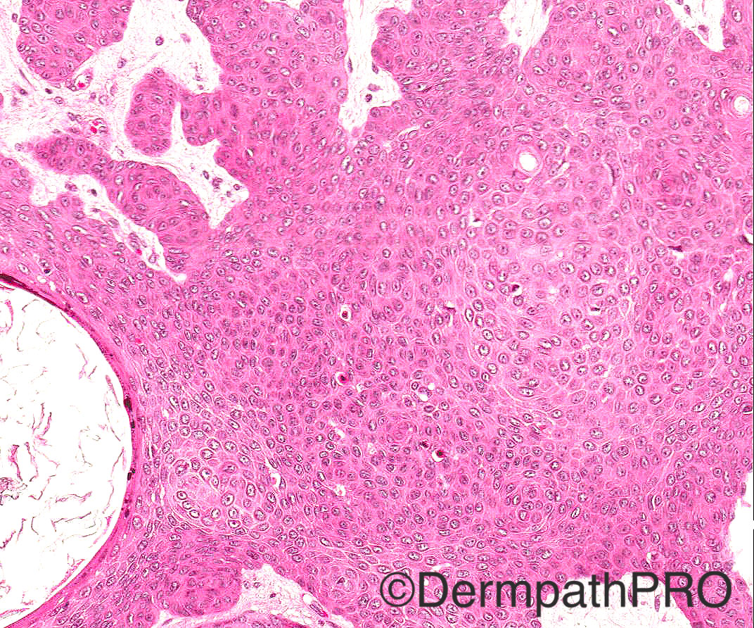
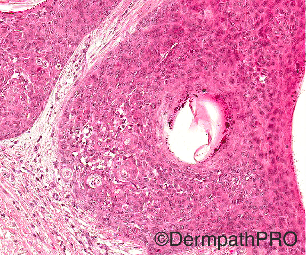
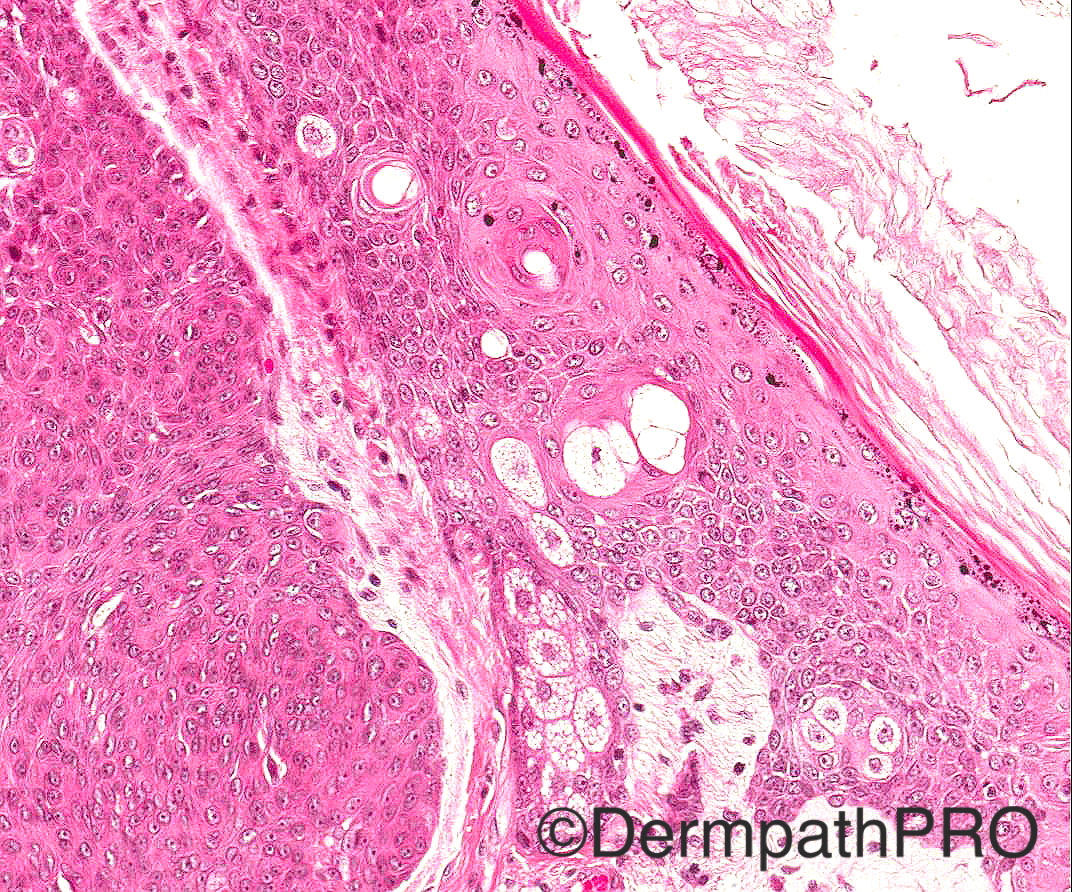
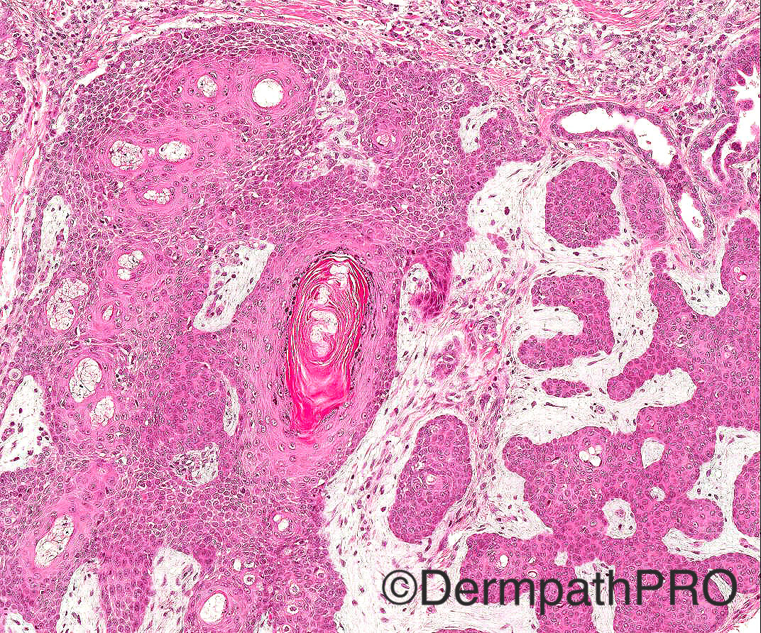
60/M odd cystic lesion face

Case posted by Dr Arti Bakshi


  Report Record

User Feedback


Dr. Mona Abdel-Halim

Posted

And also odd pathologically. To me the cells look poroid, there is ductal differentiation and glandular formations in the last image in addition to sebocytes. Appears to be mainly related to a hair follicle could it be apocrine poroma? (apocrine poroma with sebaceous differentiation)

Share this comment


Link to comment
share_externally

Raul Perret

Posted

In my differential I also considered trichofolliculoma but for me the vellus hair follicles are missing. I see infundibular keratinization, abortive hair follicles and scattered sebocytes. If this lesion is located on the upper lip I would consider pilar sheat acanthoma, if not I would consider trichofolliculoma but would perform many serials to actually see the vellus hair follicles

Share this comment


Link to comment
share_externally

Dr. Mona Abdel-Halim

Posted

Dear Raul: how u explain the structures that look like glands in the top right corner of the last image?

Share this comment


Link to comment
share_externally

Raul Perret

Posted

Dear Mona: Sincerely I just think that is a reactive duct really near the neoplasm, I would like to see that ductal morphology inside the neoplasm for considering poroma, also the prominent infundibular keratinization present in this case doesnt fit with poroma. I agree that the cells look a bit poroid in general but I think that the tinction is bothering too (everything looks way too pink without good contrast). Anyways this is just my opinion and I think that the most important point is that this lesion is benign... but lets see what the rest of the crew thinks. Have a good day

Share this comment


Link to comment
share_externally

Raul Perret

Posted

I forgot to mention the architecture of the neoplasm, If I only see this lesion in 4x I would not consider in the differential a poroma

Share this comment


Link to comment
share_externally

Dr. Richard Carr

Posted

This is a very odd follicular-sebaceous-apocrine unit hybrid:  tumour of the follicular infundibulum (isthmic epithelium in middle left - agree very pilar sheath acanthoma-like) and inverted follicular keratosis-like (middle right). Scattered sebocytic differentiation calls to mind epithelioma with sebaceous differentiation.  There are a couple of presumably trapped apocrine ducts.  Could just be an unusually large tumour of the follicular infundibulum (actually isthmus) involving the follicle.  Odd lesions always consider pre-existing naevus sebaceous. 

Share this comment


Link to comment
share_externally

Dr. Mona Abdel-Halim

Posted

I thought also as a differential diagnosis of reticulated acanthoma with sebaceous differentiation (which Dr Carr has referred to).

Share this comment


Link to comment
share_externally

proma with follicular and sebaceous differntiation  see kazakove for adenxal tumor

Share this comment


Link to comment
share_externally

Arti Bakshi

Posted

Great discussion!
Clearly the tumour shows differentiation towards outer root sheath at level of isthmus as well as infundibular differentiation. The former is represented by polygonal cells with abundant pink cytoplasm and also necrotic/apoptotic cells (middle left), thus recapitulating necrotic keratinocytes in the ORS of normal hair follicle in catagen. I regarded the glands in the last image as entrapped ducts too. But I think there is also genuine ducal differentiation ( likely sebaceous ducal) together with many sebocytes, all entirely compatible with an isthmic tumour.
Combining isthmic differentiation with the architecture of a central dilated follicular infundibulum with radiating lobules, I called this a Pilar Sheath Acanthoma ( with prominent sebaceous differentiation).
Certainly Tumour of follicular infundibulum is a good differential as it also shows isthmic differentiation. But according to my understanding, TFI has a typical silhouette of a fenestrated horizontally oriented superficial tumour, which was lacking here. Similarly reticulated acanthoma with sebaceous differentiation is a broad and superficial lesion with sebocytes attached to base of rete ridges. Neither would show a central crateriform dilated infundibulum with radiating elements. On the other hand, a trichofolliculoma does have this architecture, but would be expected to show pan follicular differentiation.
Thank you all for your contributions!

Share this comment


Link to comment
share_externally

Dr. Richard Carr

Posted

I still think there are some very TFI like features with it appearing to "hang off" the sides of the follicle.  Undoubtedly there is also a thin SEBK blending with the lesion superficially.  Pilar sheath acanthomas are very rare but in my experience usually have rather distinctive, subtly palisaded, broad rounded pushing borders and central more prominent pilar keratin than seen here.  This looks more like TFI or reticulated SEBK/IFK involving a comedonal follicle.  Lovely case though!

Share this comment


Link to comment
share_externally

Raul Perret

Posted

I agree Dr. that there are some focus of inverted follicular keratosis (the areas that you mention made me think of it when I was analizing the case for the first time) as for TFI I did not consider it mainly because of the architecture as Arti mentioned. However, my experience of TFI is limited to pictures in books and the cases that were presented in dermpathpro.... I think the discussion could go for hours with a complex lesion like this one with multiple lines of differentiation.

Share this comment


Link to comment
share_externally

Dr. Mona Abdel-Halim

Posted

I like the idea of Dr Carr that this is a follicular/seb/apocrine hybrid lesion. This is not a straight forward pilar sheath acanthoma to me due to the same reasons that Dr Carr higlighted in his last comment. I think this case shows how adnexal lesions can be complex and the most important thing here is that the lesion is benign... Nice case and enjoyable discussion... Thanks Arti..

Share this comment


Link to comment
share_externally

Dr Hurt - Ackerman regarding squamous eddies as sebaceous ducts can be found on page 200 of his "Histopathologic Diagnosis of Adnexal Epithelial Neoplasms: Atlas and Text" (2008), in a discussion on inverted follicular keratosis.

Share this comment


Link to comment
share_externally


Join the conversation

You can post now and register later. If you have an account, sign in now to post with your account.

Guest
Add a comment...

×   Pasted as rich text.   Paste as plain text instead

  Only 75 emoji are allowed.

×   Your link has been automatically embedded.   Display as a link instead

×   Your previous content has been restored.   Clear editor

×   You cannot paste images directly. Upload or insert images from URL.

Loading...

×
×
  • Create New...