Jump to content
In this section we have spot diagnoses posted on a daily basis since June 2010, now over 4000! You can review the archived cases and read the suggested diagnoses by users and the final comment by the contributors.
Case are uploaded each week day by 10 am UK time with the correct diagnosis will generally be posted at 8 pm UK time. Why not view the most recent spot diagnosis and proffer a diagnosis?

Case Number : Case 1528 - 03 May Posted By: Guest

Please read the clinical history and view the images by clicking on them before you proffer your diagnosis.
Submitted Date :
   (0 reviews)

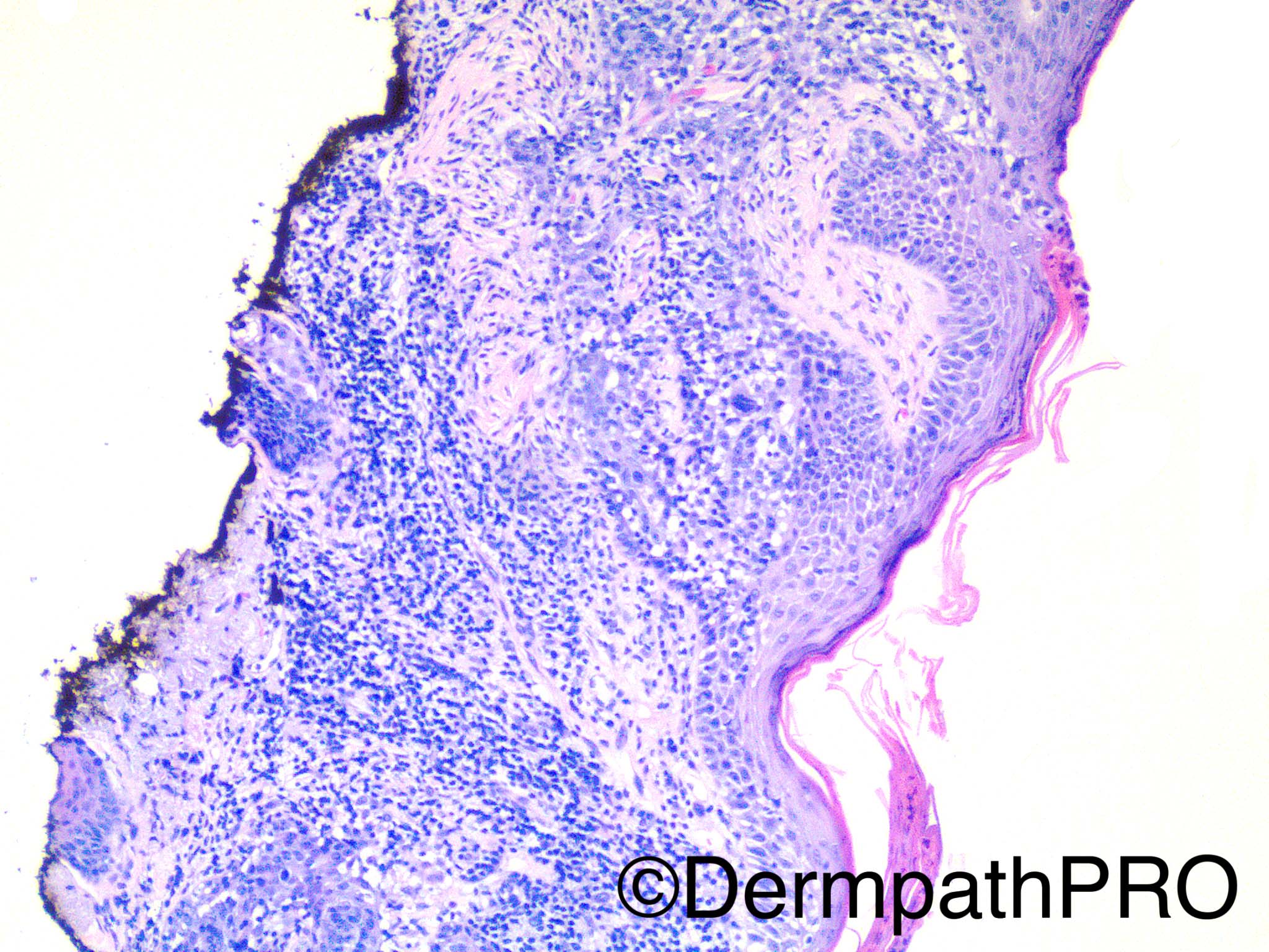
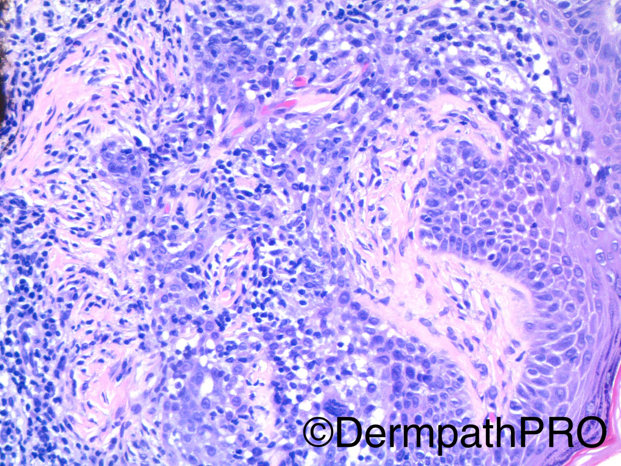
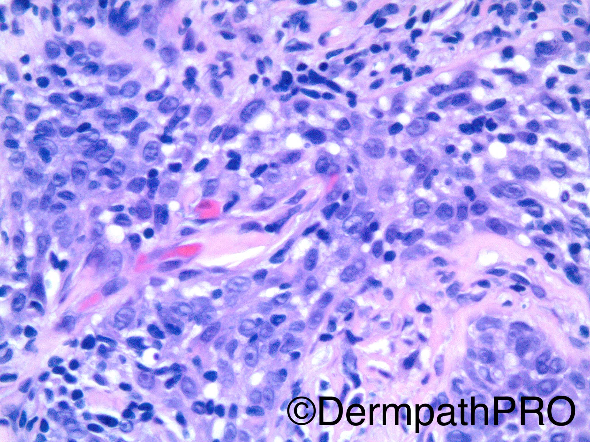
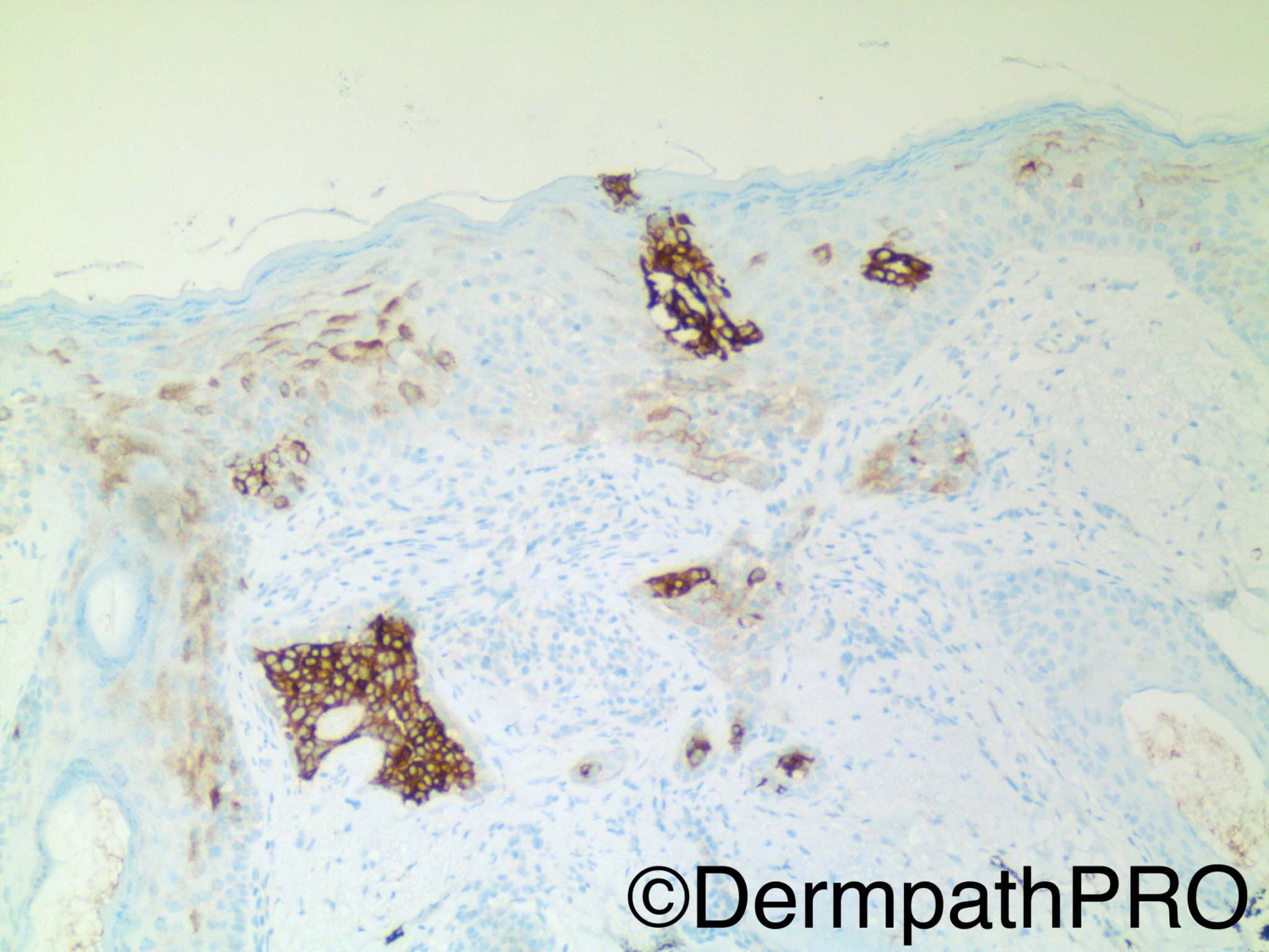
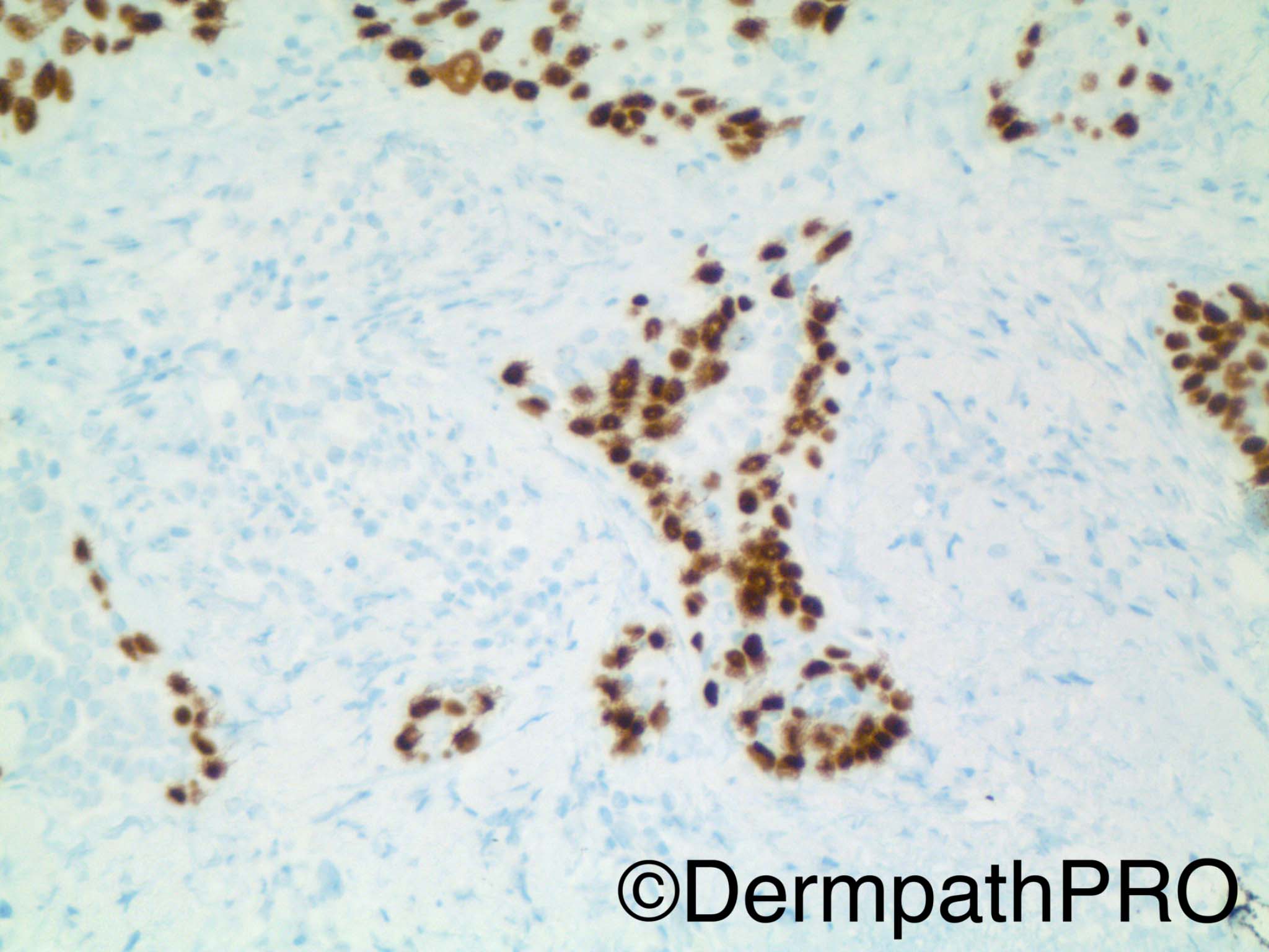
60 year old woman with lesion on the cheek. First IHC image=polykeratin, second IHC image=EMA, third IHC image=p63.

Dr Uma Sundram.


  Report Record

User Feedback


vincenzo polizzi

Posted

I think this is a syringolymphoid hyperplasia-like intraepidermal growth, with subtle sebaceous differentiation and atypical cells. there is some pagetoid spread. Favour a sebaceous carcinoma in situ, with lymphoid reaction.

Share this comment


Link to comment
share_externally

Raul Perret

Posted

I find this biopsy rather hard for interpretation. I think there are a lot of reactive changes due to the dense lymphocytic infiltrate but I am not 100% sure that the lesion is in situ (mainly due to the marked desmoplasia seen). I considered pagetoid Bowen alone or associated with invasive SCC as well as porocarcinoma (even if the cells dont look that poroid to my eyes) in my differentials. I would perform CK7 and CEA that could help in this distinction but I favor an squamous origin to this lesion. Sincerely I did not have the impression that the cells could have a sebaceous nature but who knows...

Share this comment


Link to comment
share_externally

Nitin Khirwadkar

Posted

Looks like an incisional biopsy, appearances of Bowen's. The latter is adnexotropic. Like Raul has suggested would consider an in-situ porocarcinoma. Was there an invasive SCC adjacent to this ?

Share this comment


Link to comment
share_externally

Dr. Richard Carr

Posted

Tricky because so inflamed.  I was thinking may be adnexotropic bowenoid actinic keratosis with focal pagetoid bowen's.

Share this comment


Link to comment
share_externally

Favour Bowen's.  CAM5.2, BER and EMA to confirm.  (CAM5.2/BER +ve and EMA -ve in sebaceous neopasia in-situ and CAM5.2/BER/EMA +ve in glandular neoplasia in-situ.) 

Share this comment


Link to comment
share_externally

Dr. Mona Abdel-Halim

Posted

Thought of bowenoid porocarcinoma in situ. DDx: Bowen's (pagetoid) or bowenoid AK

Share this comment


Link to comment
share_externally

Guest Arash Daryakarr

Posted

I favor bowenoid AK. Hard case.

Share this comment


Link to comment
share_externally

Uma Sundram

Posted

This was indeed a difficult case. It was a de novo lesion with no adjacent invasive squamous cell carcinoma. I had a differential diagnosis of adnexal/glandular carcinoma versus sebaceous carcinoma; an invasive component may be present. BerEp4 is positive but faint, EMA is positive, and CK7/CAM5.2 were both positive. Needless to say we recommended a re excision. It's a relatively new case and I will give follow up if I get it.

Share this comment


Link to comment
share_externally

Alice Roberts

Posted

I favor poor carcinoma in situ but not sure

Share this comment


Link to comment
share_externally


Join the conversation

You can post now and register later. If you have an account, sign in now to post with your account.

Guest
Add a comment...

×   Pasted as rich text.   Paste as plain text instead

  Only 75 emoji are allowed.

×   Your link has been automatically embedded.   Display as a link instead

×   Your previous content has been restored.   Clear editor

×   You cannot paste images directly. Upload or insert images from URL.

Loading...

×
×
  • Create New...