Jump to content
In this section we have spot diagnoses posted on a daily basis since June 2010, now over 4000! You can review the archived cases and read the suggested diagnoses by users and the final comment by the contributors.
Case are uploaded each week day by 10 am UK time with the correct diagnosis will generally be posted at 8 pm UK time. Why not view the most recent spot diagnosis and proffer a diagnosis?

Case Number : Case 1531 - 06 May Posted By: Guest

Please read the clinical history and view the images by clicking on them before you proffer your diagnosis.
Submitted Date :
   (0 reviews)

Spot Diagnosis provided by Dr Richard Carr

Additional Images to Follow at 6pm GMT


  • Thanks 1
  Report Record

User Feedback


Raul Perret

Posted

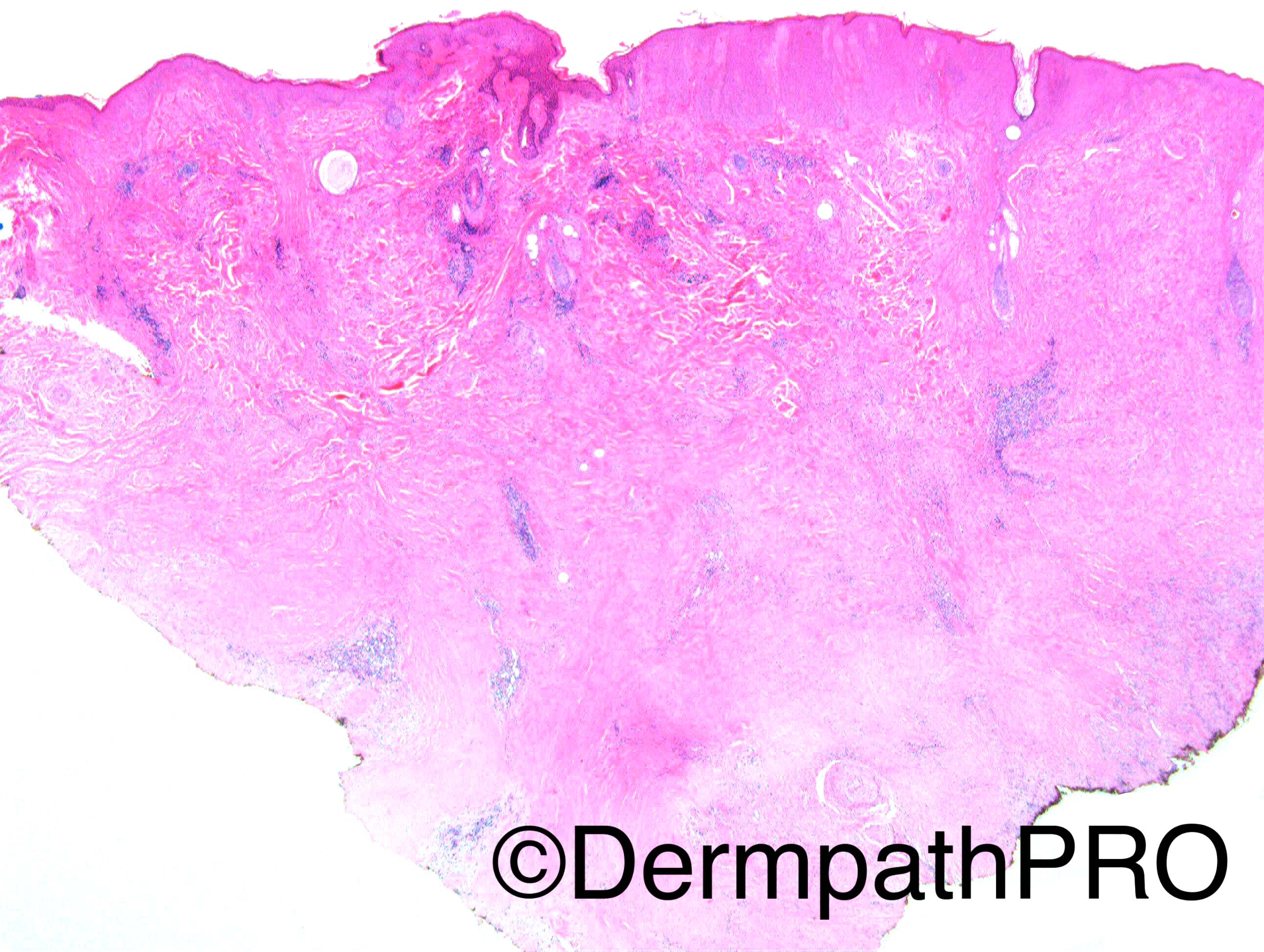
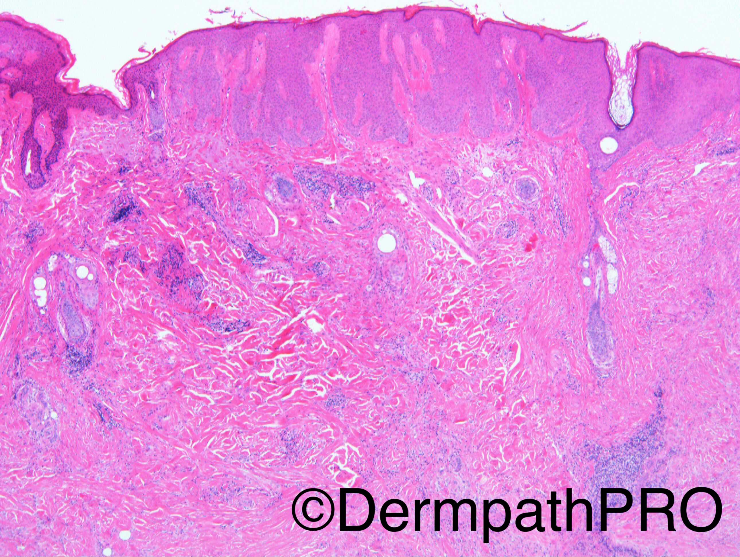
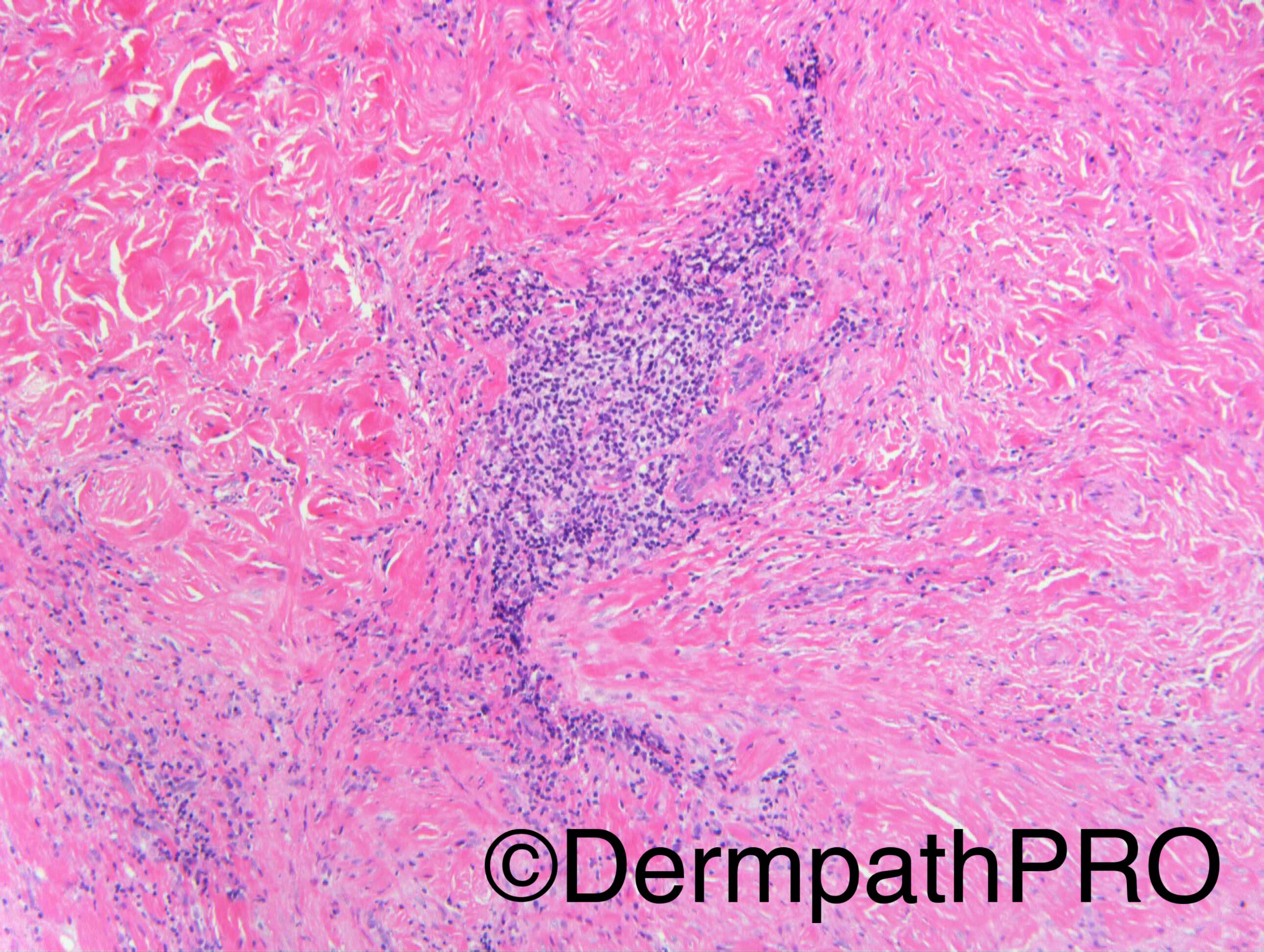
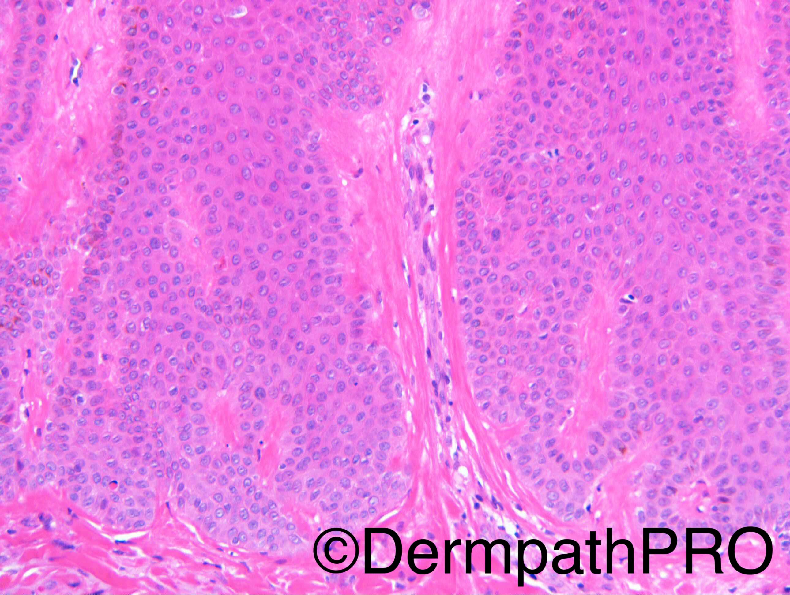
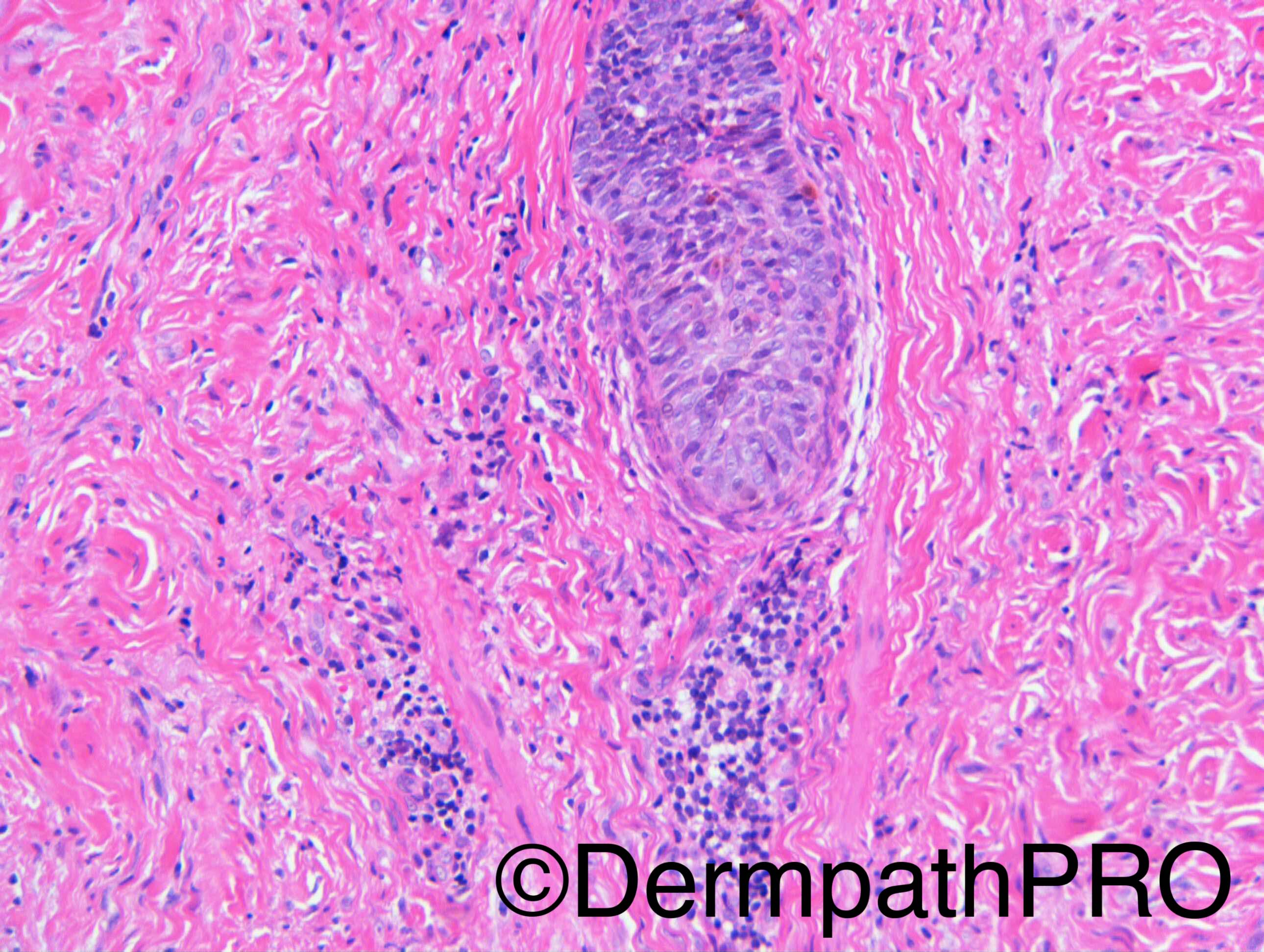
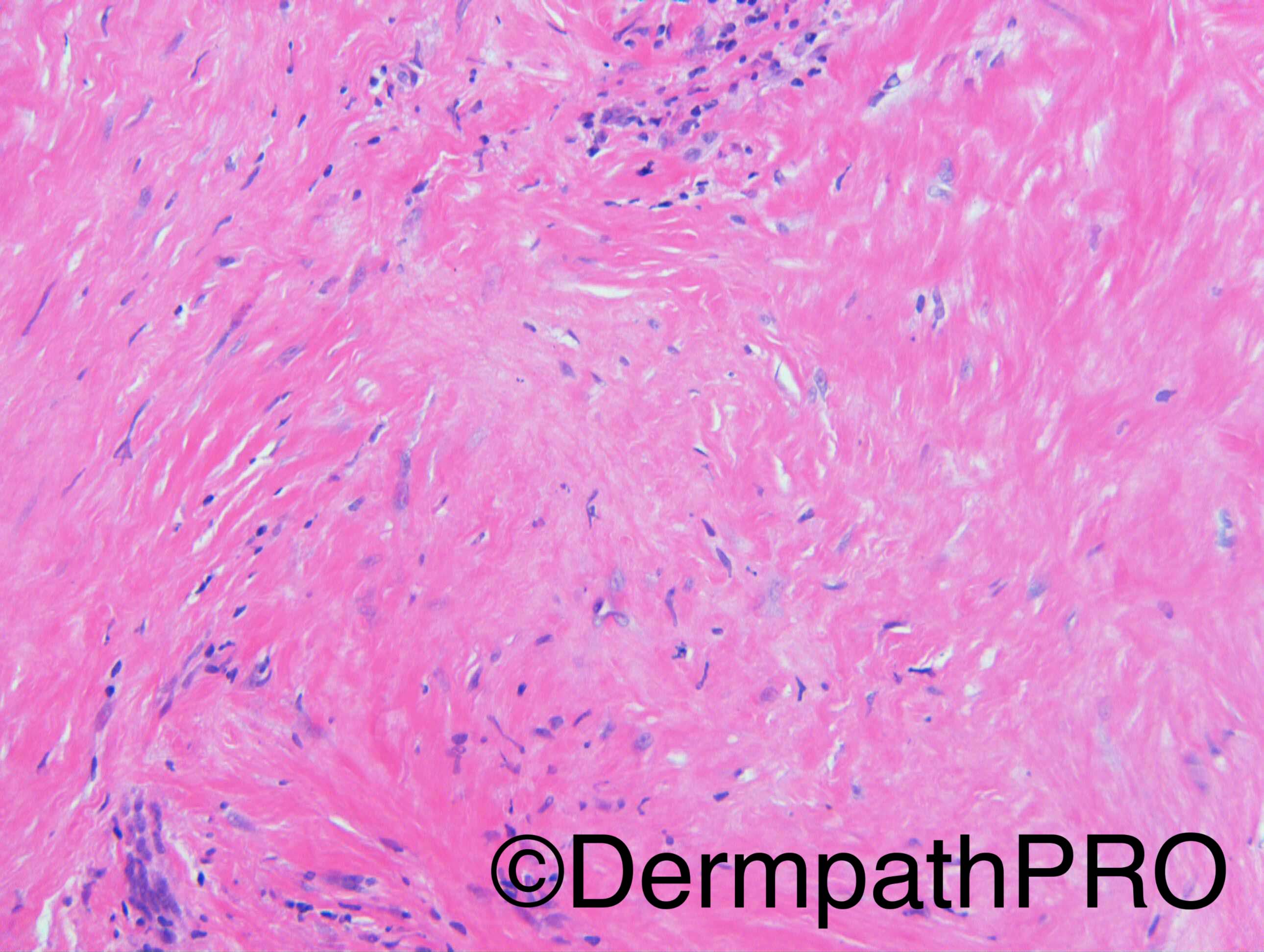
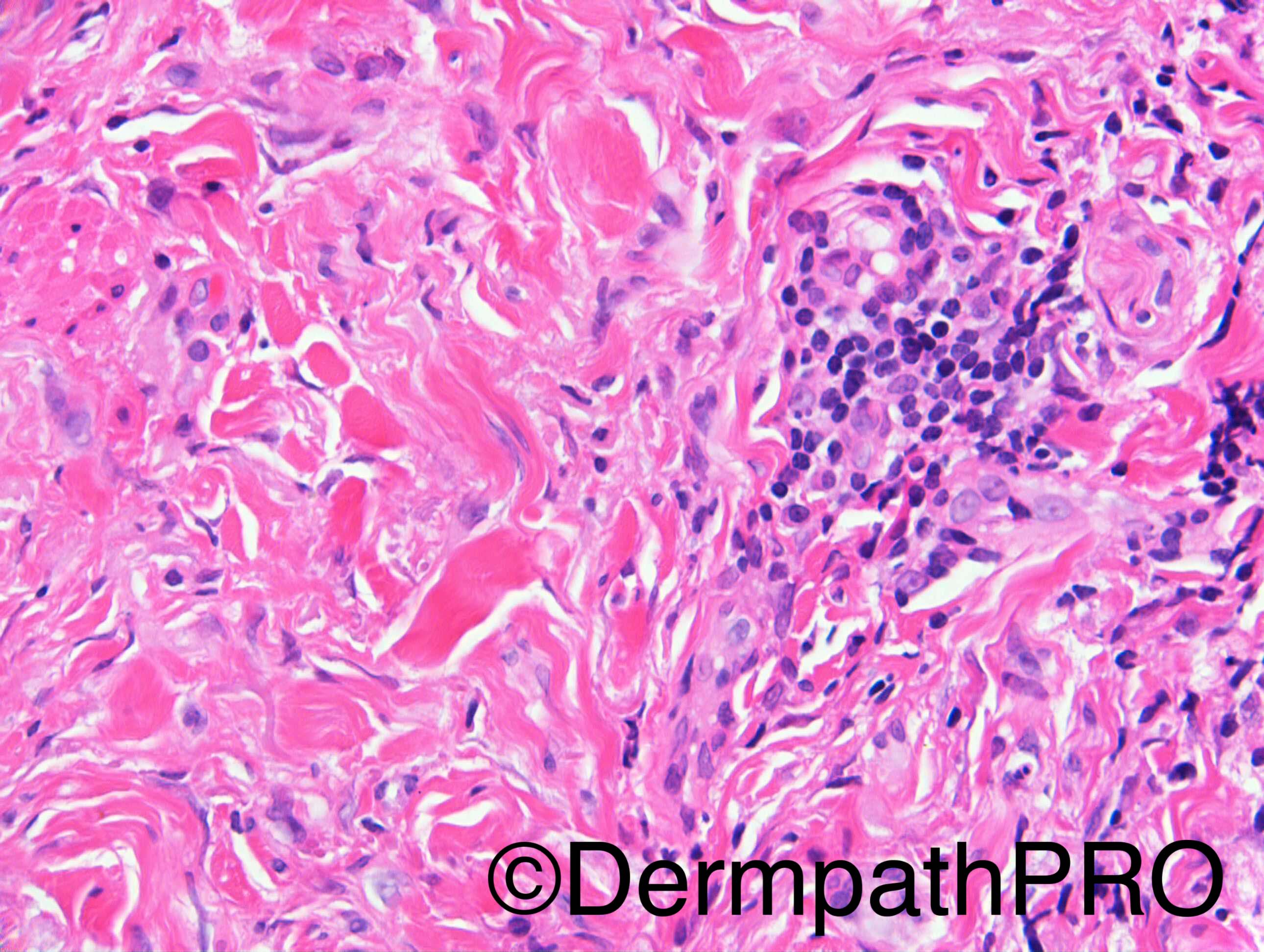
Not an easy one for me. I will wait for the next images but I considered morphea, scar tissue, I dont know how to make fit the localized acanthosis (associated acanthotic SK?) also the presence of numerous mast cells got my attention but I cannot make them fit

Share this comment


Link to comment
share_externally

vincenzo polizzi

Posted

Very very difficult for me. " I dont know how to make fit the localized acantosis (associated acanthotic SK?) also the presence of numerous mast cells got my attention but I cannot make them fit"...Completely agree. is there any perineurial lymphoid infiltration ( fig bottom left and fig 3)? This could fit to a linear scleroderma scarring alopecia...

Share this comment


Link to comment
share_externally

I thought the pictures of the epidermis showed lichen simplex chronicus and the other pictures showed chronic inflammation with mast cells in the vicinity of a telogen follicle, apart from the picture of the scar, the site of which I cannot pinpoint on the low-power picture.  I, too, cannot tie things together. 

Share this comment


Link to comment
share_externally

Arti Bakshi

Posted

Agree with the difficulties above...scleroderma and LSC, both come to mind.

Best to wait for additional images!

Share this comment


Link to comment
share_externally

Dr. Mona Abdel-Halim

Posted

Same struggles :-))
Will rather wait :-))
Wondering where is the spot diagnosis here??!!!

Share this comment


Link to comment
share_externally

IgorSC

Posted

Agree with the previous comments. Scleroderma, but having some problems when correlating with the epidermal findings.

Share this comment


Link to comment
share_externally

Nitin Khirwadkar

Posted

Agree with the comments. Lichen simplex chronicus,  Dermal scar/morphoea. Scatterred mast cells and plasma cells. No definite suggestion of amyloid, but may do a Congo red. Waiting for the answer :)

Share this comment


Link to comment
share_externally

Raul Perret

Posted

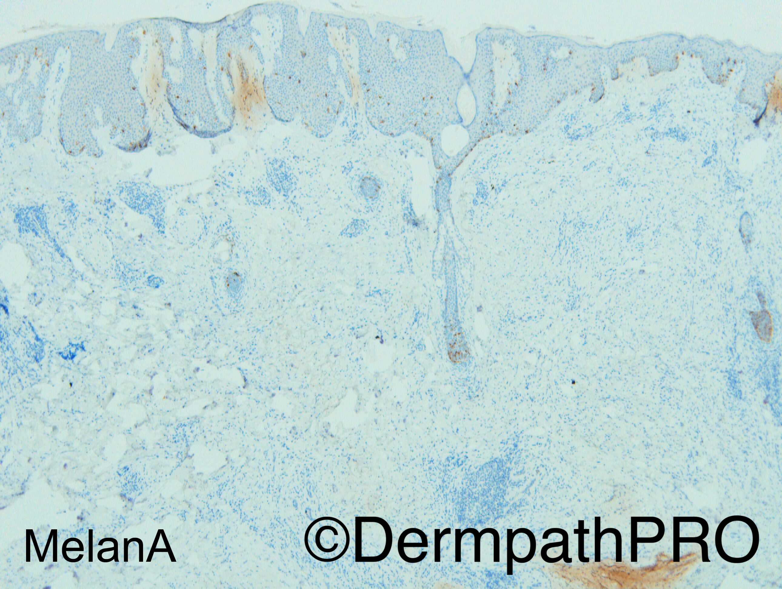
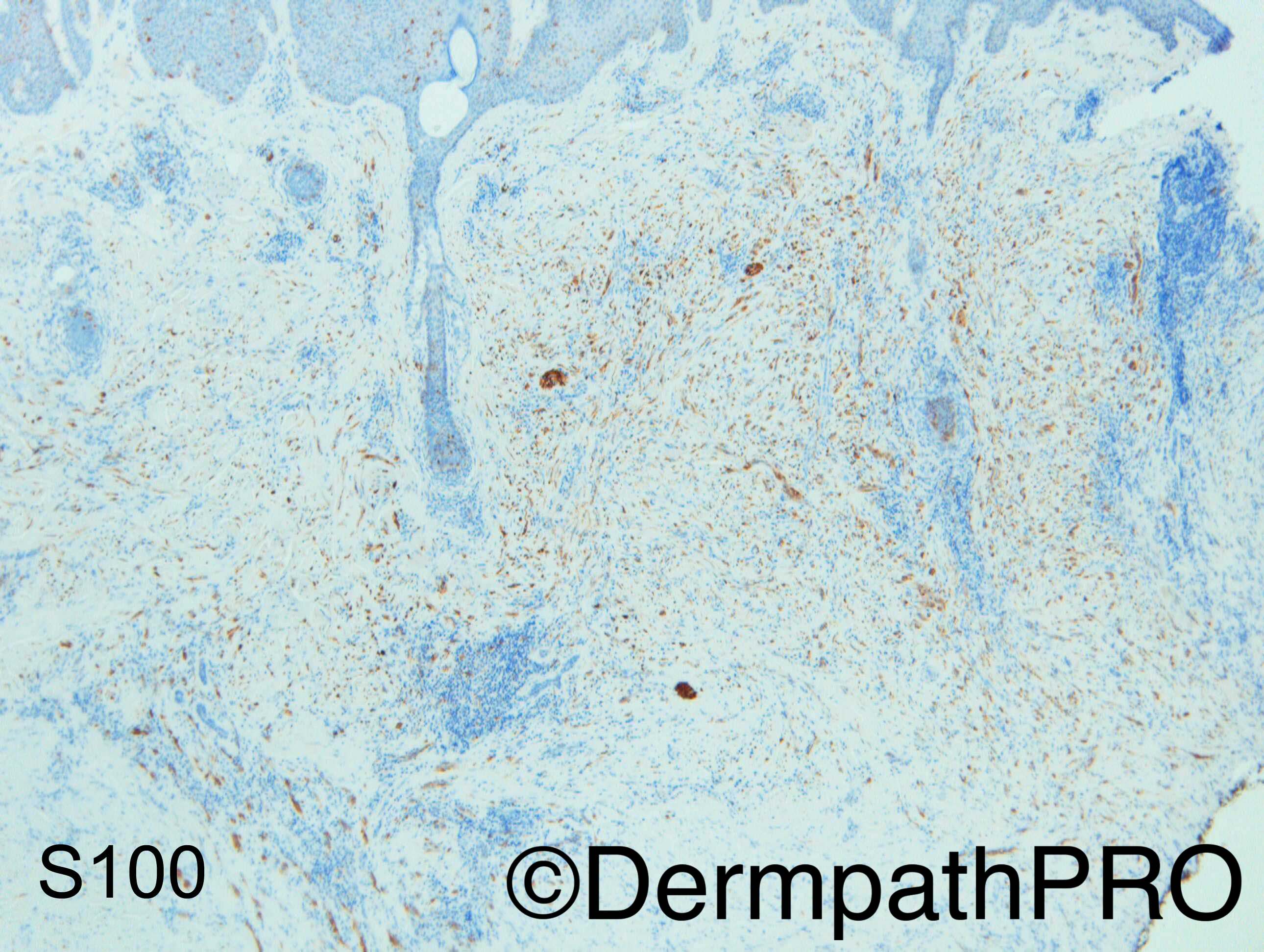
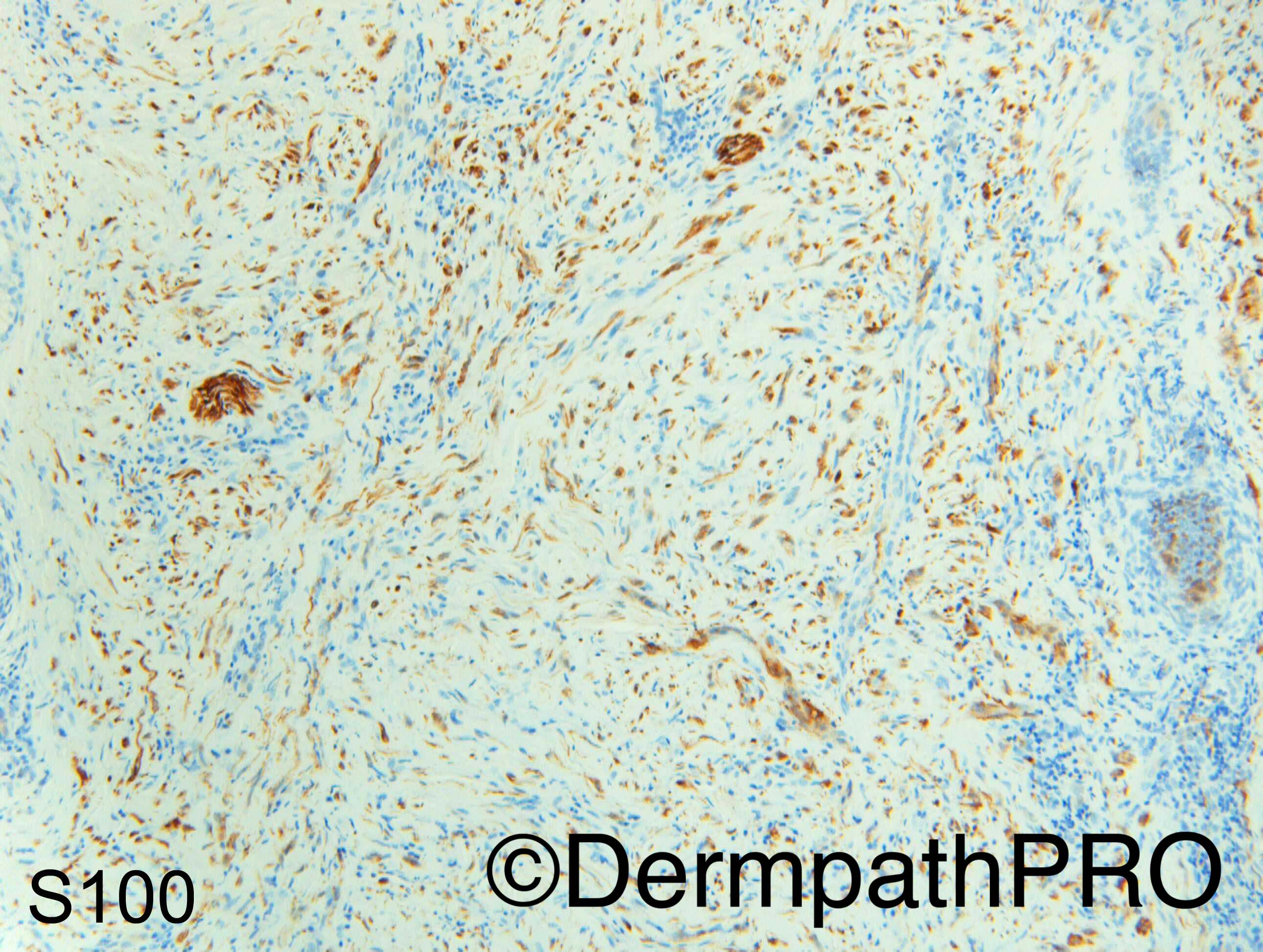
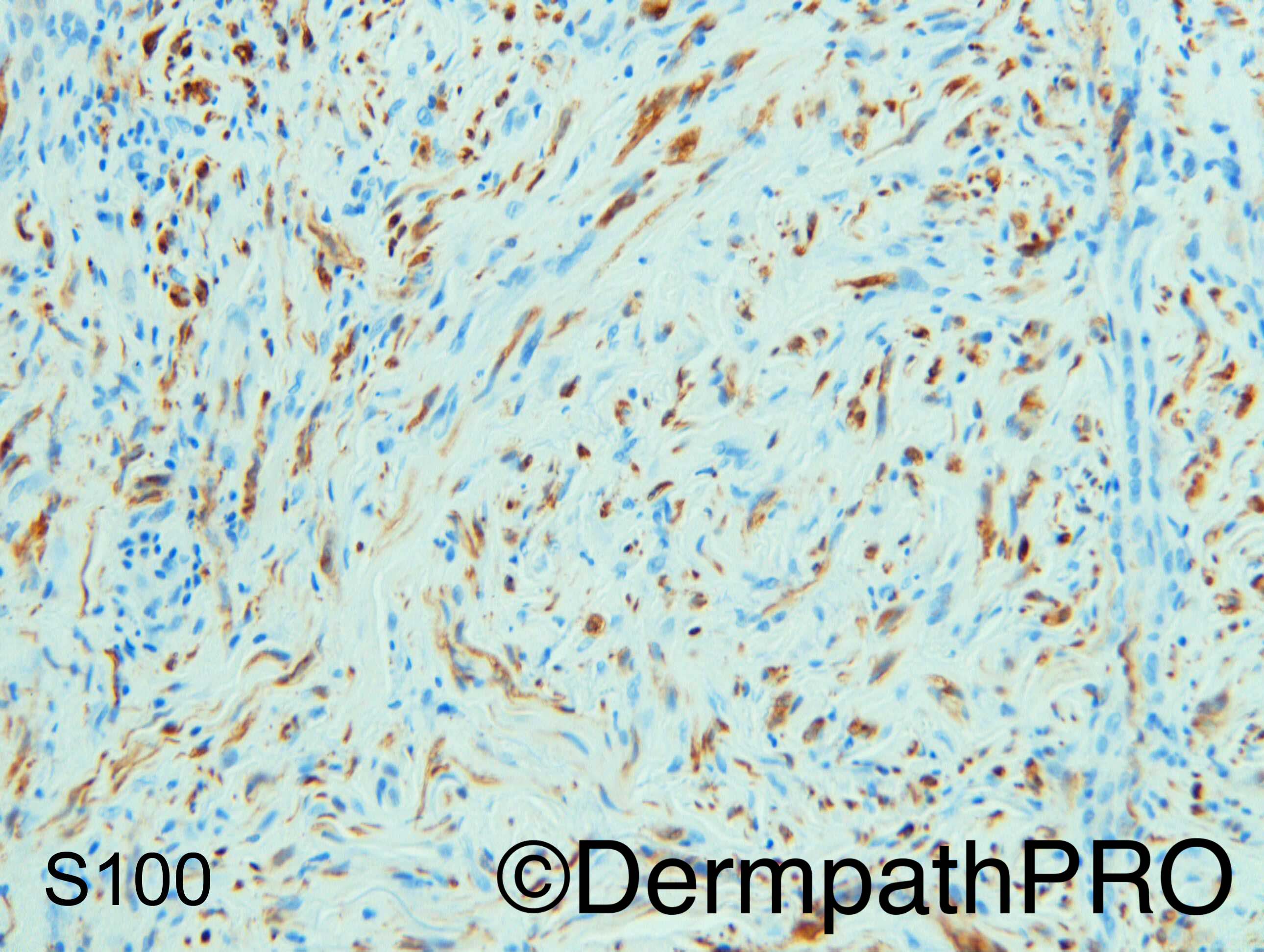
So it is desmoplastic melanoma the mimicker of scars. Not easy to suspect in these images without the immunos. I think everybody had DM in the back of their heads due to the lymphoid aggregates and sclerotic dermis but in my mind it looked too paucicellular in the images to consider it as a differential... It would be nice to perform a SOX-10 too  

Share this comment


Link to comment
share_externally

Dr. Mona Abdel-Halim

Posted

I thought of it but the LSC like changes in the overlying epidermis was confusing...

Share this comment


Link to comment
share_externally

vincenzo polizzi

Posted

Great case! Thank's Dr Carr!

Share this comment


Link to comment
share_externally

Arti Bakshi

Posted

Scary....so easily missed!!

Share this comment


Link to comment
share_externally

Dr. Richard Carr

Posted

I forgot to thank Dr Chris Allen for kindly sharing this very tricky case.


Yes it is that terrible case where you forget to think of desmoplastic melanoma.  Thankfully the clinician had queried a melanocytic macule in the clinical information and an S100 was performed.  Can't remember if a CD34 was done but it really did not look like any neural tumour (lacking the delicate collagenous stroma). Also I did note a small focus of lentigo maligna in one follicle (not illustrated).


I have to be honest and had forgotten the case completely and I did not think of the diagnosis when perusing the case on Friday!  I was also thinking along the lines of morphoea.  Phillip McKee used to say at the beginning of each sign out say to your self "desmoplastic melanoma".  My advice have a very low threshold for doing an S100 on any scar-like or funny "myofibroblastic" spindle process especially when on the sun-damaged skin in the elderly.  Fortunately the lesions are fairly indolent and the metastatic rate for pure desmoplastic melanoma is low and it is more an issue of local control.  However a delay in correct diagnosis would obviously have potentially significant issues relating to the extent of surgery.

Share this comment


Link to comment
share_externally


Join the conversation

You can post now and register later. If you have an account, sign in now to post with your account.

Guest
Add a comment...

×   Pasted as rich text.   Paste as plain text instead

  Only 75 emoji are allowed.

×   Your link has been automatically embedded.   Display as a link instead

×   Your previous content has been restored.   Clear editor

×   You cannot paste images directly. Upload or insert images from URL.

Loading...

×
×
  • Create New...