Jump to content
In this section we have spot diagnoses posted on a daily basis since June 2010, now over 4000! You can review the archived cases and read the suggested diagnoses by users and the final comment by the contributors.
Case are uploaded each week day by 10 am UK time with the correct diagnosis will generally be posted at 8 pm UK time. Why not view the most recent spot diagnosis and proffer a diagnosis?

Case Number : Case 1533 - 10 May Posted By: Guest

Please read the clinical history and view the images by clicking on them before you proffer your diagnosis.
Submitted Date :
   (0 reviews)

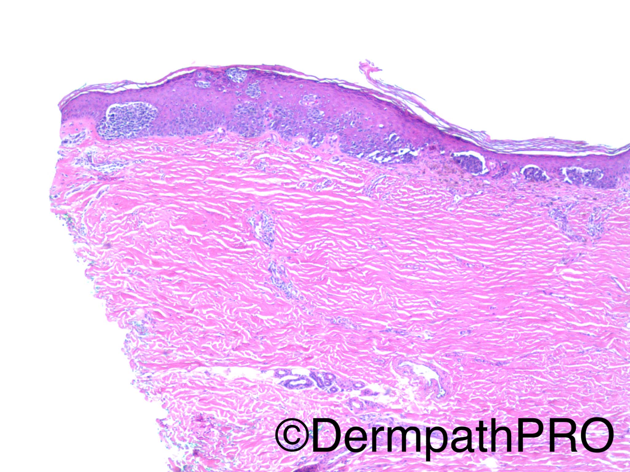
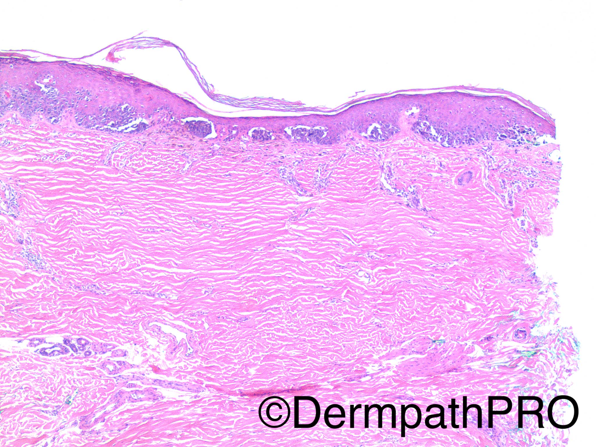
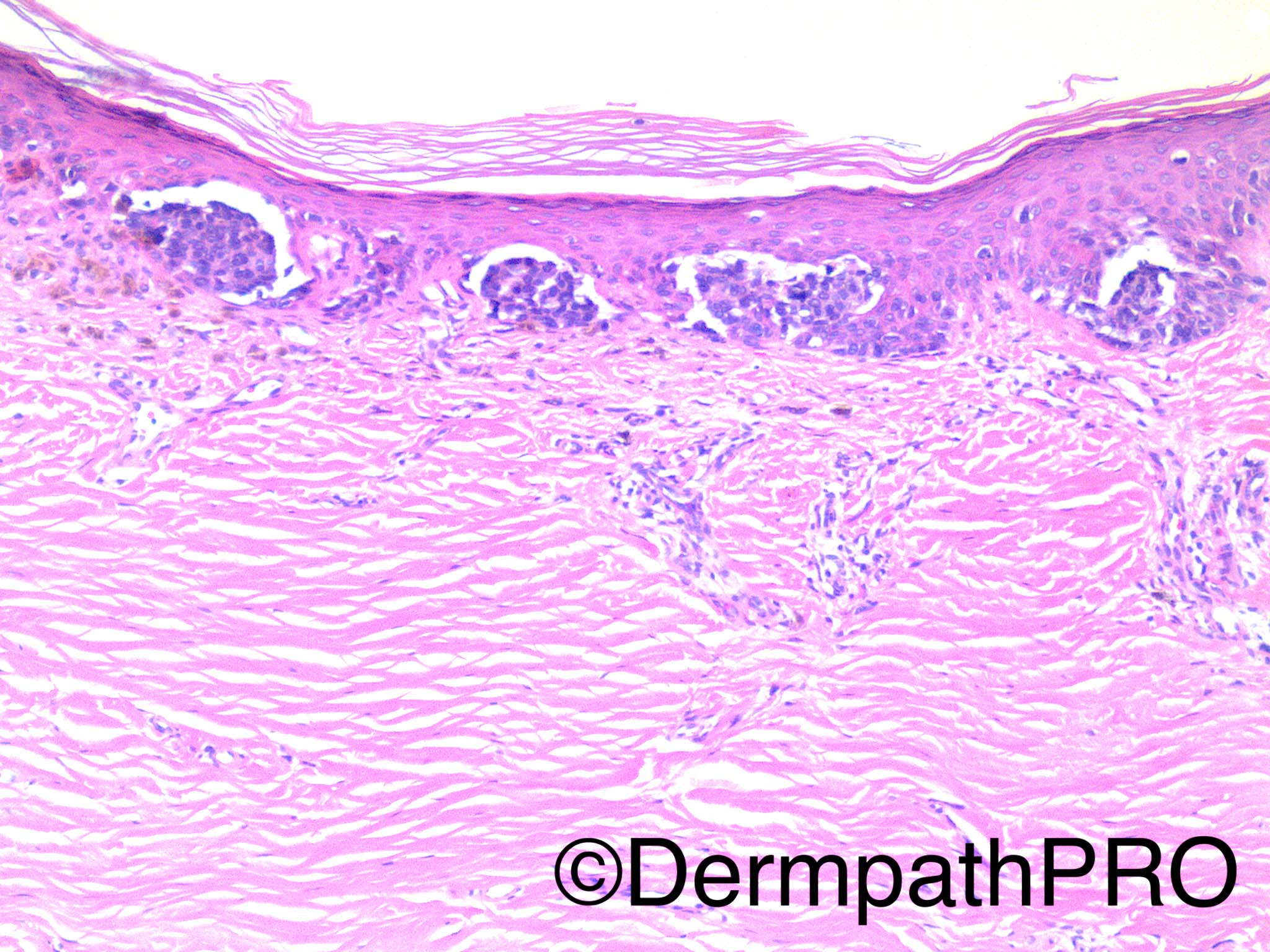
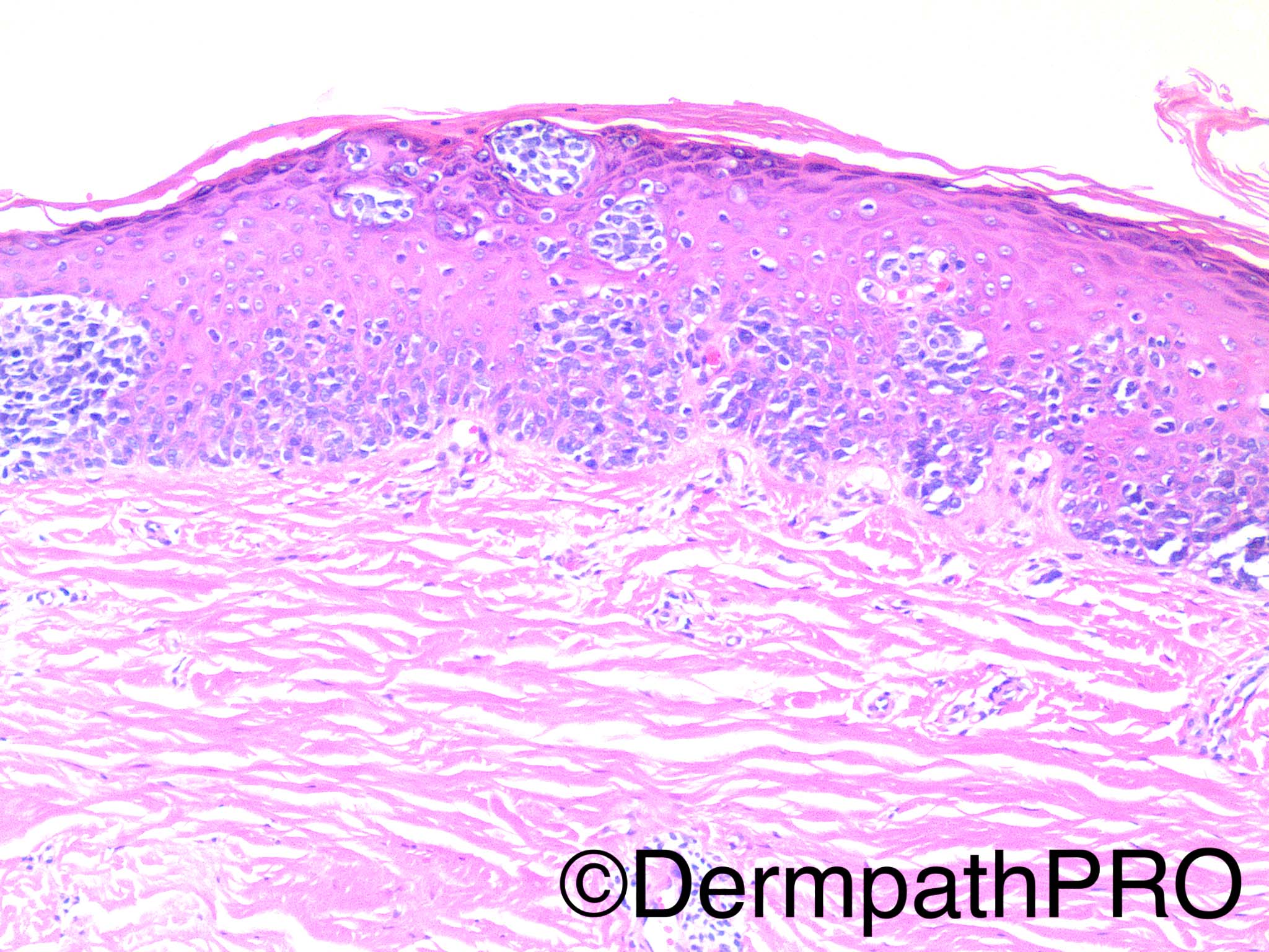
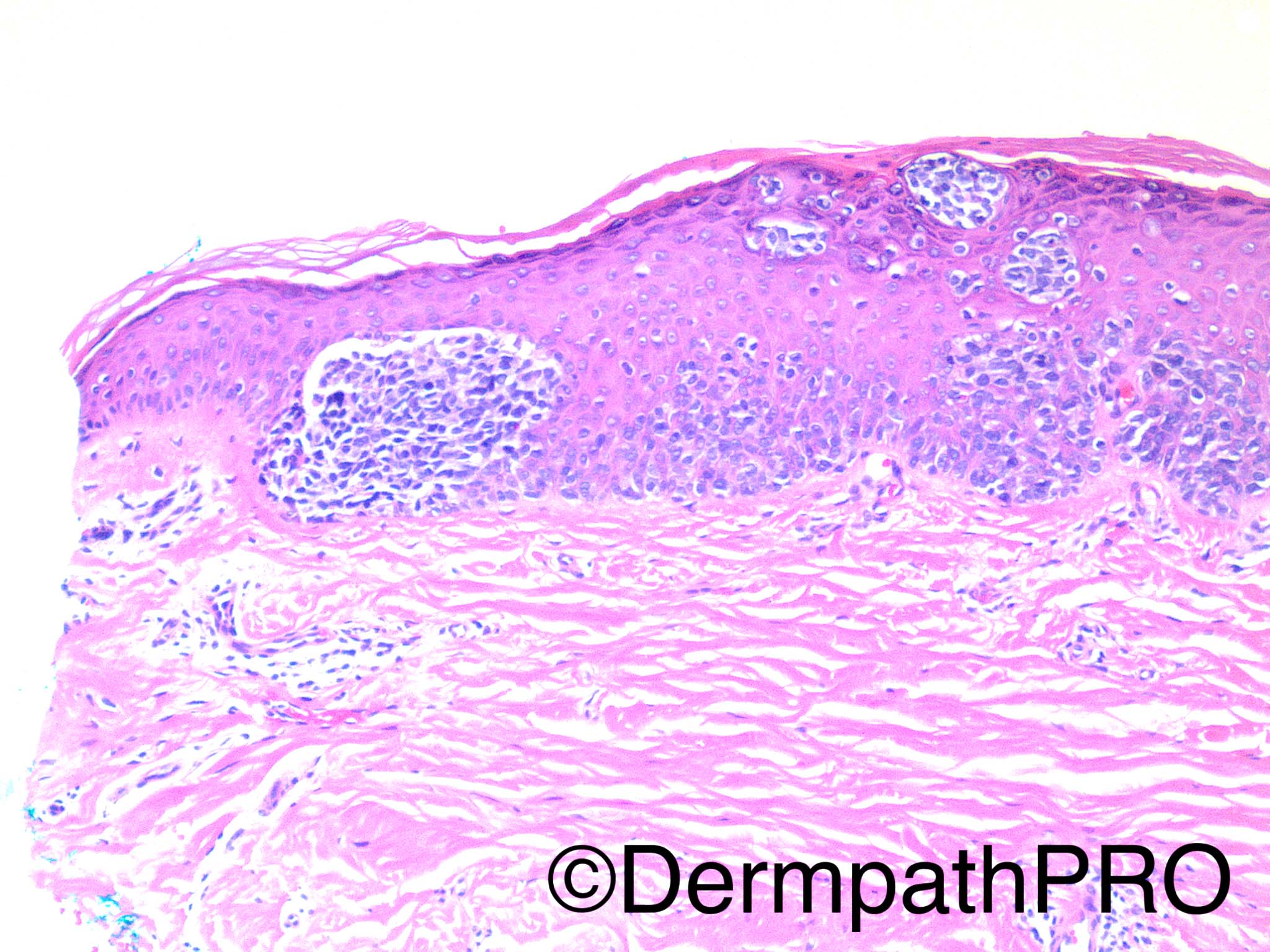
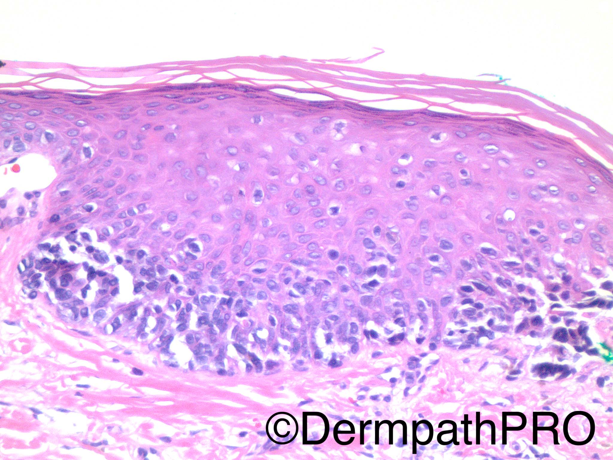
57 year old woman, history of multiple non melanoma skin cancers, with pigmented lesion on the left lower arm. No known prior biopsy.

Dr Uma Sundram


  Report Record

User Feedback


Raul Perret

Posted

I dont find this case easy, I would not go for melanoma in situ directly. The dermis looks fibrotic, I would discard a previous surgical excision in the same site. I am afraid this could be recurrent/persistent nevus, if the clinical history is not compatible then it is probably melanoma in situ. 

Share this comment


Link to comment
share_externally

IgorSC

Posted

Cases here are more difficult every week. I agree that Melanoma in situ is the first diagnosis in my mind. I don´t know how to explain the fibrosis on the dermis without a prior ressection. If there´s really no prior ressection, maybe a partial inflammatory regression with a resolving scar like tissue is the answer. I would perform some immunostains to confirm the melanocytic nature of the lesion. I think there´s a high N:C ratio, so I would like to exclude a pagetoid Merkel cell carcinoma as well.

Share this comment


Link to comment
share_externally

Nitin Khirwadkar

Posted

Tricky case! Agree with the comments above. Dermal scarring makes me think of a pseudo-melanoma (provided there has been a previous excision), otherwise an in-situ melanoma, with regression. I think the proliferation is too florid to be associated with simple trauma.

Share this comment


Link to comment
share_externally

Arti Bakshi

Posted

Pseudomelanoma overlying a scar was my first thought. But the history specifically states that there is no known previous bx, so difficult to tie together. Other cause of a dermal scar including trauma and scarring following blistering conditions etc also need to be considered. If all of this is excluded, one would have to call it a melanoma in situ. 

Share this comment


Link to comment
share_externally

Guest Arash Daryakarr

Posted

Agree with melanoma in situ if other simulating conditions like recurrent nevus ,prior trauma etc. can be excluded.By the way, i share Igor's comment about difficult cases these days! albeit we love these kind of cases.don't we?

Share this comment


Link to comment
share_externally

Raul Perret

Posted

Yes, the harder the better

Share this comment


Link to comment
share_externally

IgorSC

Posted

I learn more when I make a mistake. I think I´m learning more these days, rsrsrs... But I´m enjoying this!!

Share this comment


Link to comment
share_externally

Dr. Richard Carr

Posted

SAMPUS overlying a scar. Nice suggestion regarding in situ MCC - exceedingly rare but worth a mention on this site. In fact I was also struck be the slightly hyperchromatic nature of the cells (reminded me a little of the acral lentiginous type melanoma cells). Needs to be excised (dependent on clinical / dermatoscopic features). If confirmed to be melanocytic, punch biopsy is medico-legally suspect / unwise to make a reliable histopathological diagnosis - let the clinician know it is their call.

Share this comment


Link to comment
share_externally

vincenzo polizzi

Posted

The cells are small nevus-like ( resembling type B melanocytes ) so, if melanoma, could be a nevoid in situ melanoma... Too tricky for me. I prefer the diagnosis: "I don't know"!

Share this comment


Link to comment
share_externally

Guest Romualdo

Posted

Agree with melanoma in situ.

Share this comment


Link to comment
share_externally

Uma Sundram

Posted

Agree with all assessments, including that this is a difficult case. We finally decided on atypical intraepidermal melanocytic proliferation (the lesional cells were S100 and Melan A positive). While this could be a persistent/recurrent nevus, melanoma in situ cannot be excluded and the lesion should be completely excised. Certainly the patient/clinician could even consider a MIS-type excision with 0.5 cm margins. The possibility of a regressed melanoma was raised at our melanoma tumor board. The scar extended quite deeply and was very uniform, suggesting to me that there had been a prior surgical procedure at this site, despite the history of 'no known prior biopsy'.

Share this comment


Link to comment
share_externally


Join the conversation

You can post now and register later. If you have an account, sign in now to post with your account.

Guest
Add a comment...

×   Pasted as rich text.   Paste as plain text instead

  Only 75 emoji are allowed.

×   Your link has been automatically embedded.   Display as a link instead

×   Your previous content has been restored.   Clear editor

×   You cannot paste images directly. Upload or insert images from URL.

Loading...

×
×
  • Create New...