Jump to content
In this section we have spot diagnoses posted on a daily basis since June 2010, now over 4000! You can review the archived cases and read the suggested diagnoses by users and the final comment by the contributors.
Case are uploaded each week day by 10 am UK time with the correct diagnosis will generally be posted at 8 pm UK time. Why not view the most recent spot diagnosis and proffer a diagnosis?

Case Number : Case 1538 - 17 May Posted By: Guest

Please read the clinical history and view the images by clicking on them before you proffer your diagnosis.
Submitted Date :
   (0 reviews)

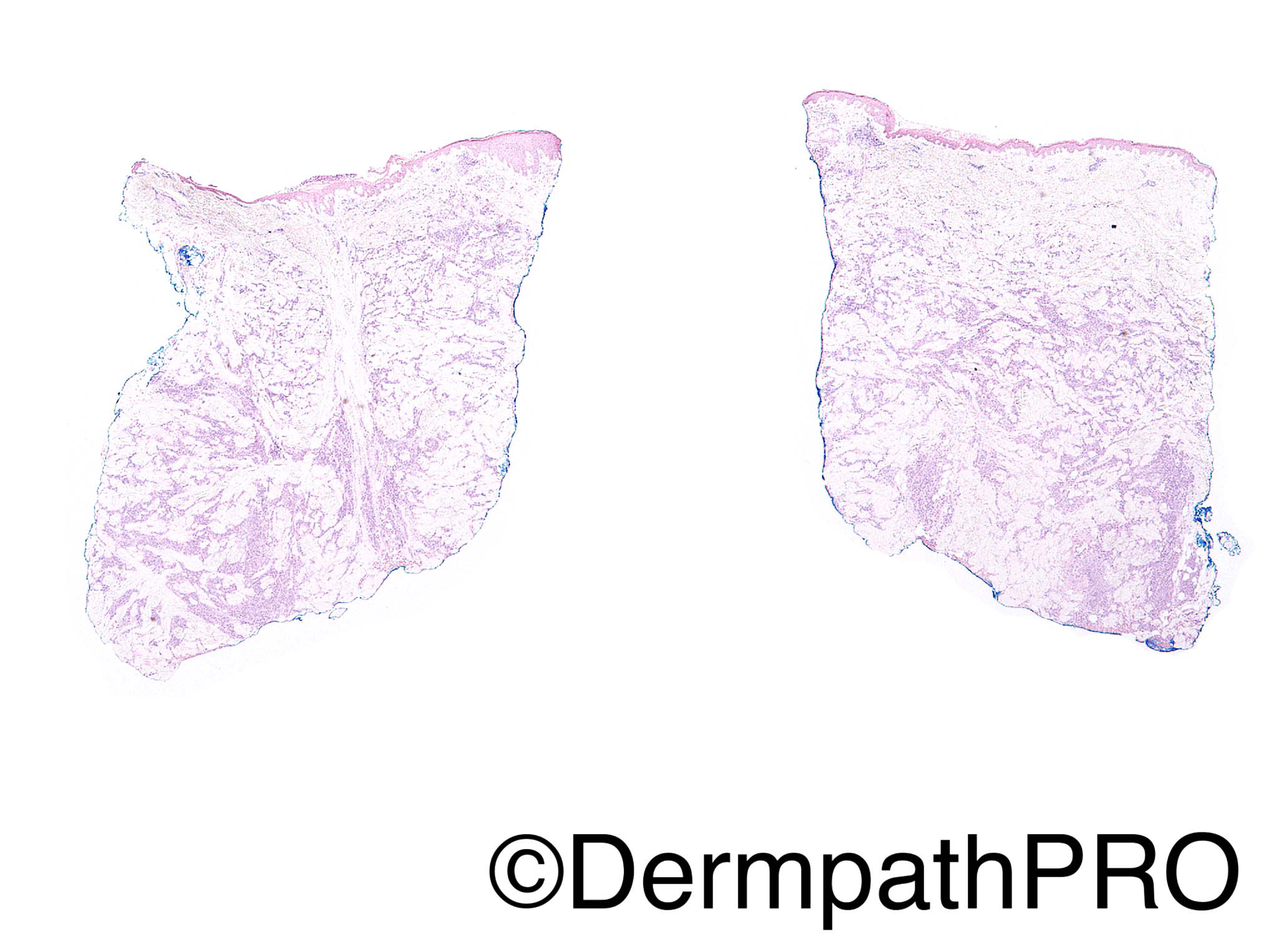
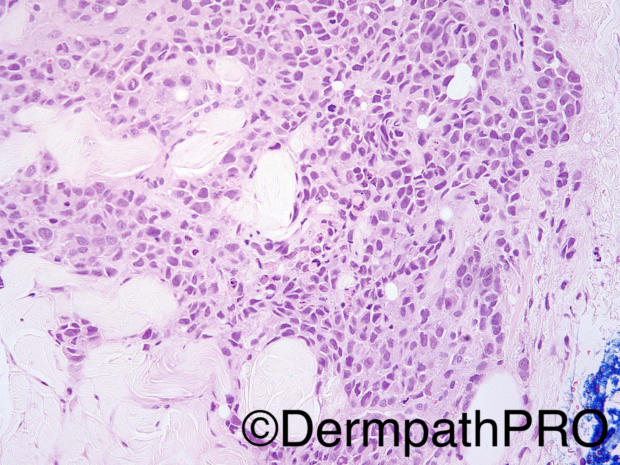
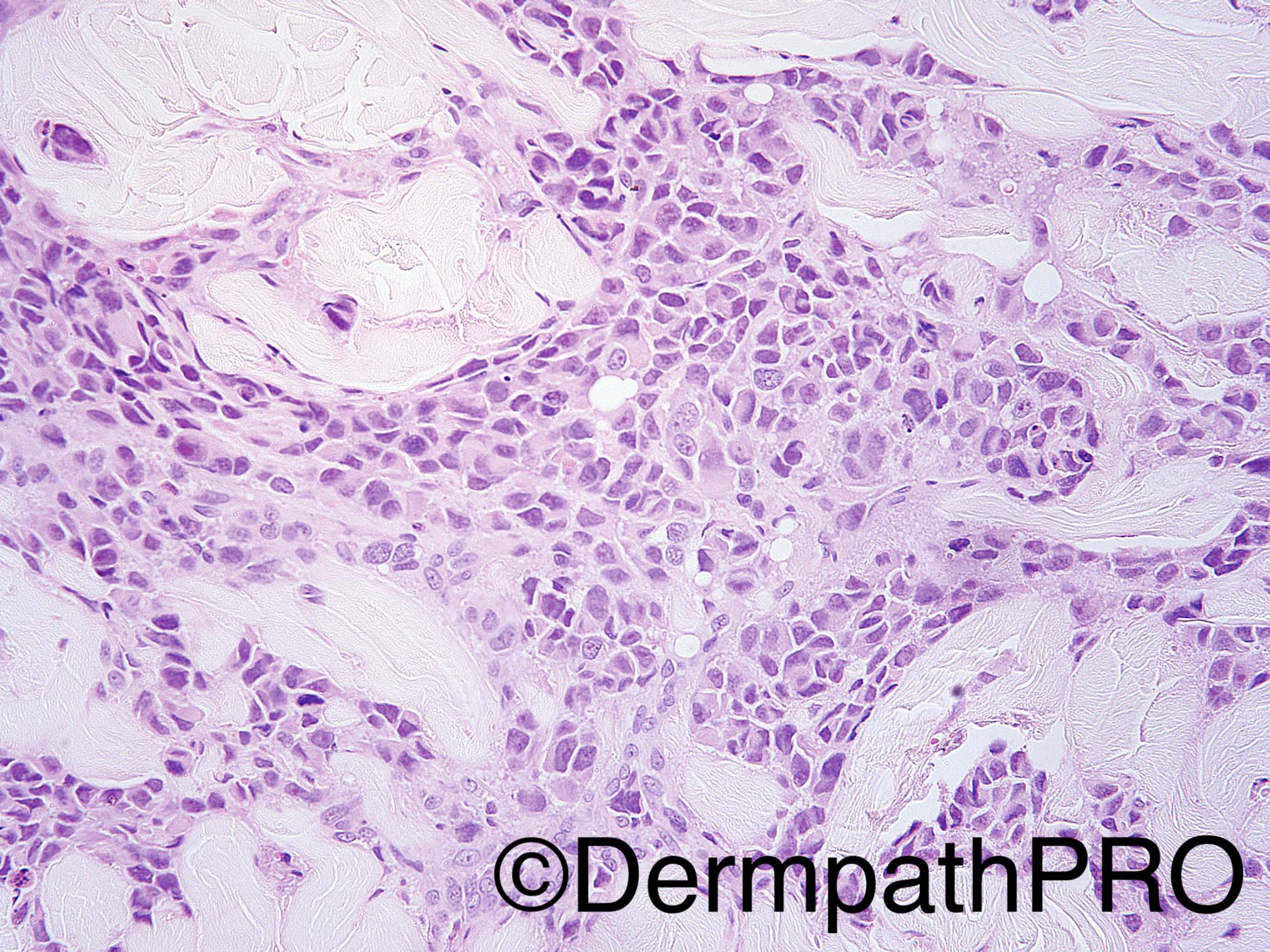
64 year old woman with back dermal lesion. The stain in image 5 is sox 10.

Dr Uma Sundram


  Report Record

User Feedback


Raul Perret

Posted

Melanoma, probably metastatic. I expect s100 and mart1 to be positive here too

Share this comment


Link to comment
share_externally

vincenzo polizzi

Posted

Agree. Metastatic "in transit" melanoma ( also intravascular maybe ).

Share this comment


Link to comment
share_externally

biswanath behera

Posted

Metastatic breast carcinoma

Share this comment


Link to comment
share_externally

Guest Arash Daryakarr

Posted

Small numbers of breast carcinomas about (10-12%) are positive for SOX10. other melanocytic markers such as S100 and melan A as well as mammaglobin or GCDFP15 worth a try.

Share this comment


Link to comment
share_externally

Arti Bakshi

Posted

Metastatic melanoma certainly likely. However important to remeber that SOX10 does stain other tumours including neural and myoepitheiomas. On high power, the cells have eccentric nuclei and plasmacytoid appearance. Whilst this morphology would do for a melanoma too, important to do a panel of markers to exclude myoepithelioma.

Share this comment


Link to comment
share_externally

Arti Bakshi

Posted

Yes, and a metastatic breast Ca too. ..thanks for the ref Raul!

Share this comment


Link to comment
share_externally

Dr. Mona Abdel-Halim

Posted

Agree with ddx, favour met breast carcinoma

Share this comment


Link to comment
share_externally

Nitin Khirwadkar

Posted

I would favour a metastasis from a breast carcinoma (ductal/pleomorphic lobular). The infiltrate has a very interstitial pattern of growth, and I think this would be unusual for a melanoma (in-transit/primary dermal). Would have expected a more nodular growth pattern with a melanoma. Requires a complete IHC panel. Thanks for the article Raul.

Share this comment


Link to comment
share_externally

vincenzo polizzi

Posted

Yes. Morphologically this tumor looks like a pleomorphic solid lobular mammary cancer, but also like a melanoma...So extraimmunos ( or clinic history) should to resolve the diagnostic puzzle...Thanks to all of you for information about SOX 10 and special types of breast cancer..

Share this comment


Link to comment
share_externally

Uma Sundram

Posted

Great job. Point here is a panel of immunohistochemical studies is important to determine lineage and sox10 staining in breast and salivary gland tumors is well worth noting. The lesional cells are positive for Melan A and S100, and negative for keratin markers. Diagnosis: metastatic melanoma. Patient has history of Stage IV melanoma.

Share this comment


Link to comment
share_externally


Join the conversation

You can post now and register later. If you have an account, sign in now to post with your account.

Guest
Add a comment...

×   Pasted as rich text.   Paste as plain text instead

  Only 75 emoji are allowed.

×   Your link has been automatically embedded.   Display as a link instead

×   Your previous content has been restored.   Clear editor

×   You cannot paste images directly. Upload or insert images from URL.

Loading...

×
×
  • Create New...