Jump to content
In this section we have spot diagnoses posted on a daily basis since June 2010, now over 4000! You can review the archived cases and read the suggested diagnoses by users and the final comment by the contributors.
Case are uploaded each week day by 10 am UK time with the correct diagnosis will generally be posted at 8 pm UK time. Why not view the most recent spot diagnosis and proffer a diagnosis?

Case Number : Case 1659 - 3 November - Dr Arti Bakshi Posted By: Guest

Please read the clinical history and view the images by clicking on them before you proffer your diagnosis.
Submitted Date :
   (0 reviews)

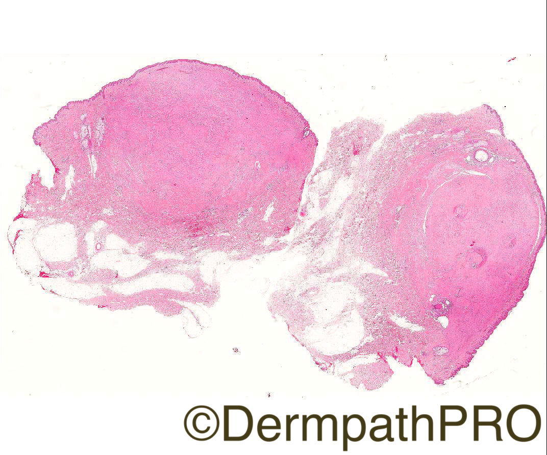
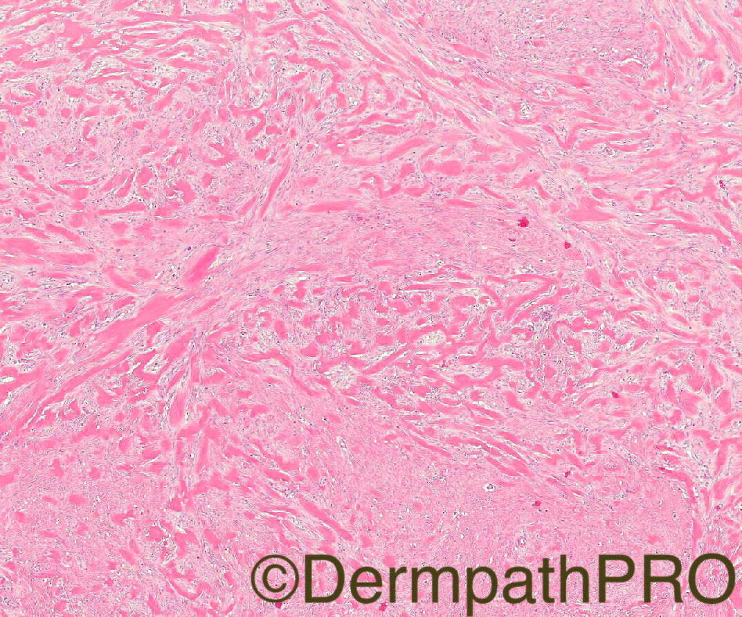
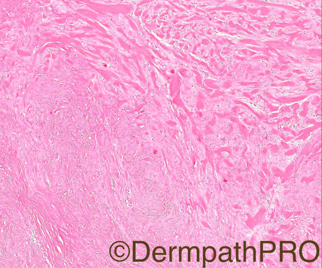
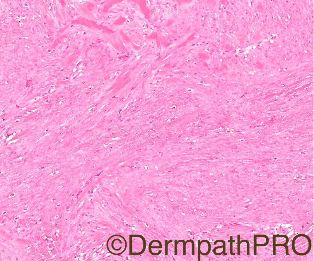
Case History: 18/M, multiple lesions on back, no history of trauma, ?leiomyomata.
 
Case Posted by Dr Arti Bakshi

Edited by Admin_Dermpath


  Report Record

User Feedback


Admin_Dermpath

Posted

Dr Arti Bakshi, who is a Consultant Histopathologist at Royal Liverpool University Hospital NHS trust will now be posting weekly Spot Diagnosis Cases, here is her post for this week.


Many thanks, Geoff

Share this comment


Link to comment
share_externally

Dr. Mona Abdel-Halim

Posted

Thinking of Elastomas (thick broad elastic fibres). Buschke-Ollendorf  Syndrome?

Share this comment


Link to comment
share_externally

Nitin Khirwadkar

Posted

What about juvenile hyaline fibromatosis? 

Share this comment


Link to comment
share_externally

Raul Perret

Posted

I thought it looks like a keloid, I found an article about multiple keloids not related to trauma (should consider syndromic association) article I think there are thick collagen bundles and cells that look fibroblastic/myofibroblastic. Other lesions that I have seen can show this pattern are keloidal dermatofibroma and more rarely sclerosing epithelioid fibrosarcoma.

Share this comment


Link to comment
share_externally

Raul Perret

Posted

I have read also some cases of nodular or keloidal scleroderma can show similar images. I think CPC is important here

Share this comment


Link to comment
share_externally

vincenzo polizzi

Posted

In rare circumstances keloid can arise spontaneously. The sites are chest or shoulder. Back is an odd site. Another option, as Raul said, is a keloidal dermatofibroma. But why not a keloid?

Share this comment


Link to comment
share_externally

Robledo F. Rocha

Posted

I appreciated Raul’s suggestion of keloidal morphea / systemic sclerosis with keloidal nodules. Although no relevant inflammatory infiltrate can be found, other findings are present, including decreased adnexal structures, increased number of fibrocytes, and narrowing of the interstitial space between homogenized collagen bundles.

Share this comment


Link to comment
share_externally

Eman El-Nabarawy

Posted

Thought of juvenile hyaline fibromatosis.

Share this comment


Link to comment
share_externally

Dr. Richard Carr

Posted

Looks like keloid. Given multiple perhaps one of the rare syndromes cited in the link from Raul.

Share this comment


Link to comment
share_externally

Arti Bakshi

Posted

Great discussion and excellent differentials!

This is a case of Multiple Spontaneous Keloids. The morphology is typical of keloid with the glassy collagen. I was stumped by the history of multiple lesions and no previous trauma too, hence considered pretty much all of the d/d considered above. Keloidal/nodular scleroderma in particular, is a very good differential, but the patient does not have any other leisons or signs/symptoms of localised scleroderma or systemic sclerosis. 

A senior dermatologist reviewed the case and felt that the lesions were clinically typical of keloid (following the excision of this lesion, the patient has developed a large 8cm keloidal scar at the site). He suggested the diagnosis of multiple spontaneous keloids (an entity, which I have to confess, I wasnt aware of at that time) and I think the histology would fit. As far as I know, the patient does not have any syndromic association or any family history. 

Share this comment


Link to comment
share_externally


Join the conversation

You can post now and register later. If you have an account, sign in now to post with your account.

Guest
Add a comment...

×   Pasted as rich text.   Paste as plain text instead

  Only 75 emoji are allowed.

×   Your link has been automatically embedded.   Display as a link instead

×   Your previous content has been restored.   Clear editor

×   You cannot paste images directly. Upload or insert images from URL.

Loading...

×
×
  • Create New...