Jump to content
In this section we have spot diagnoses posted on a daily basis since June 2010, now over 4000! You can review the archived cases and read the suggested diagnoses by users and the final comment by the contributors.
Case are uploaded each week day by 10 am UK time with the correct diagnosis will generally be posted at 8 pm UK time. Why not view the most recent spot diagnosis and proffer a diagnosis?

Case Number : Case 1661 - 7 November - Dr Richard A Carr Posted By: Guest

Please read the clinical history and view the images by clicking on them before you proffer your diagnosis.
Submitted Date :
   (0 reviews)

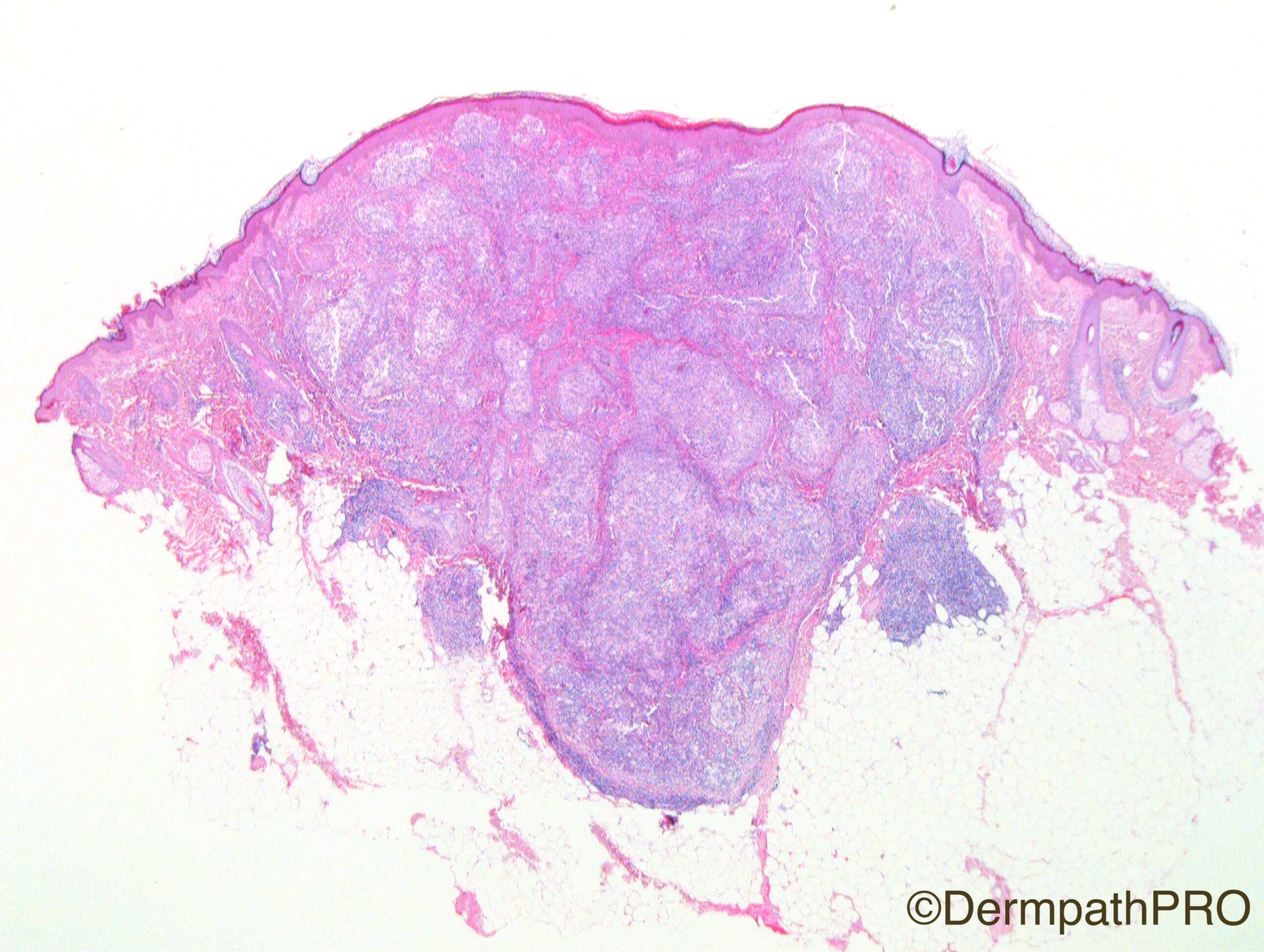
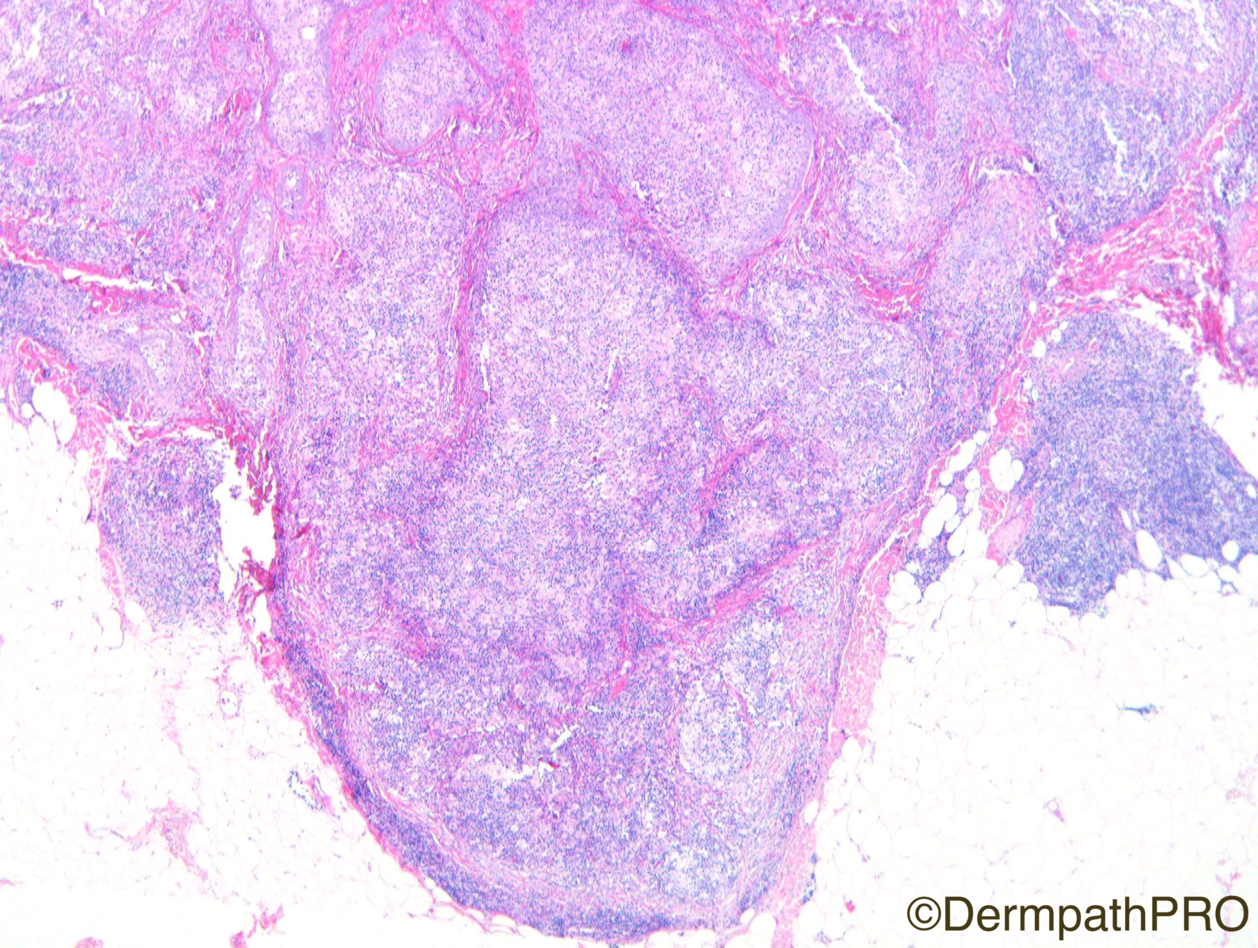
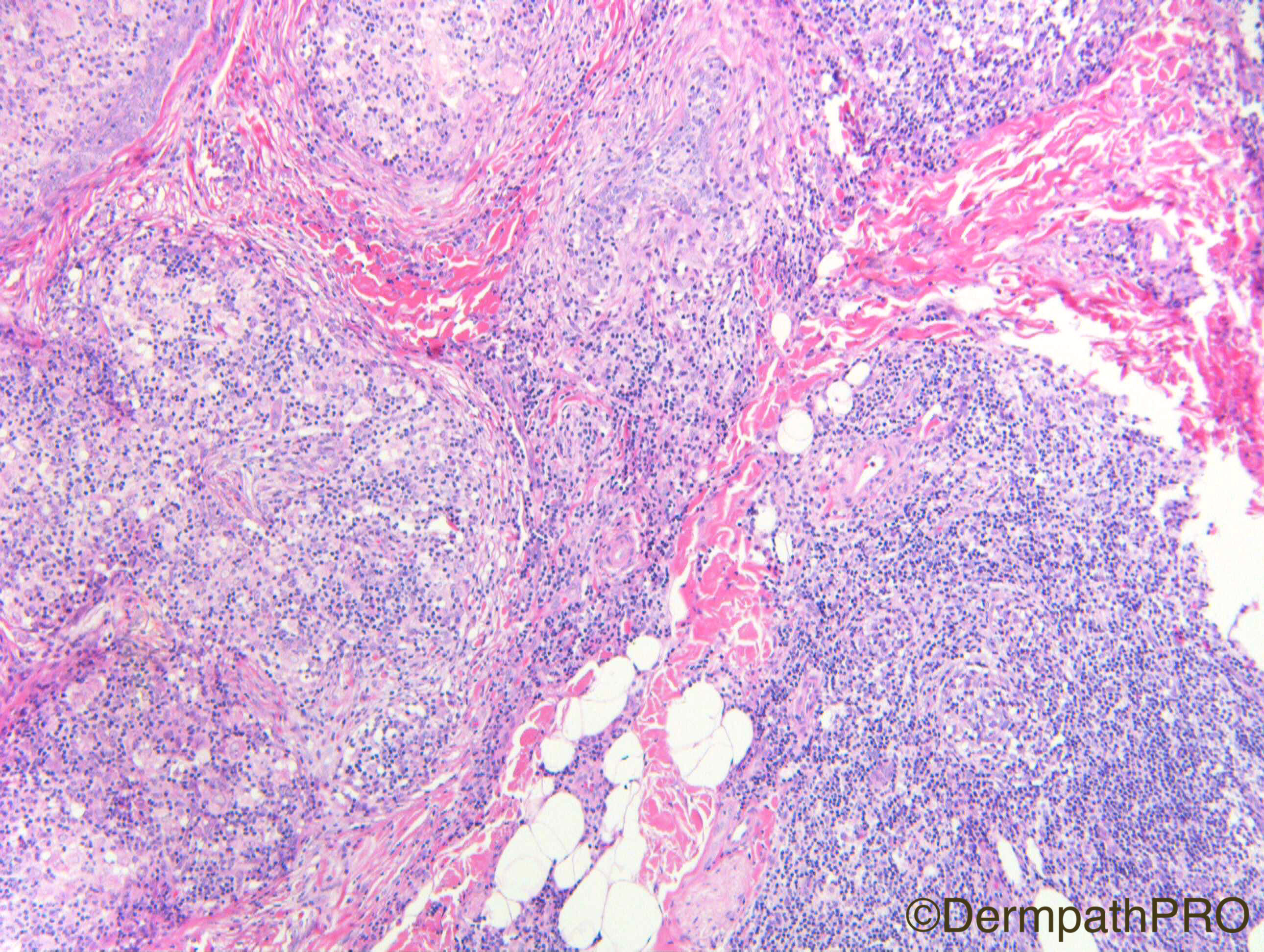
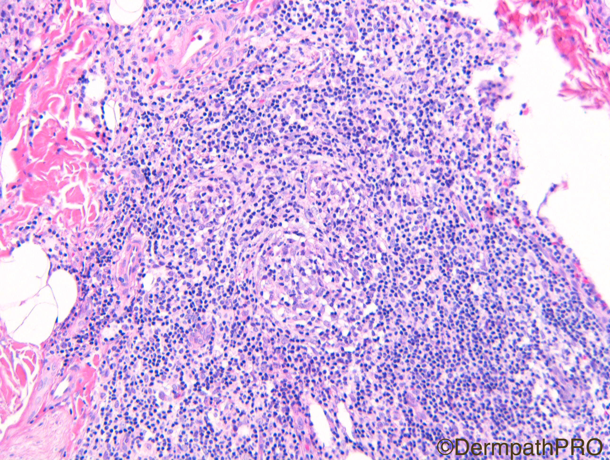
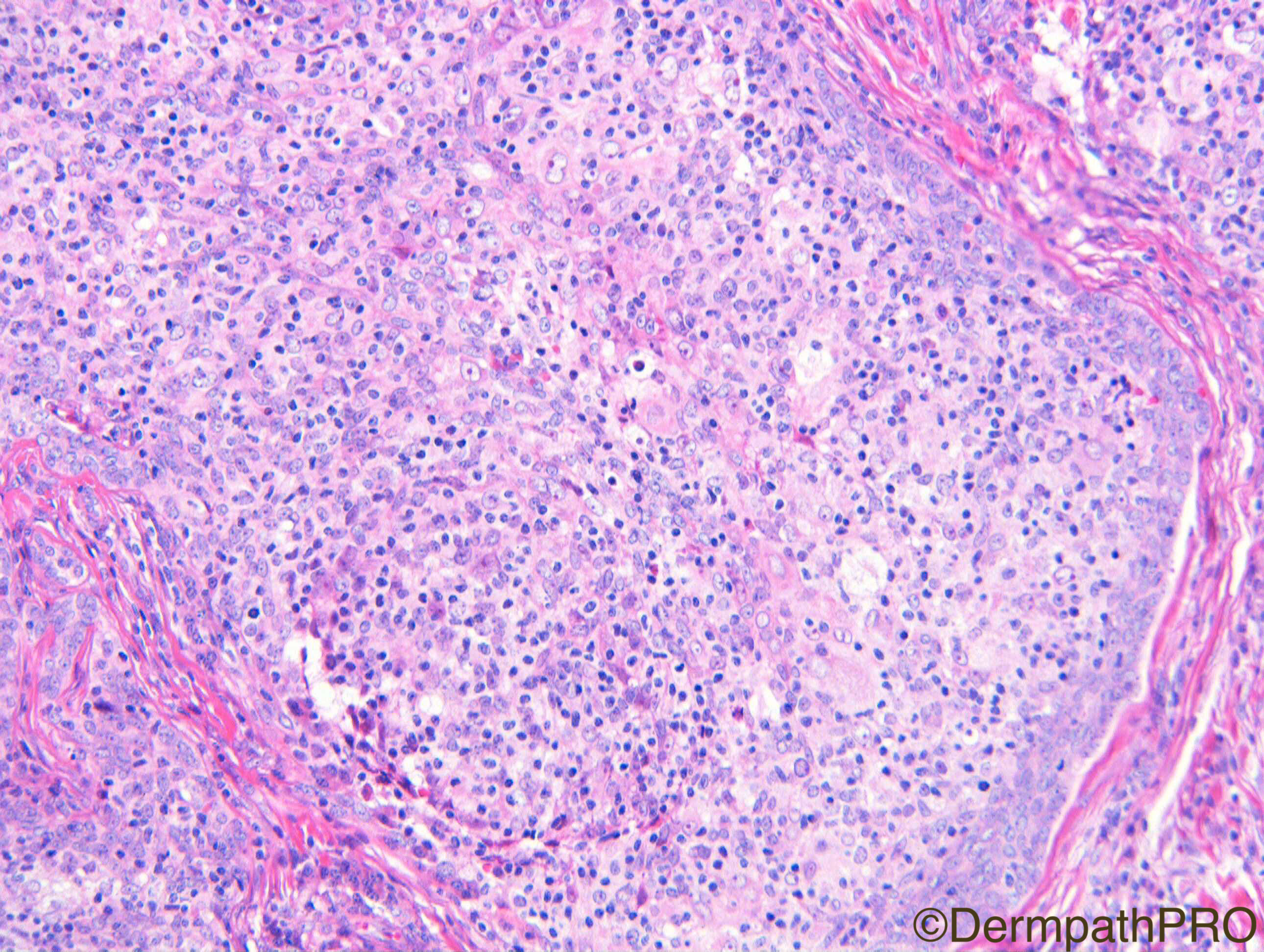
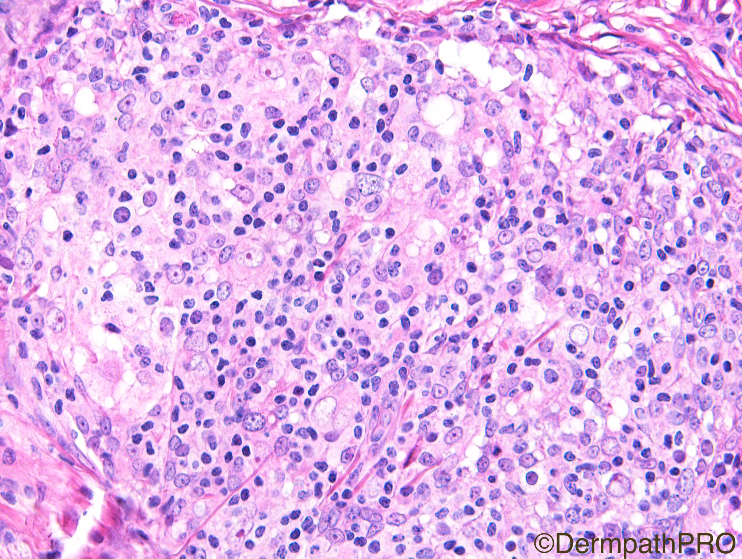
Clinical Details: Female 8 years. Multiple lesions on forehead. Present since birth.

Case Posted by Dr Richard A Carr
Edited by Admin_Dermpath


  Report Record

User Feedback


Admin_Dermpath

Posted

This looks like a great case to get your diagnostic juices running, H&E update at 6pm GMT.


Geoff

Share this comment


Link to comment
share_externally

vincenzo polizzi

Posted

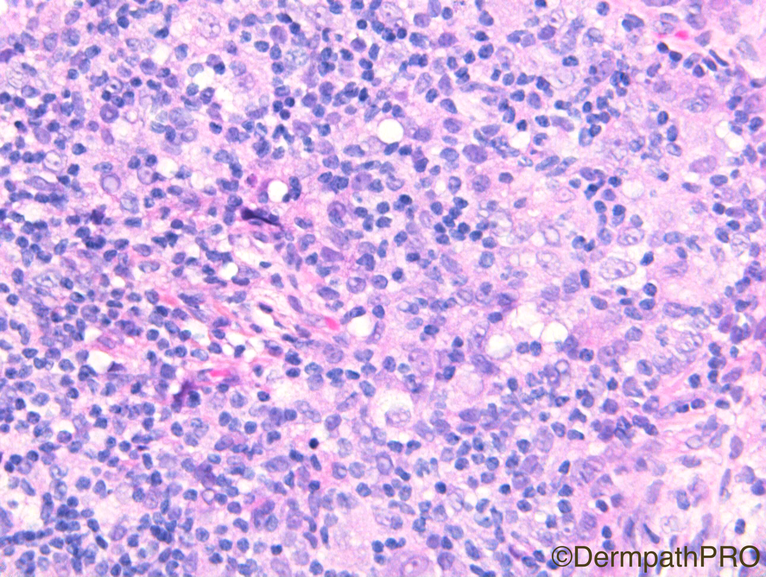
Looks like an intrafollicular RDD, but I don't know similar cases and, the most important, plasma cells poorly found here...so my first thought is of Langherans cell-rich lymphadenoma ( but is a provisional diagnosis...this case is very difficult ).

Glandular congenital lymphadenoma.


Share this comment


Link to comment
share_externally

vincenzo polizzi

Posted

I see many dendritic cells, but don't know what do these mean.

Share this comment


Link to comment
share_externally

Dr. Mona Abdel-Halim

Posted

My provisional impression is RDD, confenital cases exist. Looking for the 6.00 PM update. 

Share this comment


Link to comment
share_externally

Raul Perret

Posted

I think we need cd1a and s100. Differential between rosai dorfman and one of the manu types of langerhans cell histiocytosis

Share this comment


Link to comment
share_externally

My first thought was RDD as well. S-100, CD68 and CD1a will probably solve this case.

Share this comment


Link to comment
share_externally

vincenzo polizzi

Posted

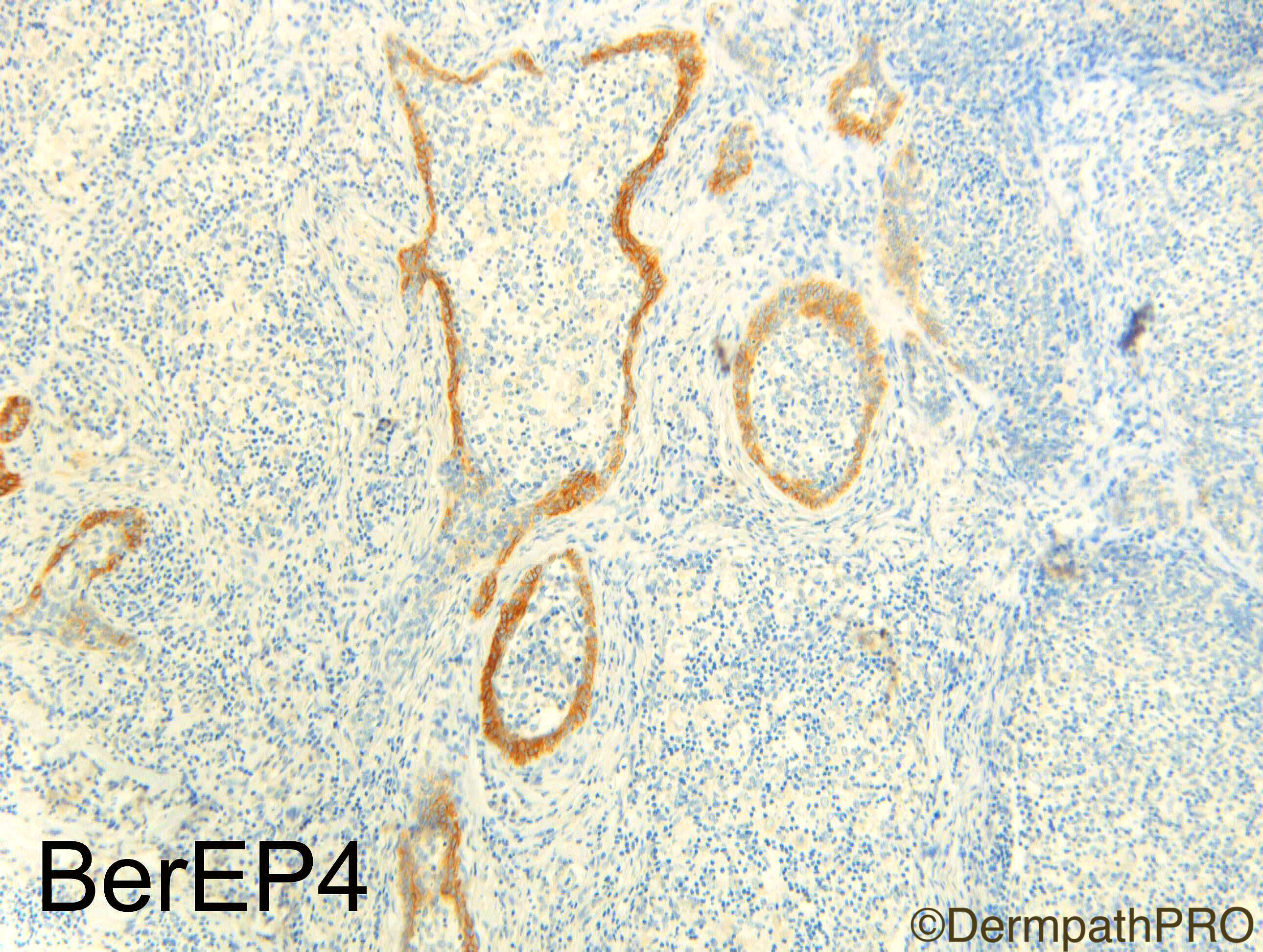
BerEp4 doesn't explain the problem, for me: because it could be a RDD that has diffusely infiltrated dermis , including preexisting follicles.   I think this is a lymphadenoma because of first fig, in which you can see an evident plurifocal follicular profile of the infiltrate, and because of I don't see any plasma cells, so characteristic of RDD...but sincerely I also think I can  be wrong!

Share this comment


Link to comment
share_externally

Robledo F. Rocha

Posted

My first impression really was cutaneous Rosai-Dorfman disease, but the clinical history militates against this hypothesis. A purely cutaneous Rosai-Dorfman disease, without lymphadenopathy, or preceding lymphadenopathy, is rare, and it is even more rare on forehead. Also, it is generally self-limited, does not last 8 years!

Adamantinoid trichoblastoma (lymphadenoma) with abundant intraepithelial lymphocytes, as Vicenzo suggested, is a good point. Peripheral palisading of basophilic basaloid cells, which can be noted in image 5, is a clue.

Share this comment


Link to comment
share_externally

Dr. Mona Abdel-Halim

Posted

I don't think you are wrong Vincenzo... BerEP4 highlighting the peripheral cells makes it lymphadenoma. Very unusual!! 

Share this comment


Link to comment
share_externally

Dr. Mona Abdel-Halim

Posted

Nice that we all thought of RDD!!

Share this comment


Link to comment
share_externally

Arti Bakshi

Posted

Wow...difficult case. Vincenzo is probably right!!...but why multiple lesions (and since birth)??


Share this comment


Link to comment
share_externally

Nitin Khirwadkar

Posted

Great case! The IHC does make this into a cutaneous lymphadenoma. These can have varying degrees of atypia/RS like cells within the central pale areas. However, not sure if these can present at birth, or have any syndromic association. Waiting to hear from Richard.

Share this comment


Link to comment
share_externally

Dr. Richard Carr

Posted

This is a cutaneous lymphadenoma.  Almost a one off case (multiple congenital lesions) but we did find the reference cited by Vincenzo who did very well again.  I also shared the case with Dmitry Kazakov by powerpoint and he agreed.  The pan-keratin stained all the lobules diffusely.  It is unusual to have the spillage of the lymphocytes in to the stroma.  I thought this case made a nice contrast one of the spiradenoma's I had placed recently.  Glad if fooled a few of you.  I would like to acknowledge Dr Anita Nagy and colleagues who made the original diagnosis and kindly shared the case with me.

Share this comment


Link to comment
share_externally


Join the conversation

You can post now and register later. If you have an account, sign in now to post with your account.

Guest
Add a comment...

×   Pasted as rich text.   Paste as plain text instead

  Only 75 emoji are allowed.

×   Your link has been automatically embedded.   Display as a link instead

×   Your previous content has been restored.   Clear editor

×   You cannot paste images directly. Upload or insert images from URL.

Loading...

×
×
  • Create New...