Jump to content
In this section we have spot diagnoses posted on a daily basis since June 2010, now over 4000! You can review the archived cases and read the suggested diagnoses by users and the final comment by the contributors.
Case are uploaded each week day by 10 am UK time with the correct diagnosis will generally be posted at 8 pm UK time. Why not view the most recent spot diagnosis and proffer a diagnosis?

Case Number : Case 1664 - 10 November - Dr Arti Bakshi Posted By: Guest

Please read the clinical history and view the images by clicking on them before you proffer your diagnosis.
Submitted Date :
   (0 reviews)

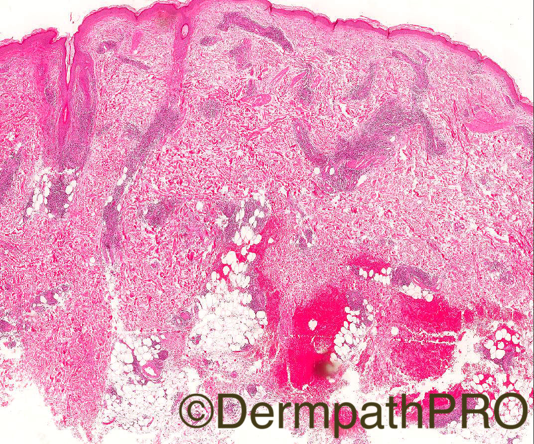
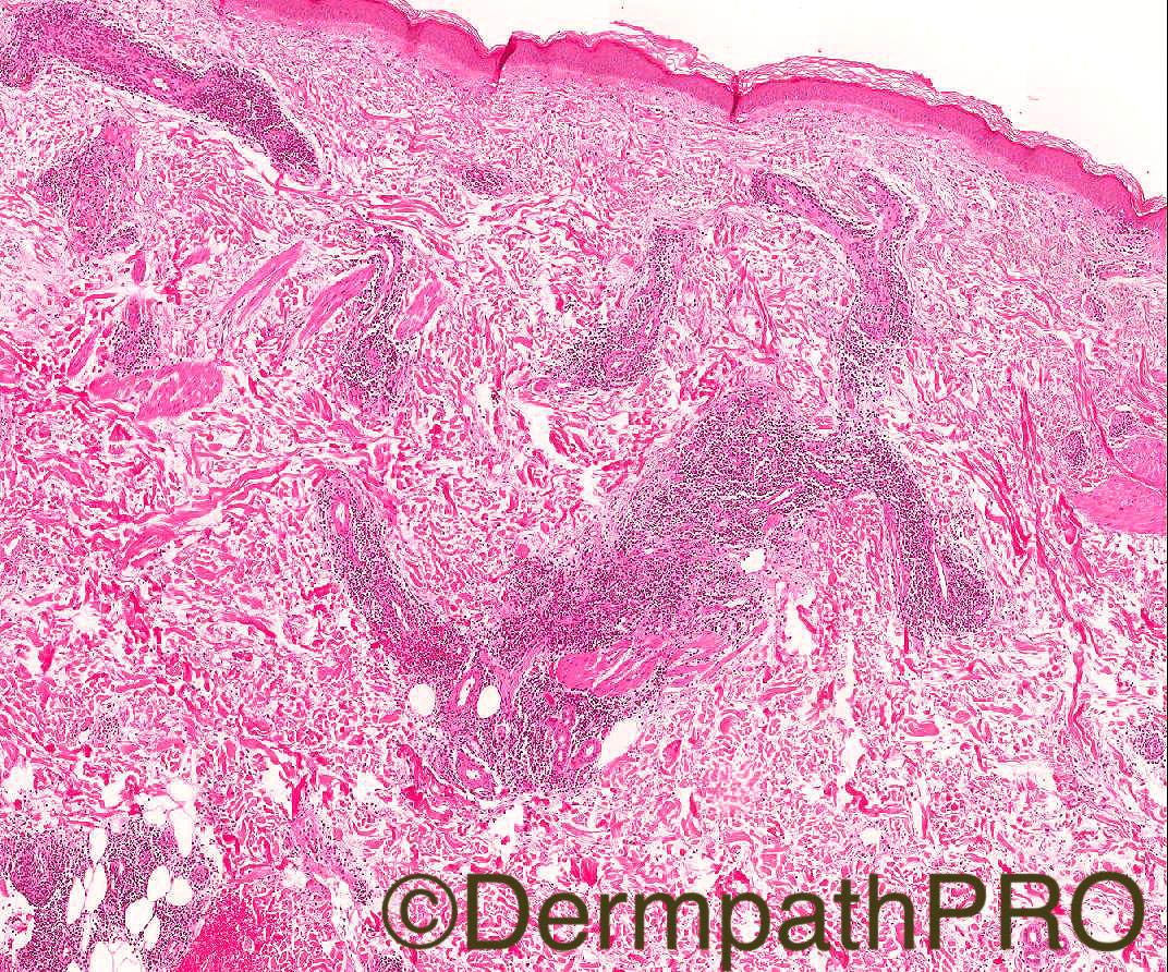
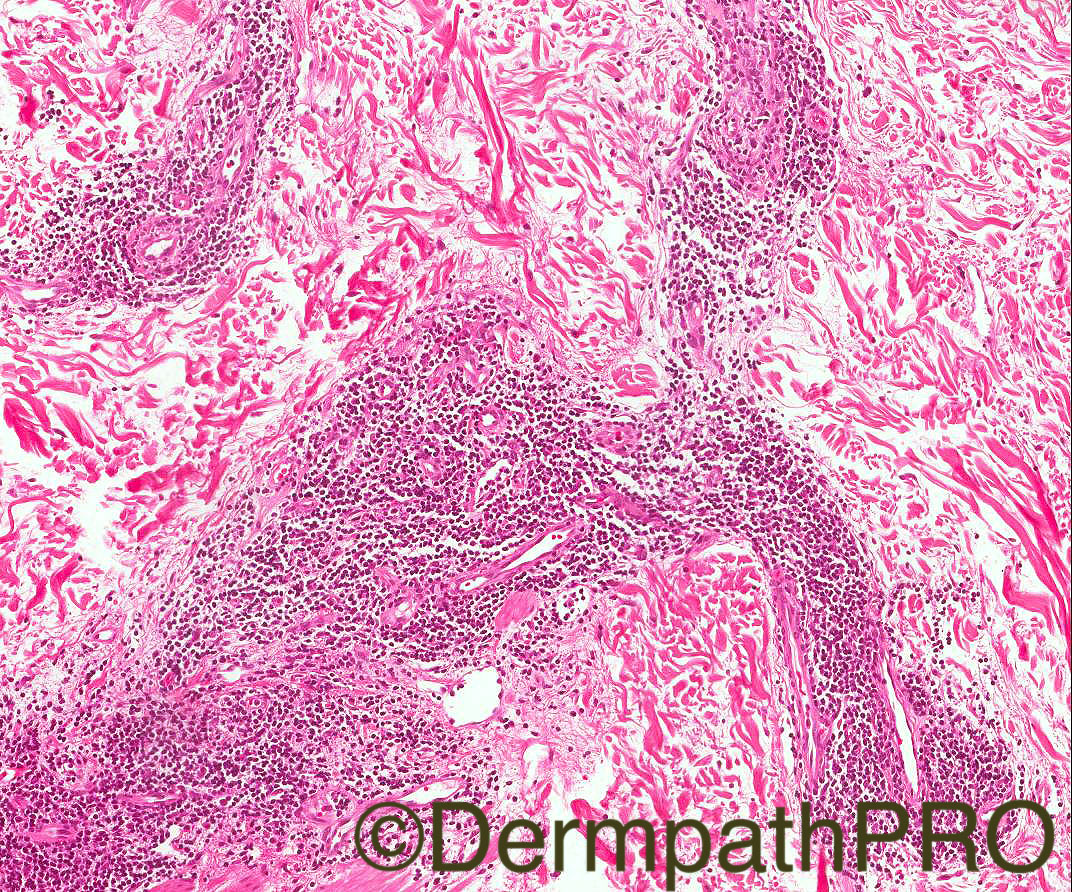
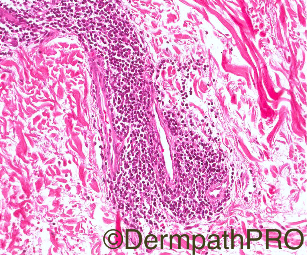
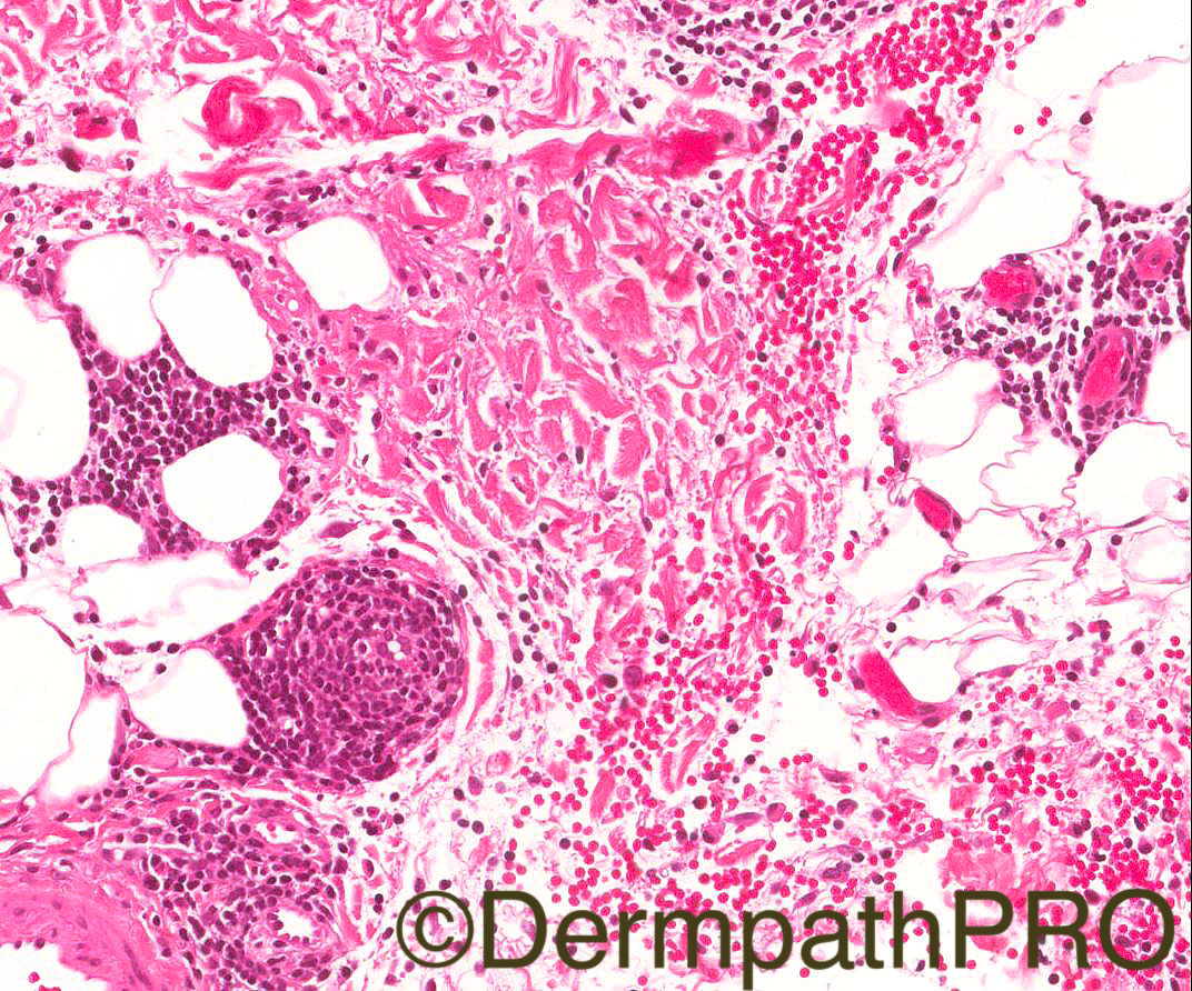
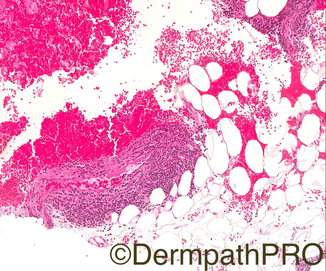
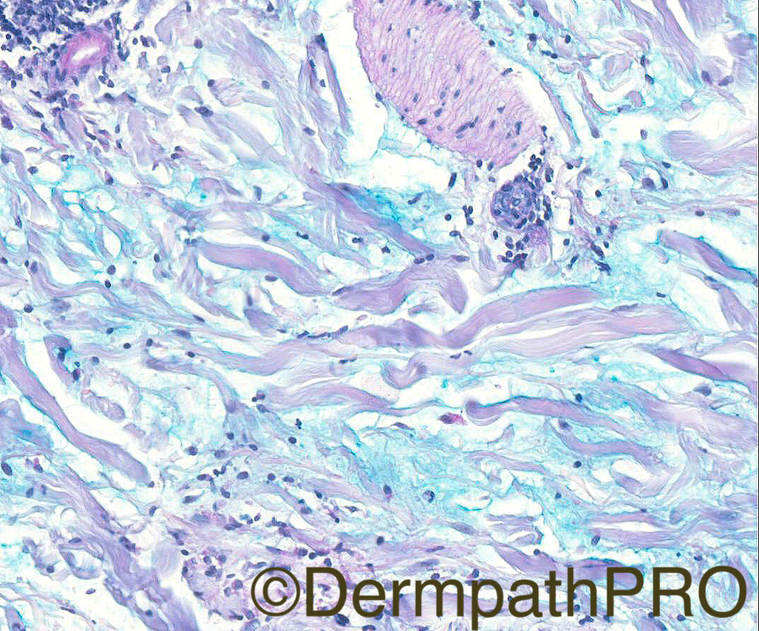
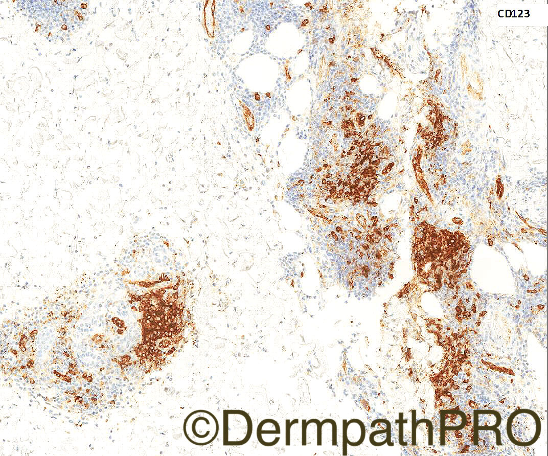
Case History: 51/F, erythematous inflamed patch on right upper arm (approximately 5”). Thickened/indurated initially, but more macular now. No other lesions. ANA negative. ?granuloma annulare

Case Posted by Dr Arti Bakshi

Edited by Admin_Dermpath


  Report Record

User Feedback


Admin_Dermpath

Posted

Pretty in Pink, but is it? What do you think Dr Arti Bakshi's Case is?


Cheers, Geoff

DermpathPRO Projects

Share this comment


Link to comment
share_externally

vincenzo polizzi

Posted

Lupus diagnosis is always a challenge. But I agree with Hazem.   

Share this comment


Link to comment
share_externally

Raul Perret

Posted

Agree with the impression of tumid lupus/jessner. I thought that the clinical picture was funny though: isolated lesion in a non photoexposed area in an adult older than 45. I would still perform immunos: cd20, 3, 4, 8 and 123. The clinical picture and histology looks rather benign but I would not like to miss a lymphoma

Share this comment


Link to comment
share_externally

Clara Jaramillo

Posted (edited)

Lupus erythematosus tumidus

Edited by Clara Jaramillo

Share this comment


Link to comment
share_externally

Robledo F. Rocha

Posted

Superficial and deep perivascular and periappendageal lymphocytic infiltrate accompanied by prominent dermal mucin deposition. Epidermal changes are lacking, or there is only slight epidermal atrophy. Agree with lupus erythematosus tumidus.

Interestingly enough the intermittent firmness of the lesion.

Share this comment


Link to comment
share_externally

Arti Bakshi

Posted

Yes, this is a case of tumid lupus erythematosus.

Raul, we did do some immunos and it was a T cell rich infiltrate with a mixed CD4 and CD8 population. Have asked Geoff to put an image of CD123, which shows nice clusters of palsmacytoid dendritic cells. 

For the newbies, tumid lupus erythematosus is a distinct subtype of chronic lupus erythematosus which differs clinically from other forms of lupus in the absence of surface epidermal changes, lack of scarring on resolution and low risk of association with systemic lupus (and often negative serology).  Here is a link to a nice review article:

http://www.actasdermo.org/en/lupus-erythematosus-tumidus-a-clinical/articulo/S1578219011001053/

Share this comment


Link to comment
share_externally

Arti Bakshi

Posted

Great to see some new names in the contributors...welcome to all!

Share this comment


Link to comment
share_externally

Raul Perret

Posted

Thank you Arti for this interesting case and big welcome to the new participants

Share this comment


Link to comment
share_externally


Join the conversation

You can post now and register later. If you have an account, sign in now to post with your account.

Guest
Add a comment...

×   Pasted as rich text.   Paste as plain text instead

  Only 75 emoji are allowed.

×   Your link has been automatically embedded.   Display as a link instead

×   Your previous content has been restored.   Clear editor

×   You cannot paste images directly. Upload or insert images from URL.

Loading...

×
×
  • Create New...