Jump to content
In this section we have spot diagnoses posted on a daily basis since June 2010, now over 4000! You can review the archived cases and read the suggested diagnoses by users and the final comment by the contributors.
Case are uploaded each week day by 10 am UK time with the correct diagnosis will generally be posted at 8 pm UK time. Why not view the most recent spot diagnosis and proffer a diagnosis?

Case Number : Case 1665 - 11 November - Dr Richard Carr Posted By: Guest

Please read the clinical history and view the images by clicking on them before you proffer your diagnosis.
Submitted Date :
   (0 reviews)

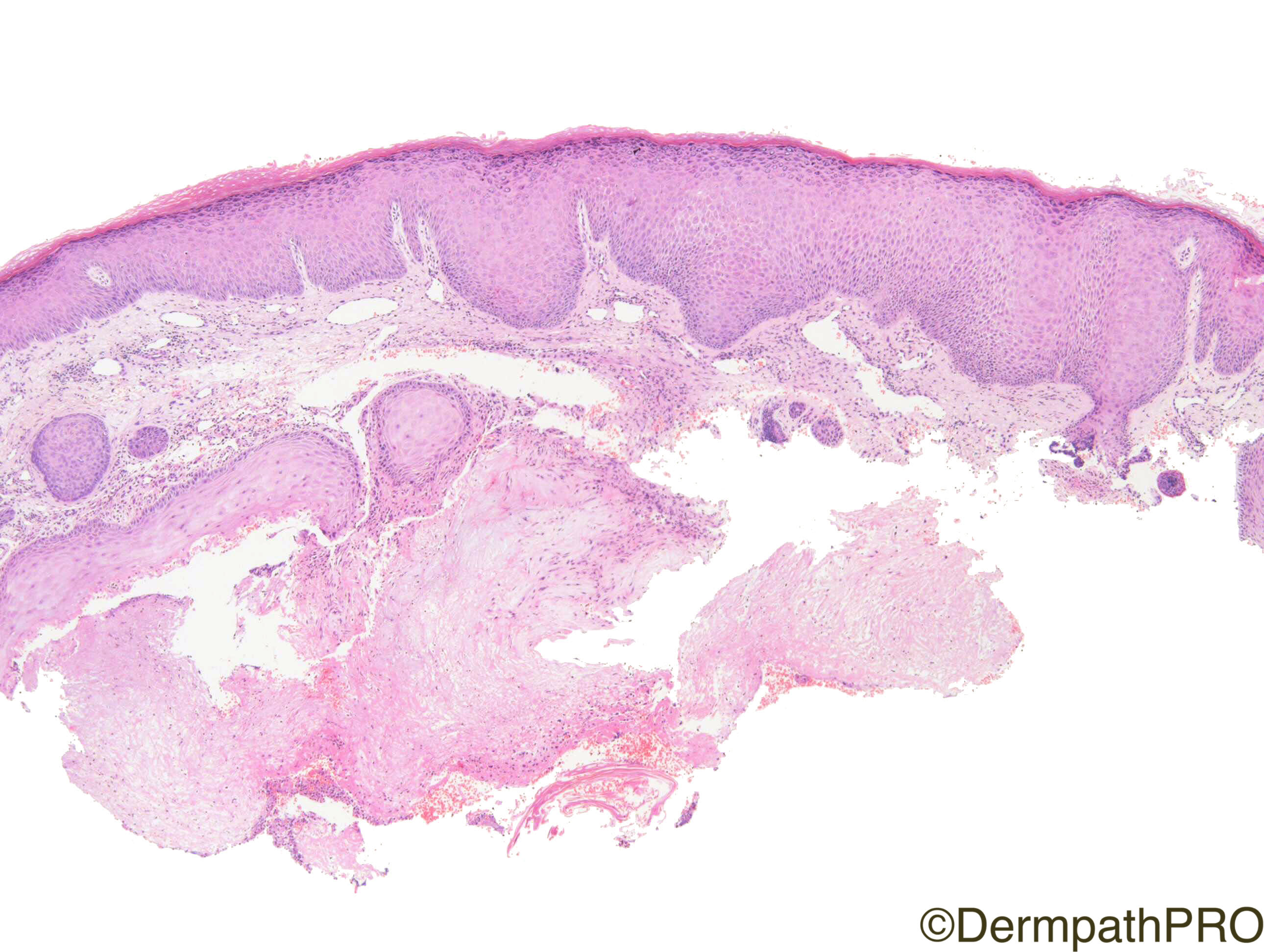
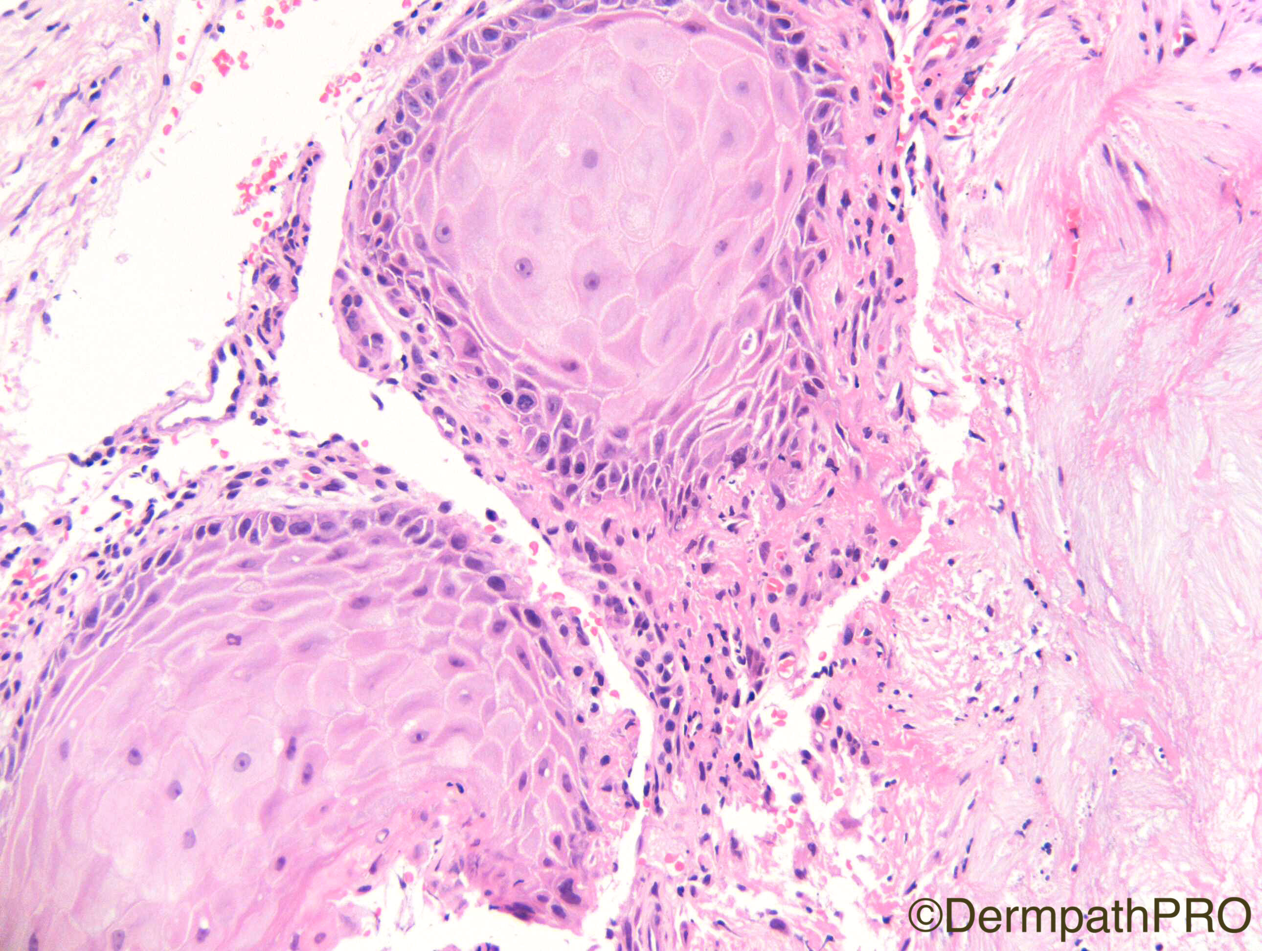
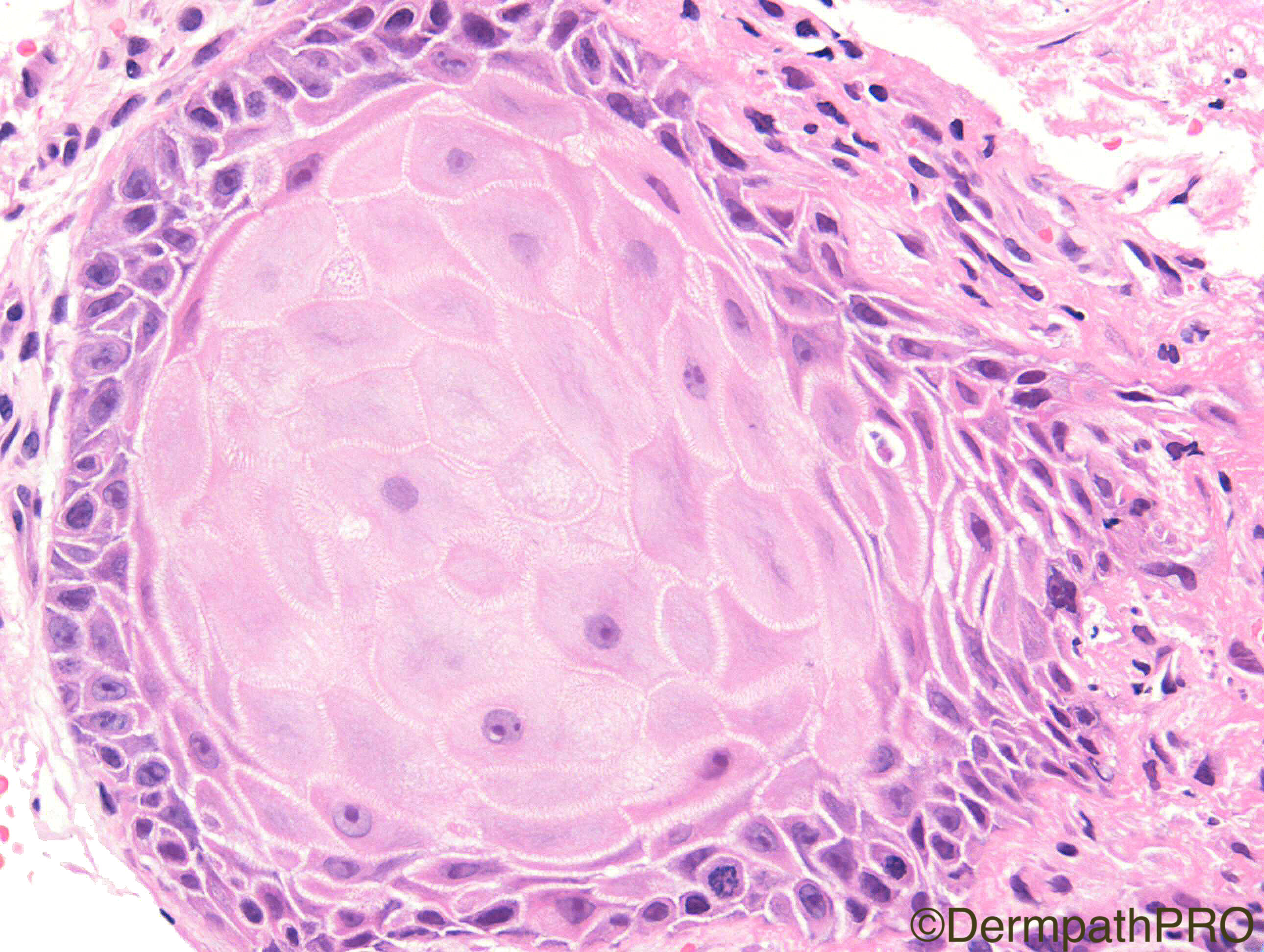
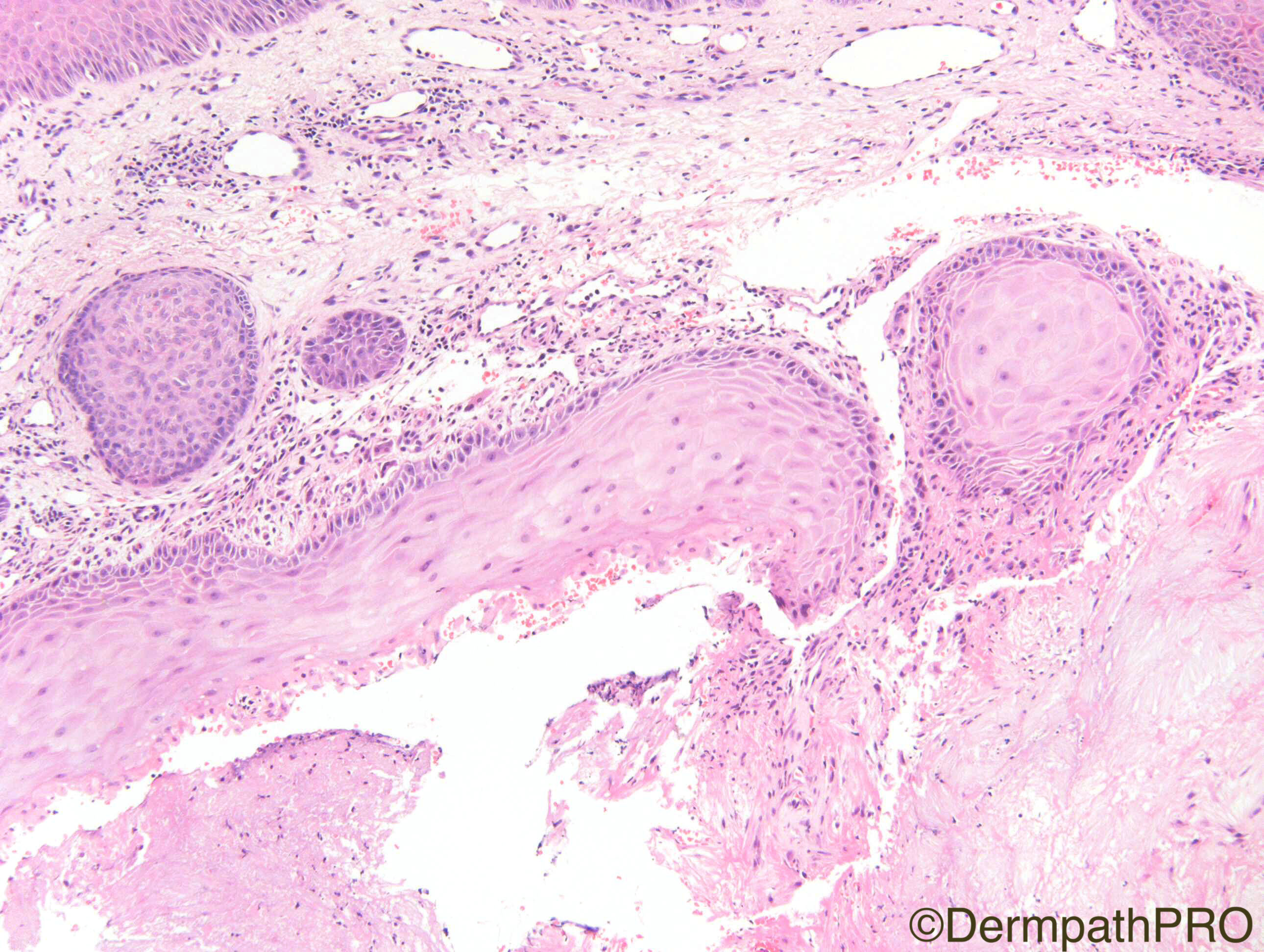
Clinical Details: Pimple on ear 1 year. Now growing after local trauma. ?SCC
H&Ex4

Case Posted by Dr Richard Carr

  Report Record

User Feedback


Admin_Dermpath

Posted

Richard's cases always take us up a notch or two, really top class images.

Cheers, Geoff - DermpathPRO Projects

Share this comment


Link to comment
share_externally

Guest Hazem Hamed

Posted

Reactive squamous proliferation (pseudocarcinomatous hyperplasia in association with gouty tophi).

Share this comment


Link to comment
share_externally

Guest Hazem Hamed

Posted

Case Report- Pseudocarcinomatous hyperplasia involving the ear from gout: a diagnostic pitfall. JCP 2013

Share this comment


Link to comment
share_externally

Raul Perret

Posted

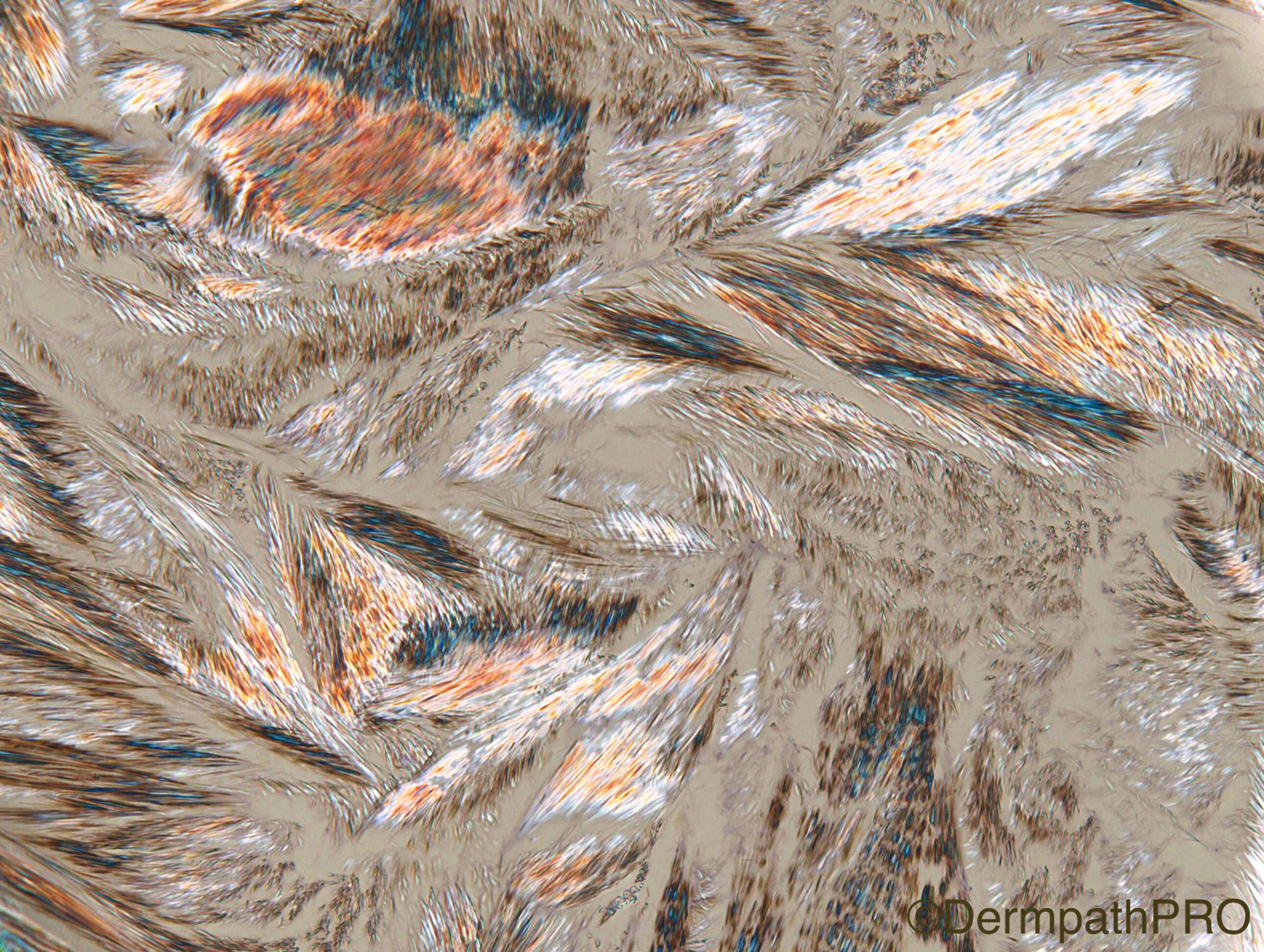
Agree, crystal spaces better seen in third picture. I guess that other differential to consider in low power is chondrodermatitis nodularis helicis but higher power clears the picture.

Share this comment


Link to comment
share_externally

vincenzo polizzi

Posted

My first impression was of a broken off epidermal cyst, but I think it's a misleading image...There is a pseudoepitheliomatous hyperplasia around degenerate collagen with slit like spaces and fibrin : what make me favor a CHONDRODERMATITIS NODULARIS HELICIS.

Share this comment


Link to comment
share_externally

Robledo F. Rocha

Posted

I was looking for granulomatous reaction with macrophages and multinucleated foreign body giant cells that typically surrounds the urate crystals deposits. Assuming that granulomatous reaction can be found in the deepest part of the first image, this my be gout. I have never seen pseudoepitheliomatous hyperplasia of the epidermis related with gout tophus.

Share this comment


Link to comment
share_externally

Admin_Dermpath

Posted

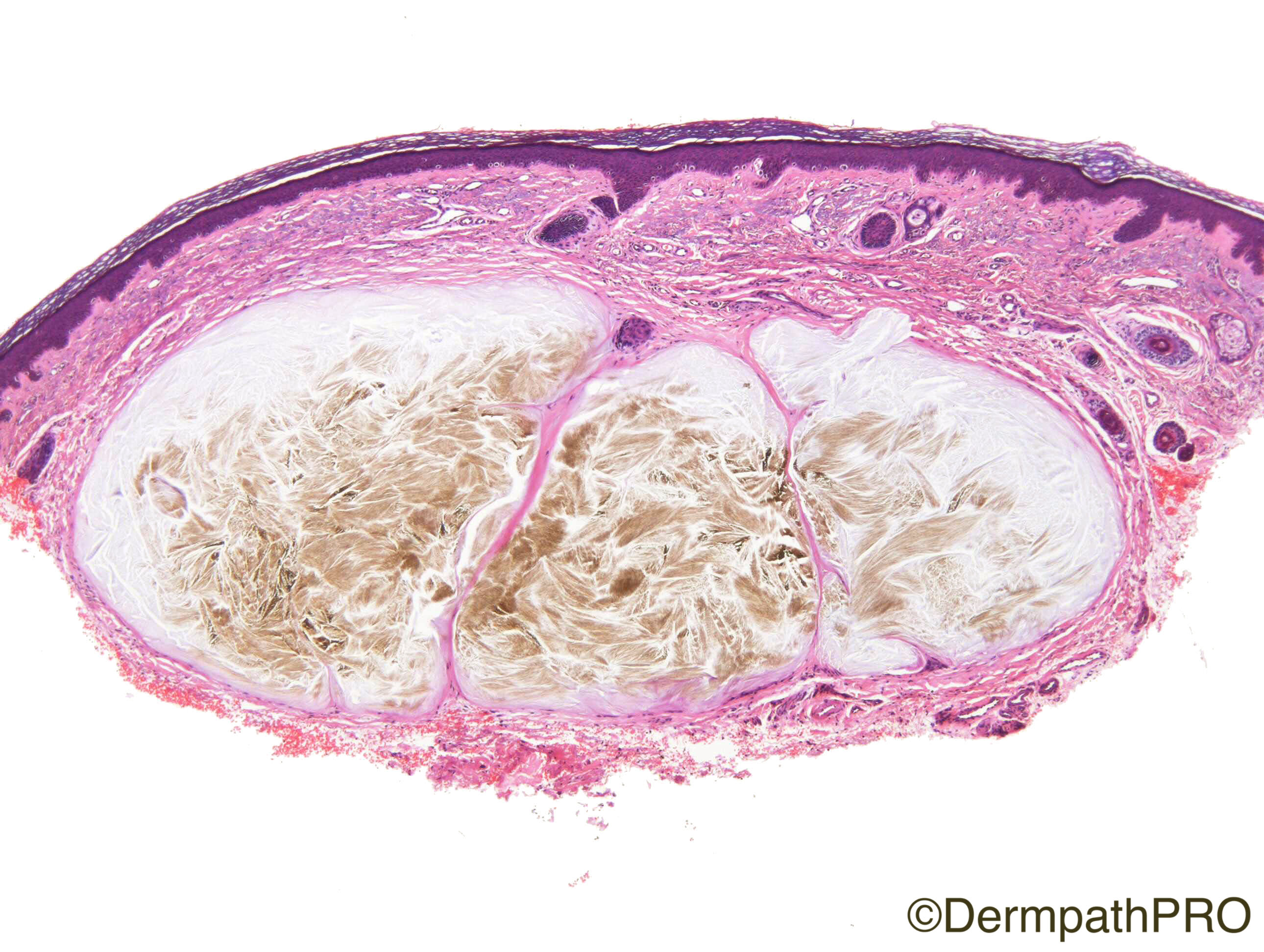
Igor et al, you will like the images that I have just uploaded to add to this case.

Cheers, Geoff

Share this comment


Link to comment
share_externally

Wow!! Amazing!!! The brown color in H&E and the polirized light are great!

Share this comment


Link to comment
share_externally

vincenzo polizzi

Posted

Well done Hazem and Raul!!! Never seen a similar case. Very nice and didactically useful. 

I should have perceived the cristal spaces, as Raul did!!! 


Share this comment


Link to comment
share_externally

Dr. Richard Carr

Posted

Yes well done Hazem et al and thanks for the reference too.  I've seen a few cases on the ear now.  The uric acid crystals usually dissolve in processing as they are water soluble.  The second two images were from a new lesion on the opposite year and luckily the crystals were evident.  I've now diagnosed tophi on the ear several times and i'm pretty sure another also had pseudoepitheliomatous hyperplasia. 

Share this comment


Link to comment
share_externally


Join the conversation

You can post now and register later. If you have an account, sign in now to post with your account.

Guest
Add a comment...

×   Pasted as rich text.   Paste as plain text instead

  Only 75 emoji are allowed.

×   Your link has been automatically embedded.   Display as a link instead

×   Your previous content has been restored.   Clear editor

×   You cannot paste images directly. Upload or insert images from URL.

Loading...

×
×
  • Create New...