Jump to content
In this section we have spot diagnoses posted on a daily basis since June 2010, now over 4000! You can review the archived cases and read the suggested diagnoses by users and the final comment by the contributors.
Case are uploaded each week day by 10 am UK time with the correct diagnosis will generally be posted at 8 pm UK time. Why not view the most recent spot diagnosis and proffer a diagnosis?

Case Number : Case 1666 - 14 November - Dr Uma Sundram Posted By: Guest

Please read the clinical history and view the images by clicking on them before you proffer your diagnosis.
Submitted Date :
   (0 reviews)

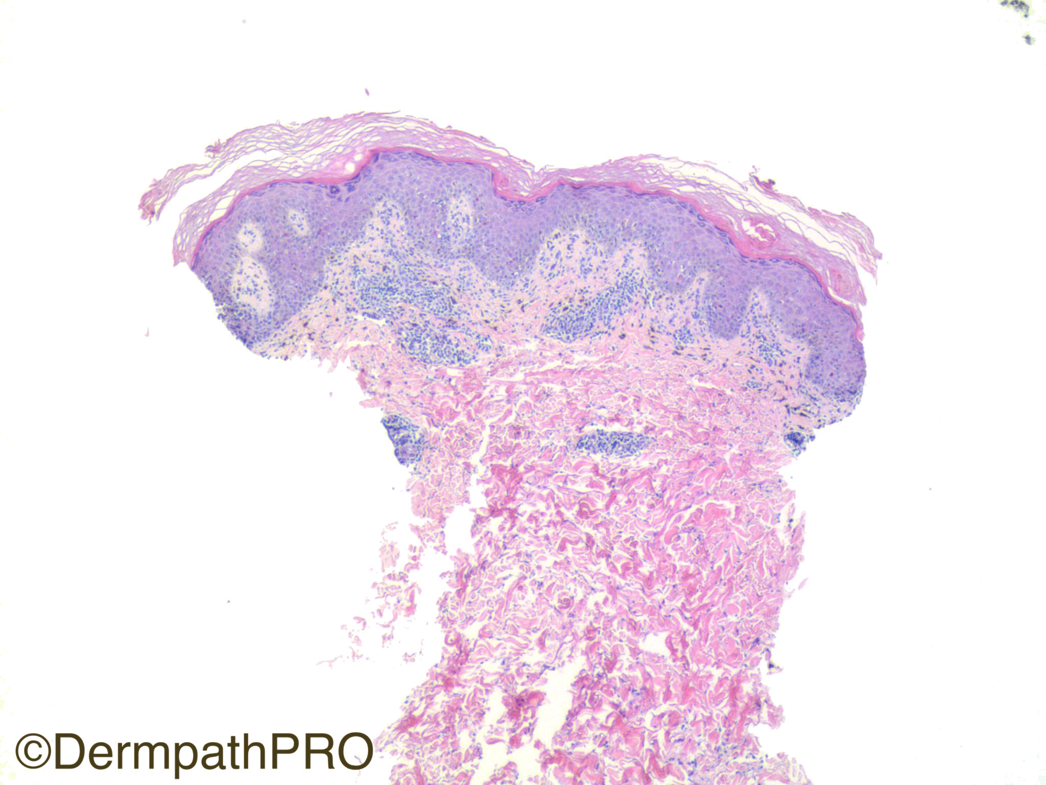
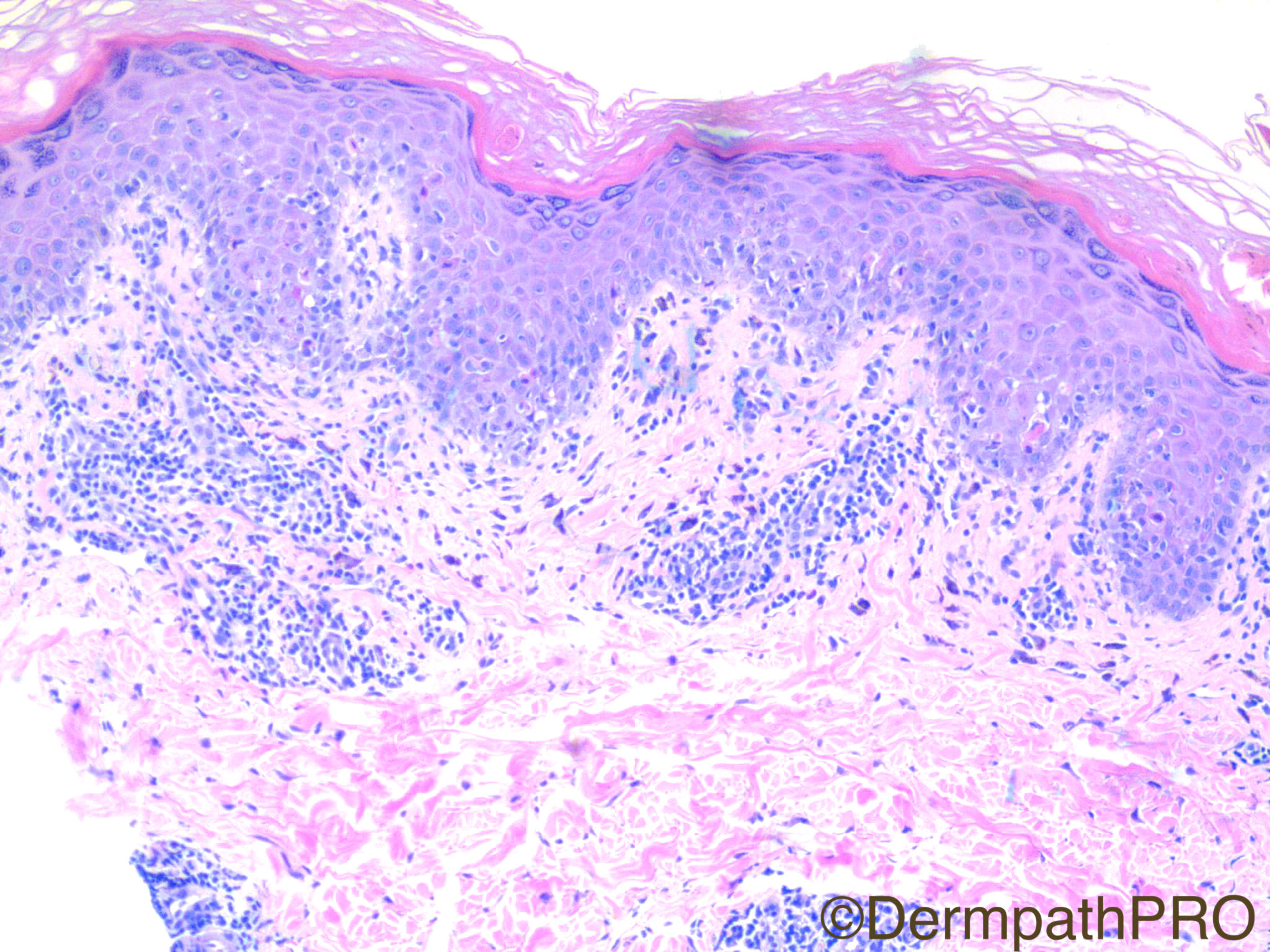
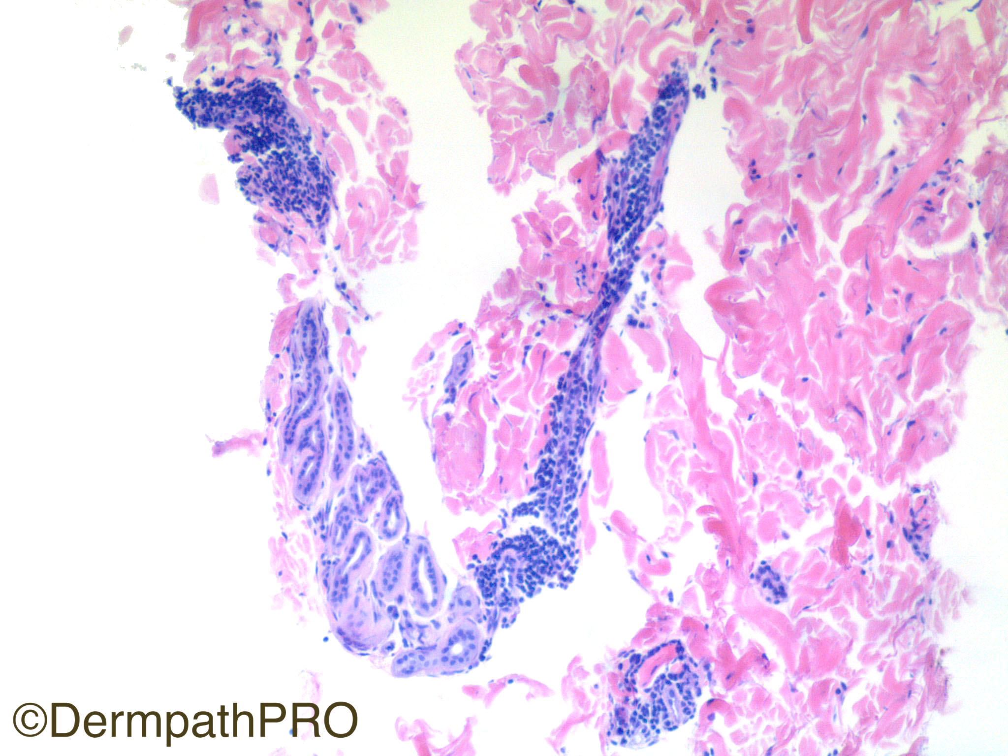
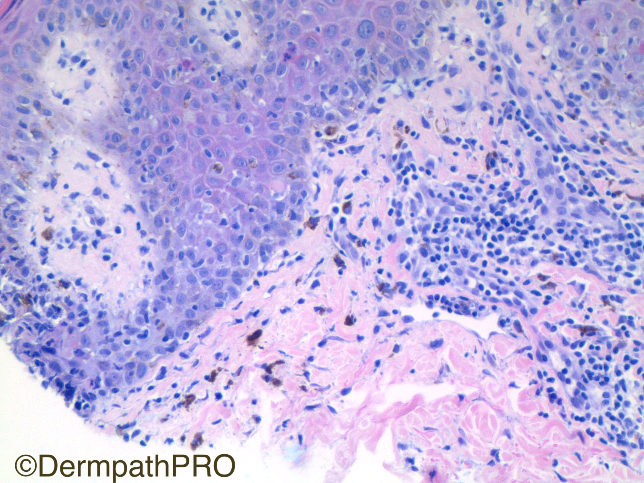
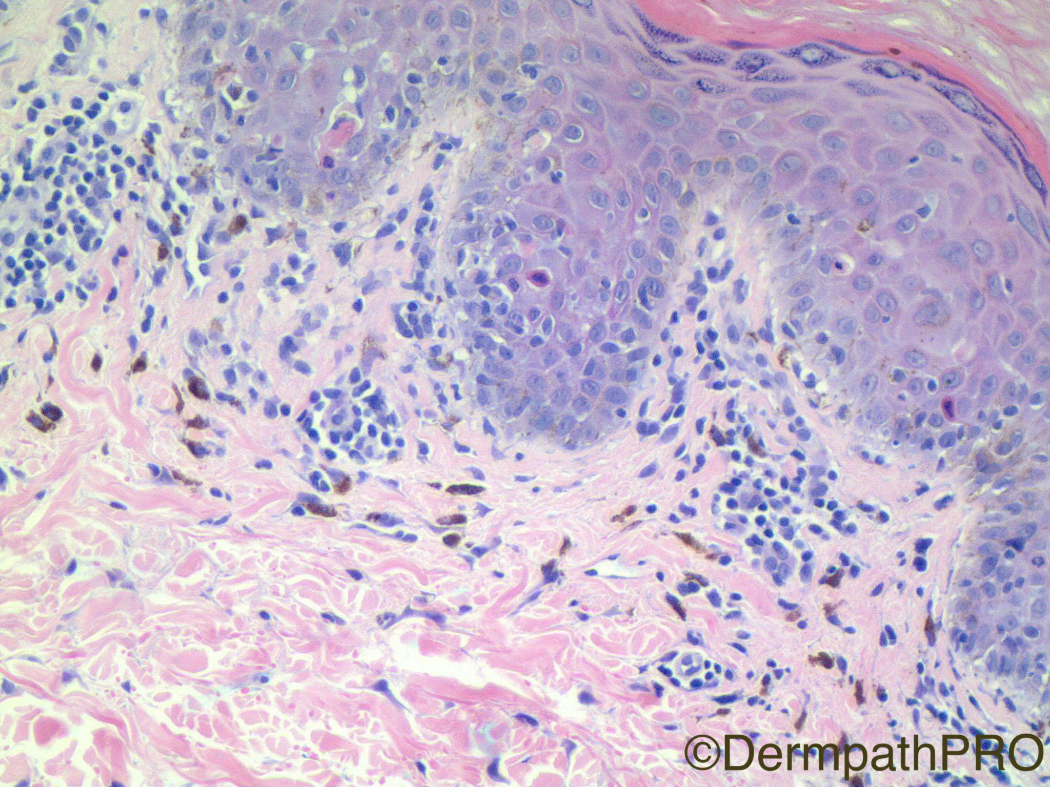
Clinical History: 42 year old woman with solitary annular ecchymotic plaque with central crusting.

Case Posted by Dr Uma Sundram


  Report Record

User Feedback


Admin_Dermpath

Posted

The first of two cases this week from our new Editor in Chief Dr Uma Sundram.

Geoff - DermpathPRO Projects

Share this comment


Link to comment
share_externally

Guest Hazem Hamed

Posted

Interface reaction pattern with scattered apoptotic keratinocytes (mainly at the rete ridges), pigment incontinence and mild predominantly superficial perivascular lymphocytic infiltrate. Could not see plasma cells or eosinophils. No parakeratosis. No significant lymphoid atypia. No RBC extravasation.  There is somewhat squaring of the rete ridges. Given the clinical history of a solitary annular lesion I think we could exclude most of the entities that can show interface reaction pattern including subacute lupus, GVHD, EM,…………… I would think of annular lichenoid dermatitis which was described initially in ''Youth'' but reported also in older age groups and can present as a solitary lesion. Fixed drug reaction is in the differential diagnosis.  Hope history is not missing an important clue !!!

Share this comment


Link to comment
share_externally

vincenzo polizzi

Posted

What about an annular variant of Lichen striatus, in adult?

Share this comment


Link to comment
share_externally

Dr. Mona Abdel-Halim

Posted

Agree with Hazem's Ddx. CPC is needed. Lesions of FDR usually have a dusky hue (echymotic like) and may develop surface crustations so I think it is FDR. 

Share this comment


Link to comment
share_externally

Robledo F. Rocha

Posted

Superficial perivascular and syringocentric lymphocytic infiltrate with foci of basal layer liquefaction and dyskeratotic keratinocytes make me think of lichen striatus. Annular configuration of the lesion in an adult is an unusual, but known, clinical presentation of lichen striatus.

Share this comment


Link to comment
share_externally

vincenzo polizzi

Posted

I favor LS, but I don't know if central crusting as described can fit well on it...CPC are mandatory here(as always). 

Share this comment


Link to comment
share_externally

Arti Bakshi

Posted

My first thought was Fixed drug eruption, which can be annular and solitary. But agree with other d/d too. Needs CPC. 

Share this comment


Link to comment
share_externally

Raul Perret

Posted (edited)

I agree with most of the appreciations, there is a lichenoid reaction pattern with perivascular and focally perieccrine lymphocytic infiltrate. From my point of view, there is too much pigment incontinence and civatte bodies which make me favour in first place fixed drug eruption. I would not have thought of lichen striatus if picture 3 was not present. In addition, I dont see parakeratosis and Civatte bodies are too abundant for this diagnosis. For ALDY i think there is not enough elongation of the rete and acanthosis. I dont like doing an specific diagnosis with this kind of pattern in real life (except if the clinical is clear) so should be careful. 

Edited by Raul Perret

Share this comment


Link to comment
share_externally

vincenzo polizzi

Posted

May be I'm wrong, but I see focal parakeratosis ( top right picture 1-2 ), many melanophages in papillary dermis, lymphocytic exocytosis and many dyskeratotic cells also over the basal layers, as well described in lichen striatus. And if I take a look at the first picture, really it seems to me to see a lichen striatus...But I wouldn't do this diagnosis without clinical support.

Share this comment


Link to comment
share_externally

Uma Sundram

Posted

Super job everyone! I like showing these types of cases to elicit responses, which demonstrate how difficult dermatopathology really is :)

Clinically the lesion was characteristic for fixed drug eruption (clinical setting and drug used). I thought the lesion had features of both lichen striatus and early erythema multiforme but the clinical really favored FDE.

Share this comment


Link to comment
share_externally


Join the conversation

You can post now and register later. If you have an account, sign in now to post with your account.

Guest
Add a comment...

×   Pasted as rich text.   Paste as plain text instead

  Only 75 emoji are allowed.

×   Your link has been automatically embedded.   Display as a link instead

×   Your previous content has been restored.   Clear editor

×   You cannot paste images directly. Upload or insert images from URL.

Loading...

×
×
  • Create New...