Jump to content
In this section we have spot diagnoses posted on a daily basis since June 2010, now over 4000! You can review the archived cases and read the suggested diagnoses by users and the final comment by the contributors.
Case are uploaded each week day by 10 am UK time with the correct diagnosis will generally be posted at 8 pm UK time. Why not view the most recent spot diagnosis and proffer a diagnosis?

Case Number : Case 1668 - 16 November - Dr Hafeez Diwan Posted By: Guest

Please read the clinical history and view the images by clicking on them before you proffer your diagnosis.
Submitted Date :
   (0 reviews)

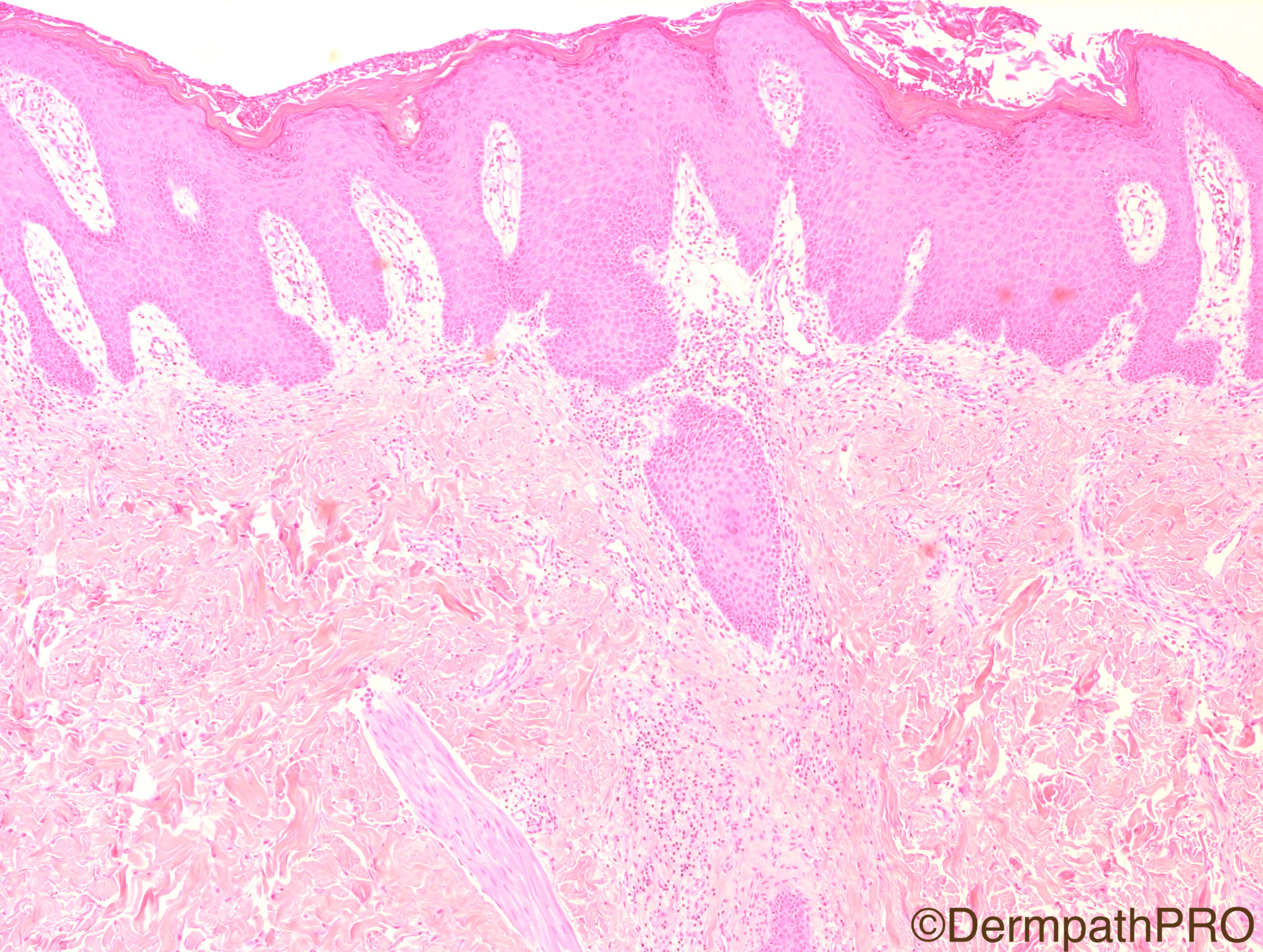
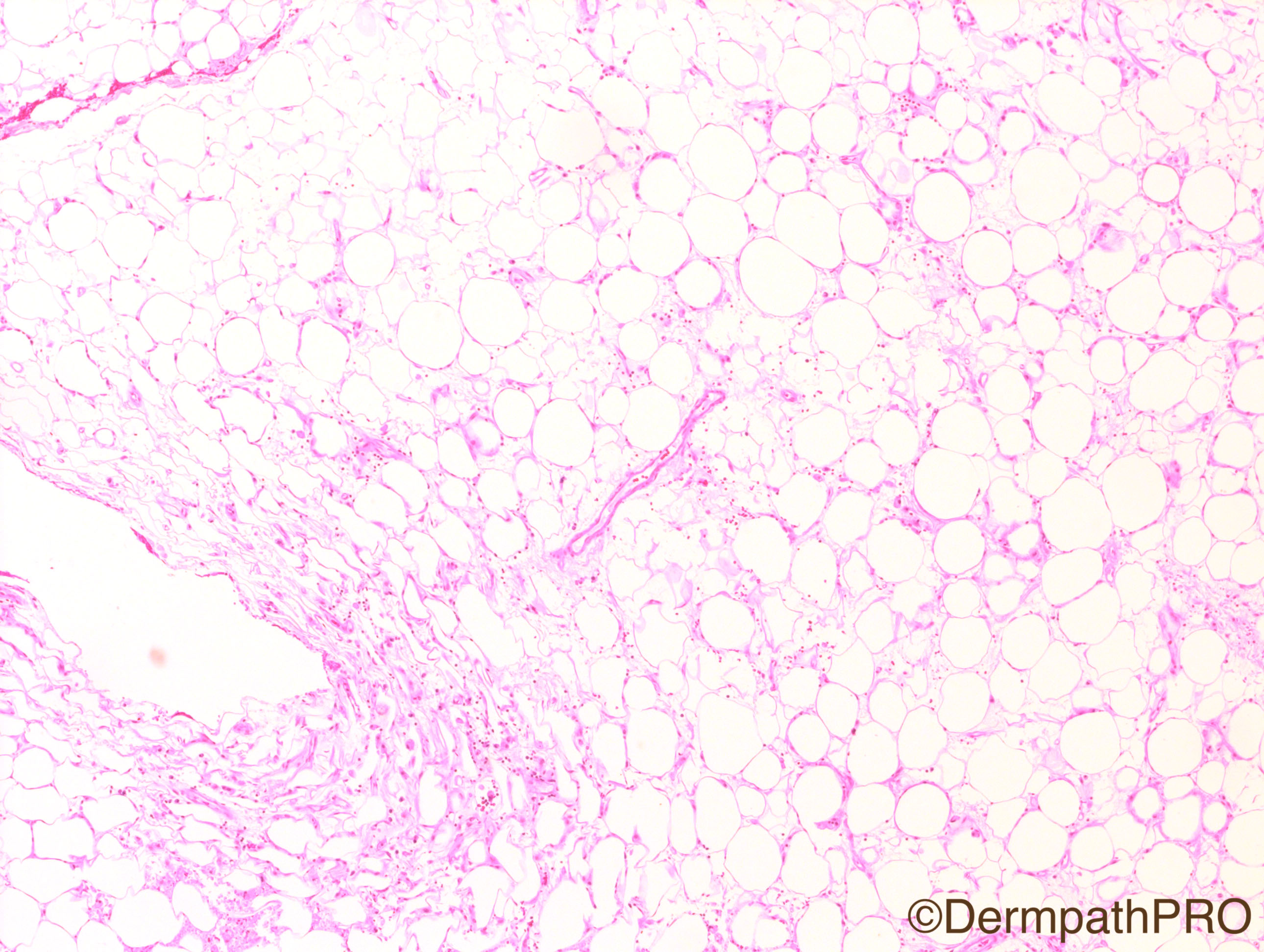
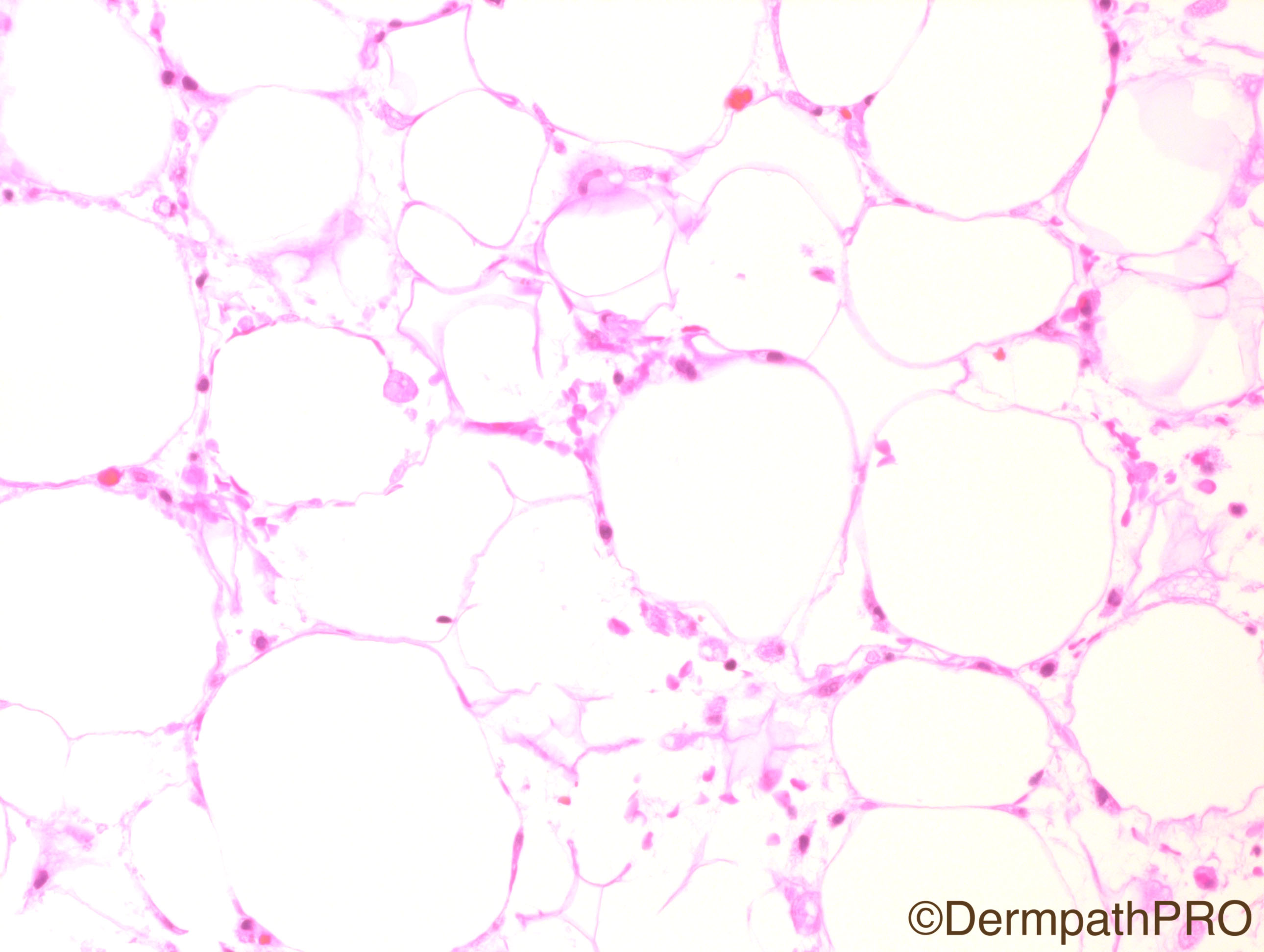
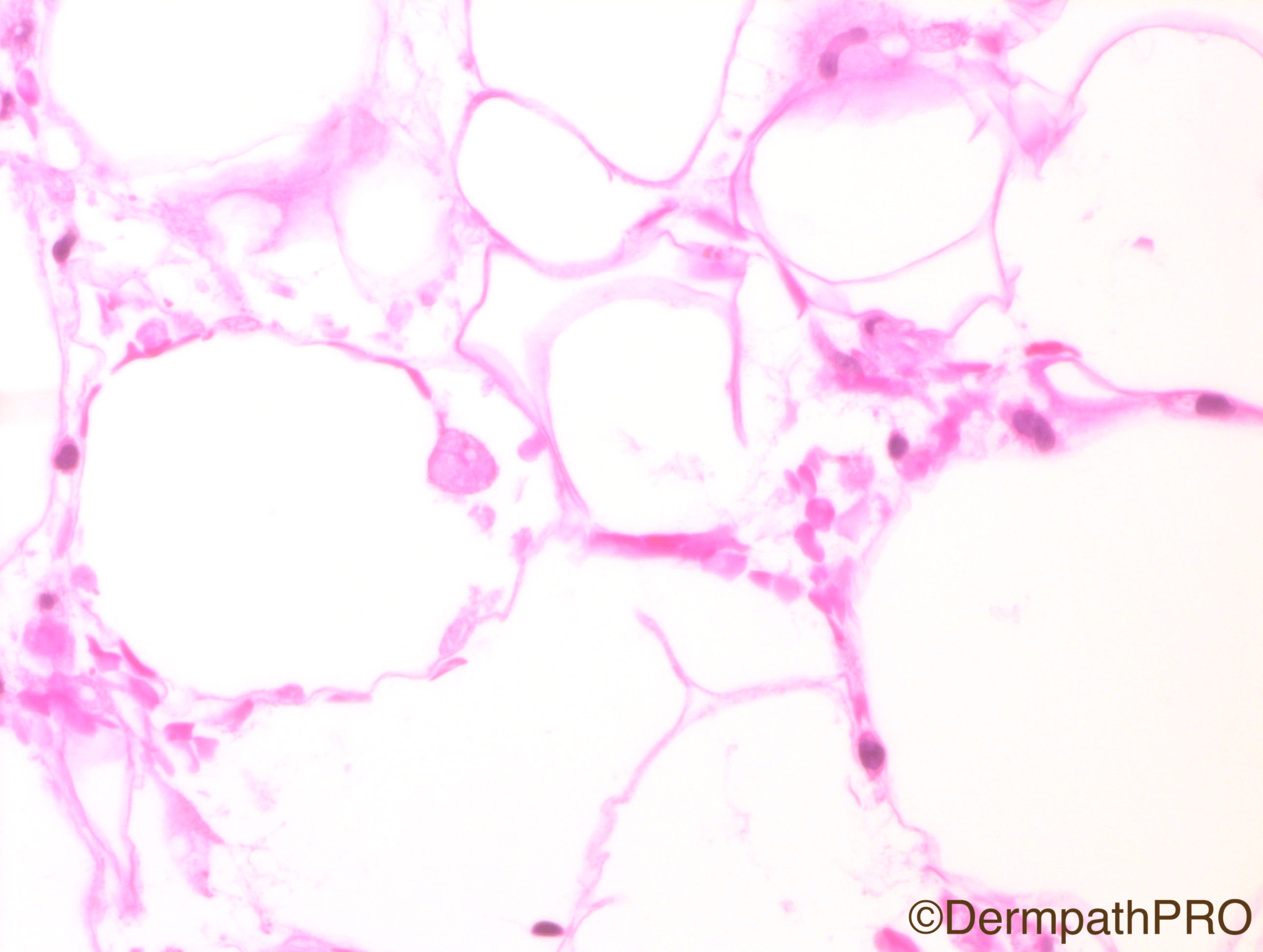
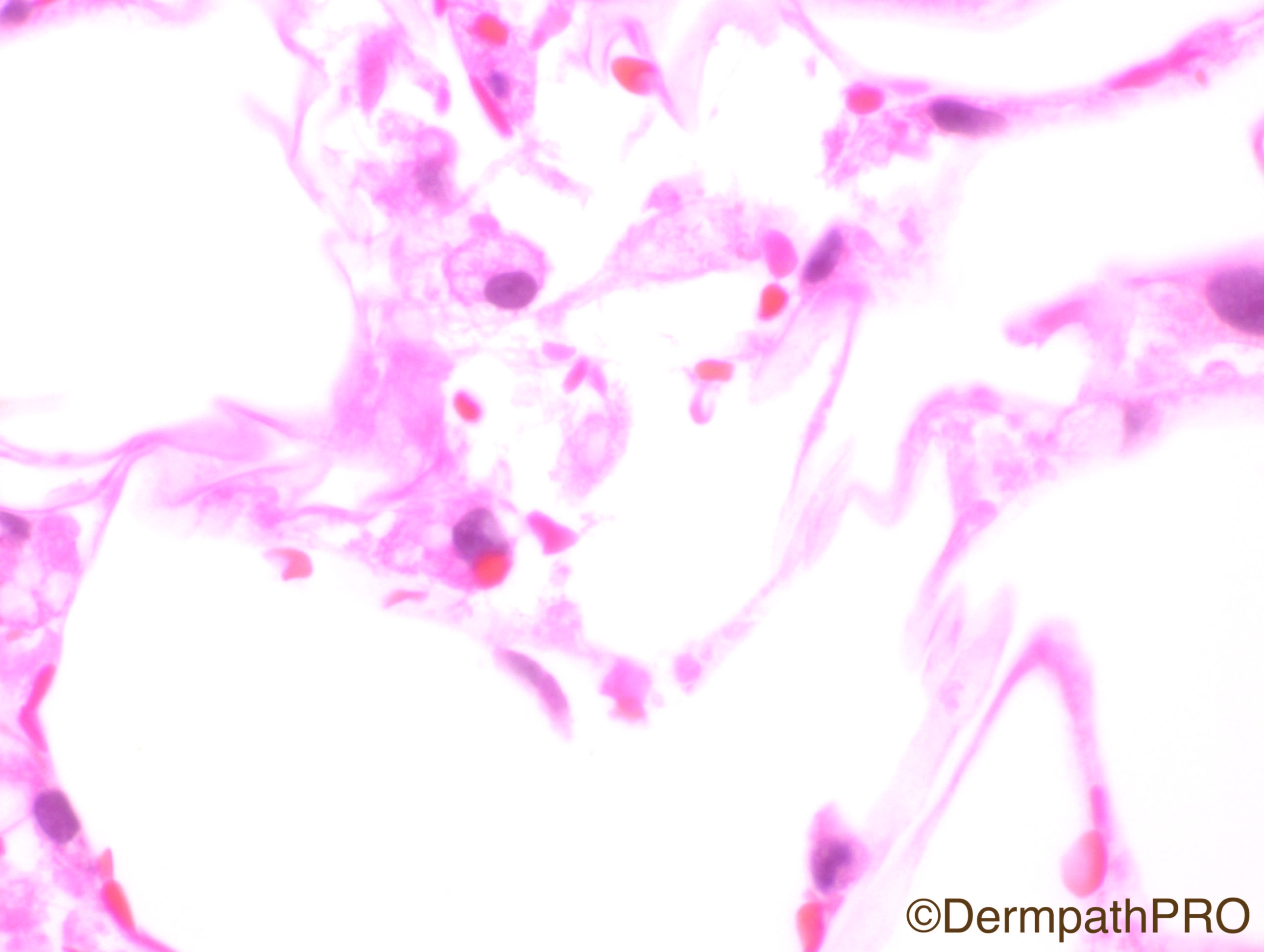
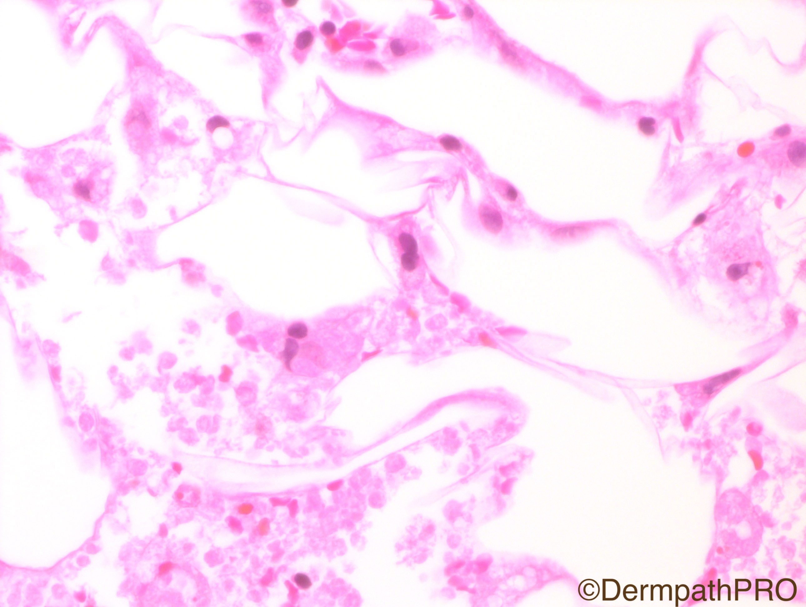
Clinical History: 17 year-old male with purpuric lesions. This biopsy is from the calf.

Case Posted by Dr Hafeez Diwan

Edited by Admin_Dermpath


  Report Record

User Feedback


vincenzo polizzi

Posted

Yes. Agree. Hemocytophagic  Sindrome (mononucleosis?)

Share this comment


Link to comment
share_externally

Robledo F. Rocha

Posted

Subcutaneous foamy macrophages with phagocytosed erythrocytes, but no bean bag cells showing lymphophagocytosis. Maybe higher power view of the first image will reveal characteristic findings of lupus erythematosus (on low-power view, I suspect there is an interface lymphocytic infiltrate with perifollicular involvement). I favor lupus panniculitis.

Alternatively, all the cells on the superficial dermis I suspect to be lymphocytes are, in reality, extravasated erythrocytes. In this case, bleeding diatheses and other forms of non-inflammatory purpura are good suggestions.

Share this comment


Link to comment
share_externally

Dr. Mona Abdel-Halim

Posted

Actually I am facing difficulty in interpretating the photos :-(

Share this comment


Link to comment
share_externally

Telangectasia macularis eruptiva perstan.Aform of mastocytosis.

Share this comment


Link to comment
share_externally

Nitin Khirwadkar

Posted

As Robledo has mentioned there is a superficial dermal infiltrate, probably all lymphocytic. Focally, it is in a peri-follicular location. However, I am not sure about an interface pattern. There is fat necrosis. Within foci of latter are large histiocytes, with big nuclei, and some with erythrophagocytosis. I think I would favour cytophagic histiocytic panniculitis. No vasculitis. Require CPC.

Share this comment


Link to comment
share_externally

Raul Perret

Posted

I find this case challenging and hard to interpretate in these pictures. But I agree with the great suggesiton of cytophagic histiocytic panniculitis for CPC.

Share this comment


Link to comment
share_externally

Dr. Hafeez Diwan

Posted

I have tried to photograph hemophagocytosis.  I called this hemophagocytic lymphohistiocytosis.

Share this comment


Link to comment
share_externally

Dr. Richard Carr

Posted

Great case Hafeez. I'm a bit late.  I enjoyed this challenging case that I also struggled with!  Well done to those who thought of the diagnosis. I wanted to pass on a little anecdote.  We had a neighbour who whose child was wasting away and had had a diagnosis of HLH made on bone marrow biopsy (at another centre). He continued to deteriorate and things were looking bleak until fortunately (all be it late in the day) some clever pathologist when taking a further look at the bone marrow spotted the Leishmaniasis followed by appropriate treatment and a full recovery.

Share this comment


Link to comment
share_externally


Join the conversation

You can post now and register later. If you have an account, sign in now to post with your account.

Guest
Add a comment...

×   Pasted as rich text.   Paste as plain text instead

  Only 75 emoji are allowed.

×   Your link has been automatically embedded.   Display as a link instead

×   Your previous content has been restored.   Clear editor

×   You cannot paste images directly. Upload or insert images from URL.

Loading...

×
×
  • Create New...