Jump to content
In this section we have spot diagnoses posted on a daily basis since June 2010, now over 4000! You can review the archived cases and read the suggested diagnoses by users and the final comment by the contributors.
Case are uploaded each week day by 10 am UK time with the correct diagnosis will generally be posted at 8 pm UK time. Why not view the most recent spot diagnosis and proffer a diagnosis?

Case Number : Case 1692 - 22 November - Dr Uma Sundram Posted By: Guest

Please read the clinical history and view the images by clicking on them before you proffer your diagnosis.
Submitted Date :
   (0 reviews)

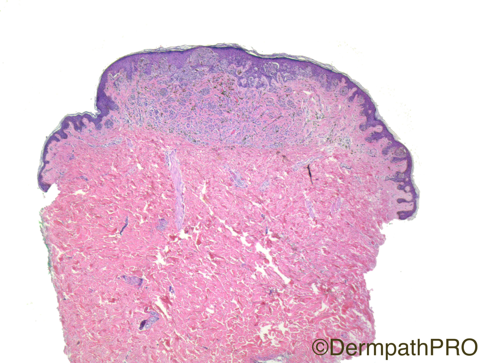
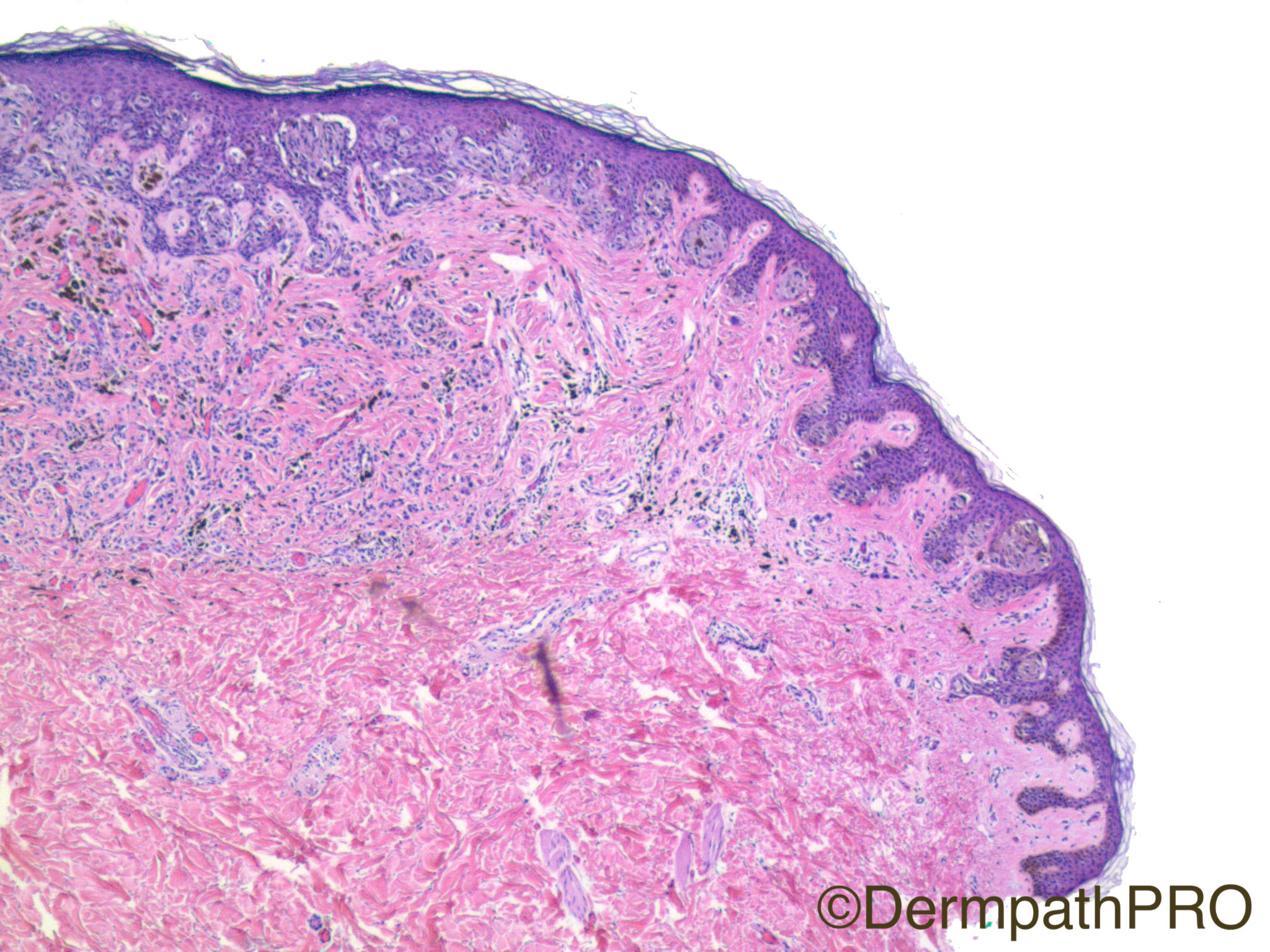
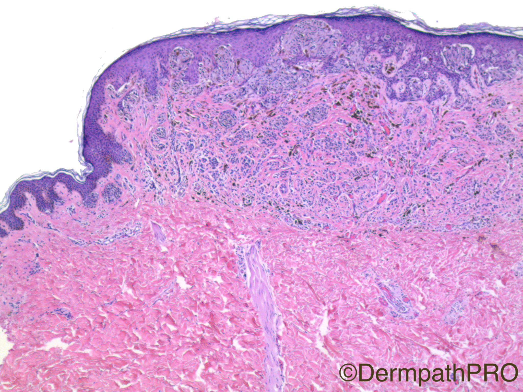
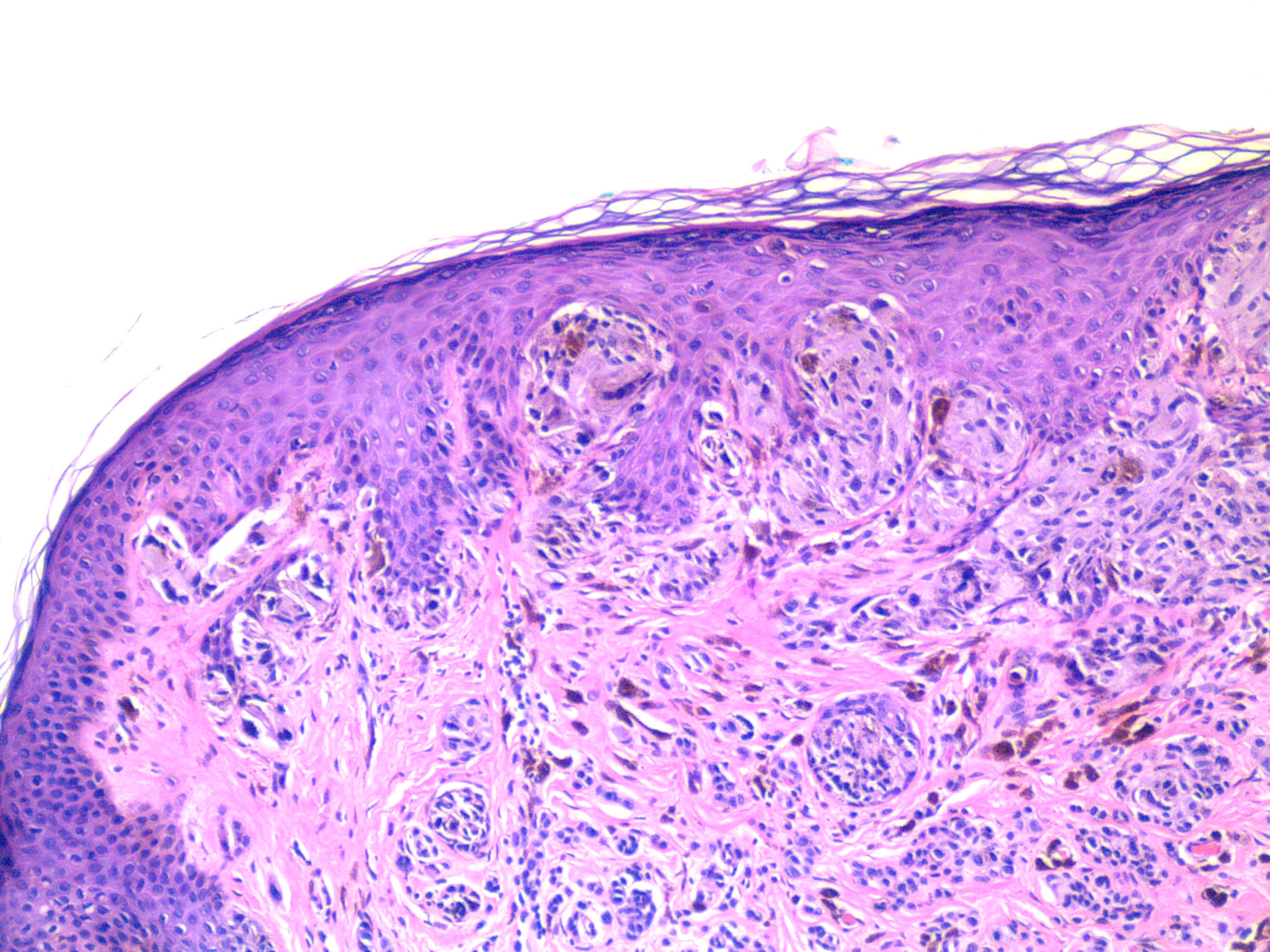
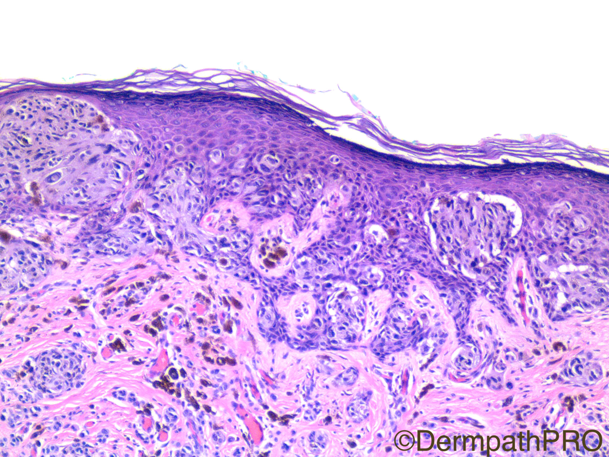
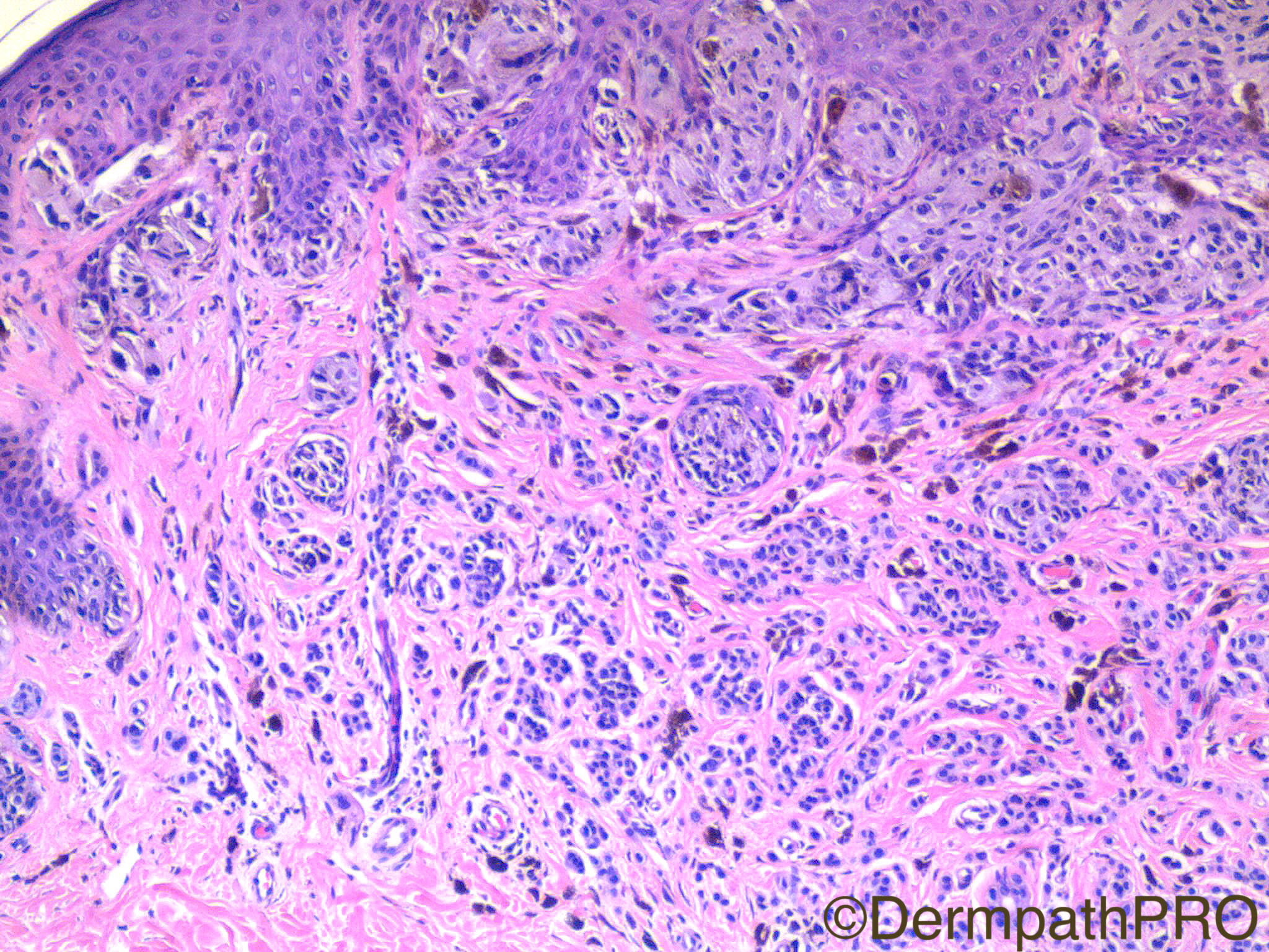
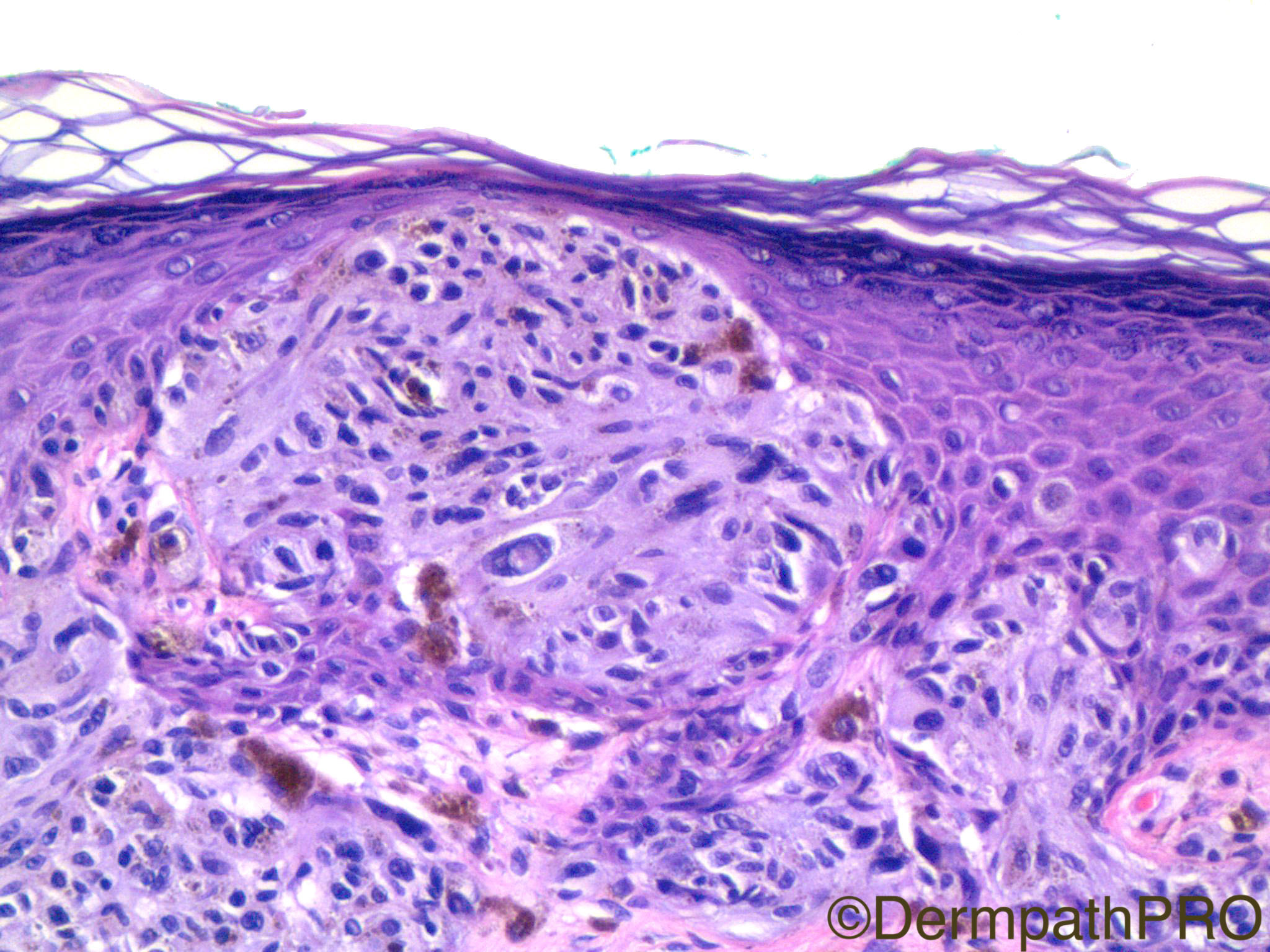
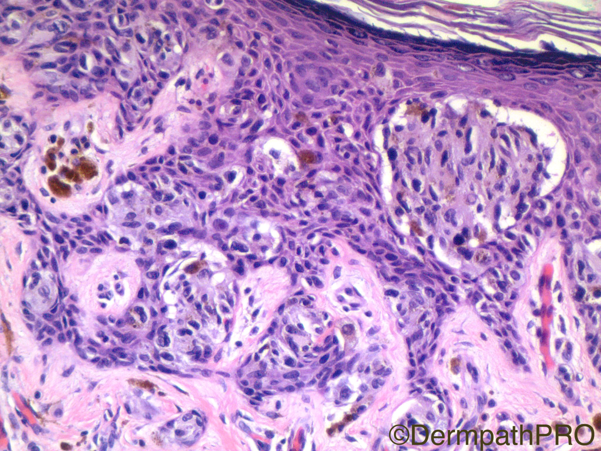
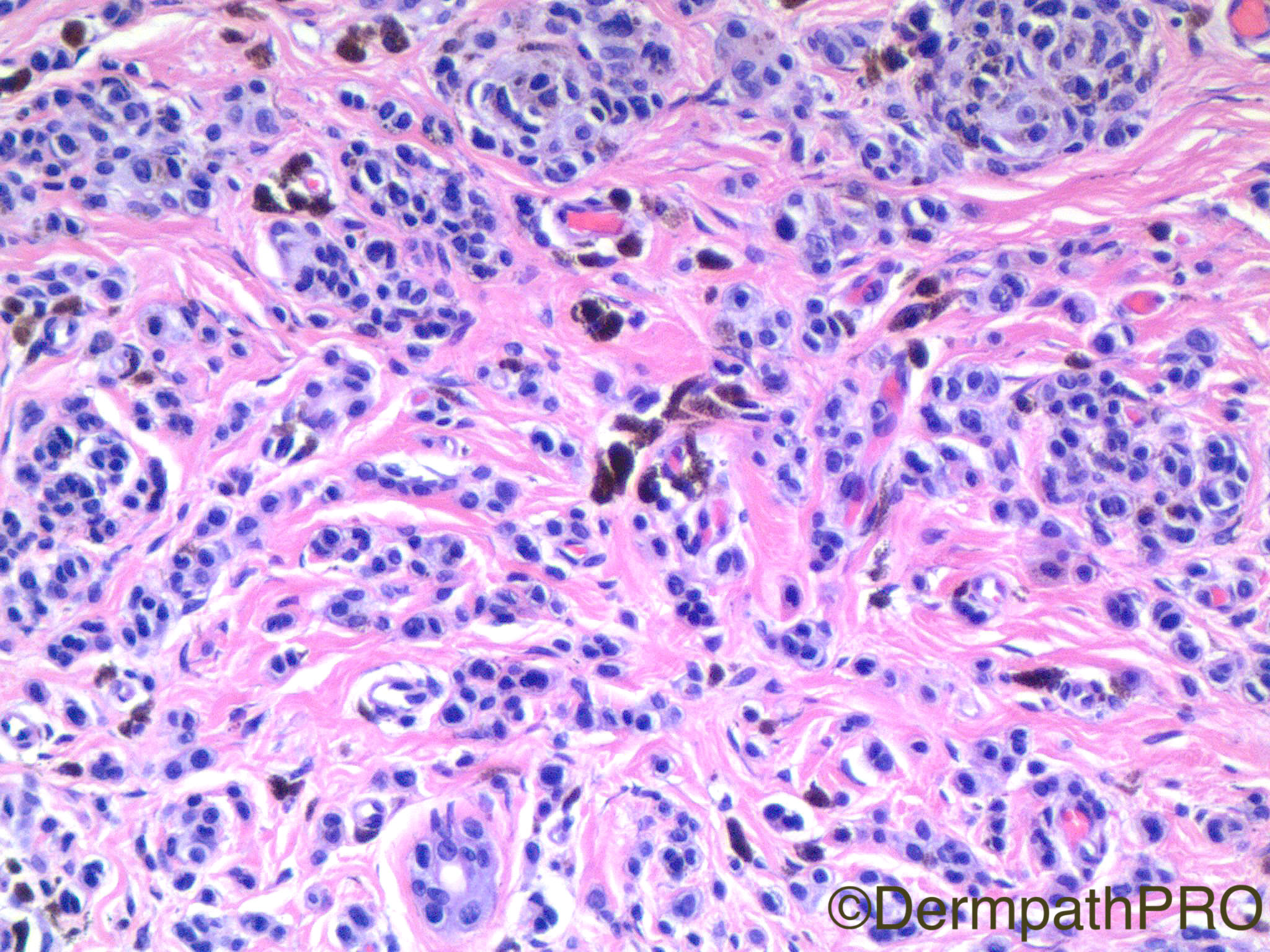
Clinical History: 26 year old woman with erythematous papule on left buttock.

Case Posted by Dr Uma Sundram
Edited by Admin_Dermpath


  Report Record

User Feedback


Admin_Dermpath

Posted

A lovely set of images for you today from Dr Uma Sundram.

Geoff Cross - DermpathPRO Projects

Share this comment


Link to comment
share_externally

Victor Delgado

Posted

my opinion is for Spitz nevus; is almost well circumscribed with shouldering, irregular acanthosis, thinned rete ridges, melanocitic confluence, little bit of asymetrical nested and epidermal consumption, kamino bodies, discohesive nests and pagetoid spread. The dermal component is made by cords and nests of melanocytes that shows deep maduration.

Share this comment


Link to comment
share_externally

vincenzo polizzi

Posted

The more atypical features of this lesion are in the right half ( pagetoid spreading of melanocytes ), where dermis looks like sclerosing/cicatricial, so I think of a Spitz Nevus, with sclerosing/recurrent-like features.

Share this comment


Link to comment
share_externally

Raul Perret

Posted

I have a different impression. I would call this lesion a compound nevus with architectural and cytological atypia of the junctional component (Dysplastic or atypia associated with a flexural site), margins free. Although there are some areas of the junctional component reminiscent of Spitz nevi (spaces around nests, pagetoid spread) I dont find them enough for this dx. In addition the intadermal component looks completely different (banal nevus) with really small melanocytes. I ultimately agree that this lesion does not look like melanoma

Share this comment


Link to comment
share_externally

Robledo F. Rocha

Posted

Combined nevus (common melanocytic nevus beneath to Spitz nevus), or melanocytic nevus with phenotypic heterogeneity, according to Barnhill’s terminology.

Share this comment


Link to comment
share_externally

Clara Jaramillo

Posted

Melanocytic nevus (combined) > Spitz nevus 

Share this comment


Link to comment
share_externally

Dr. Mona Abdel-Halim

Posted

Combined spitz and banal nevus is my impression.

Share this comment


Link to comment
share_externally

Nitin Khirwadkar

Posted

Spitz naevus. Has some features of a SPARK. There is some confluence of nests in parts of the junctional proliferation. The low power images show foci of fine dermal scarring, right half of the lesion, possibly secondary to trauma. 

Share this comment


Link to comment
share_externally

Dr. Richard Carr

Posted

Spitz naevus or combined Spitz/naevocellular naevus with focal dermal scarring. Margin a little close but could adopt watchful waiting I think.

Share this comment


Link to comment
share_externally

Uma Sundram

Posted

We too went with combined Spitz and ordinary nevus. We made no outright recommendations and left it in the hands of the clinician/patient; ultimately the patient decided to have the lesion re excised.

Share this comment


Link to comment
share_externally


Join the conversation

You can post now and register later. If you have an account, sign in now to post with your account.

Guest
Add a comment...

×   Pasted as rich text.   Paste as plain text instead

  Only 75 emoji are allowed.

×   Your link has been automatically embedded.   Display as a link instead

×   Your previous content has been restored.   Clear editor

×   You cannot paste images directly. Upload or insert images from URL.

Loading...

×
×
  • Create New...