Jump to content
In this section we have spot diagnoses posted on a daily basis since June 2010, now over 4000! You can review the archived cases and read the suggested diagnoses by users and the final comment by the contributors.
Case are uploaded each week day by 10 am UK time with the correct diagnosis will generally be posted at 8 pm UK time. Why not view the most recent spot diagnosis and proffer a diagnosis?

Case Number : Case 1698 - 30 November - Dr Hafeez Diwan Posted By: Guest

Please read the clinical history and view the images by clicking on them before you proffer your diagnosis.
Submitted Date :
   (0 reviews)

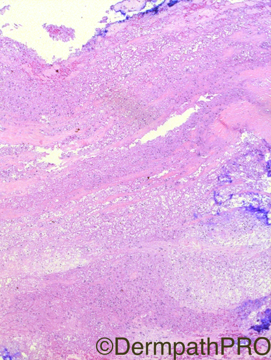
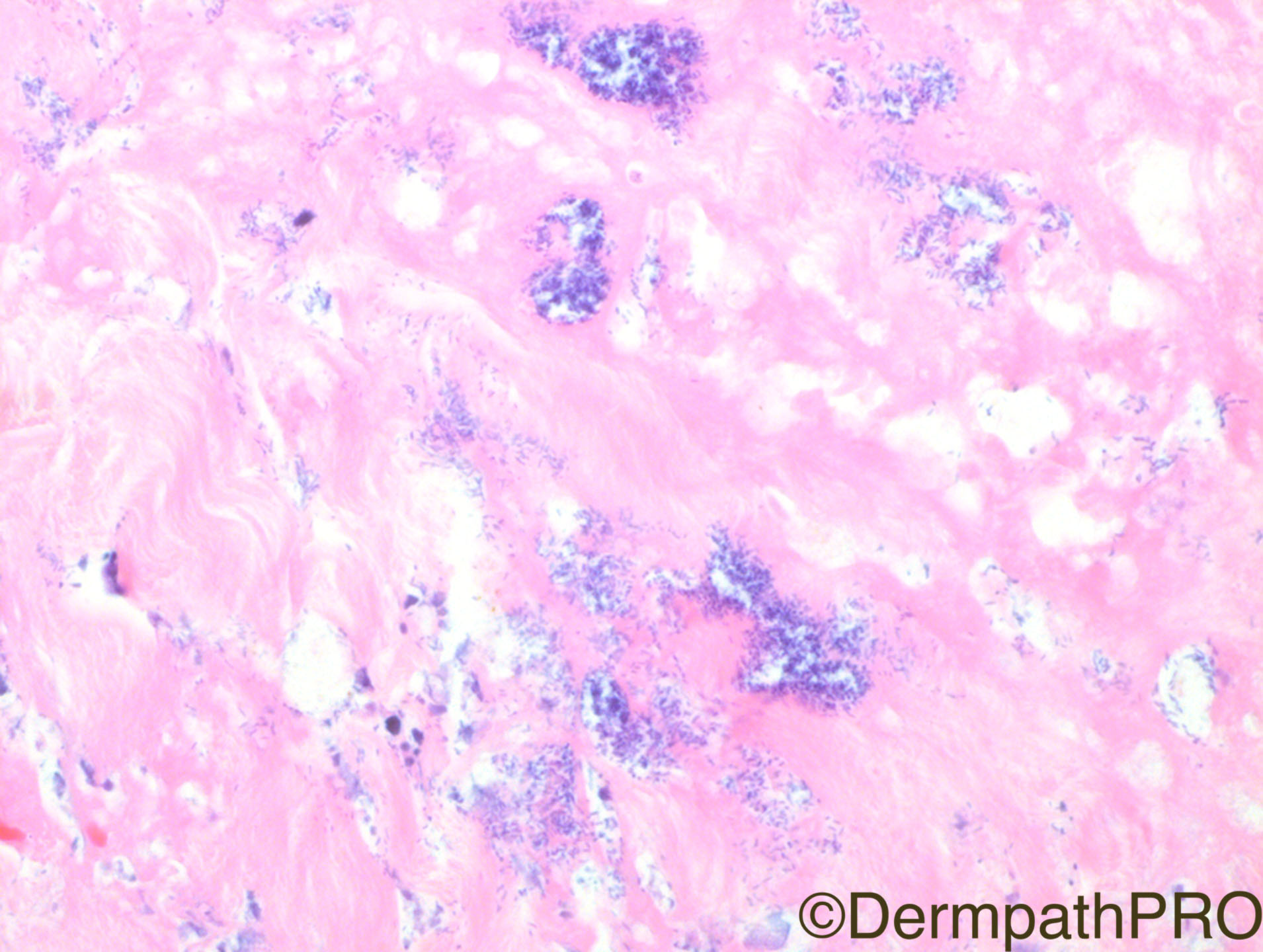
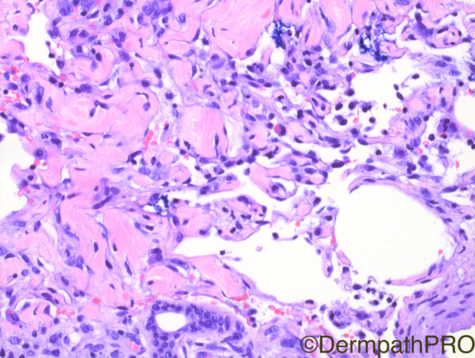
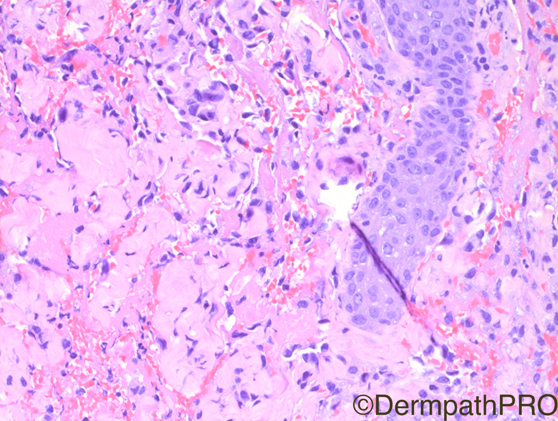
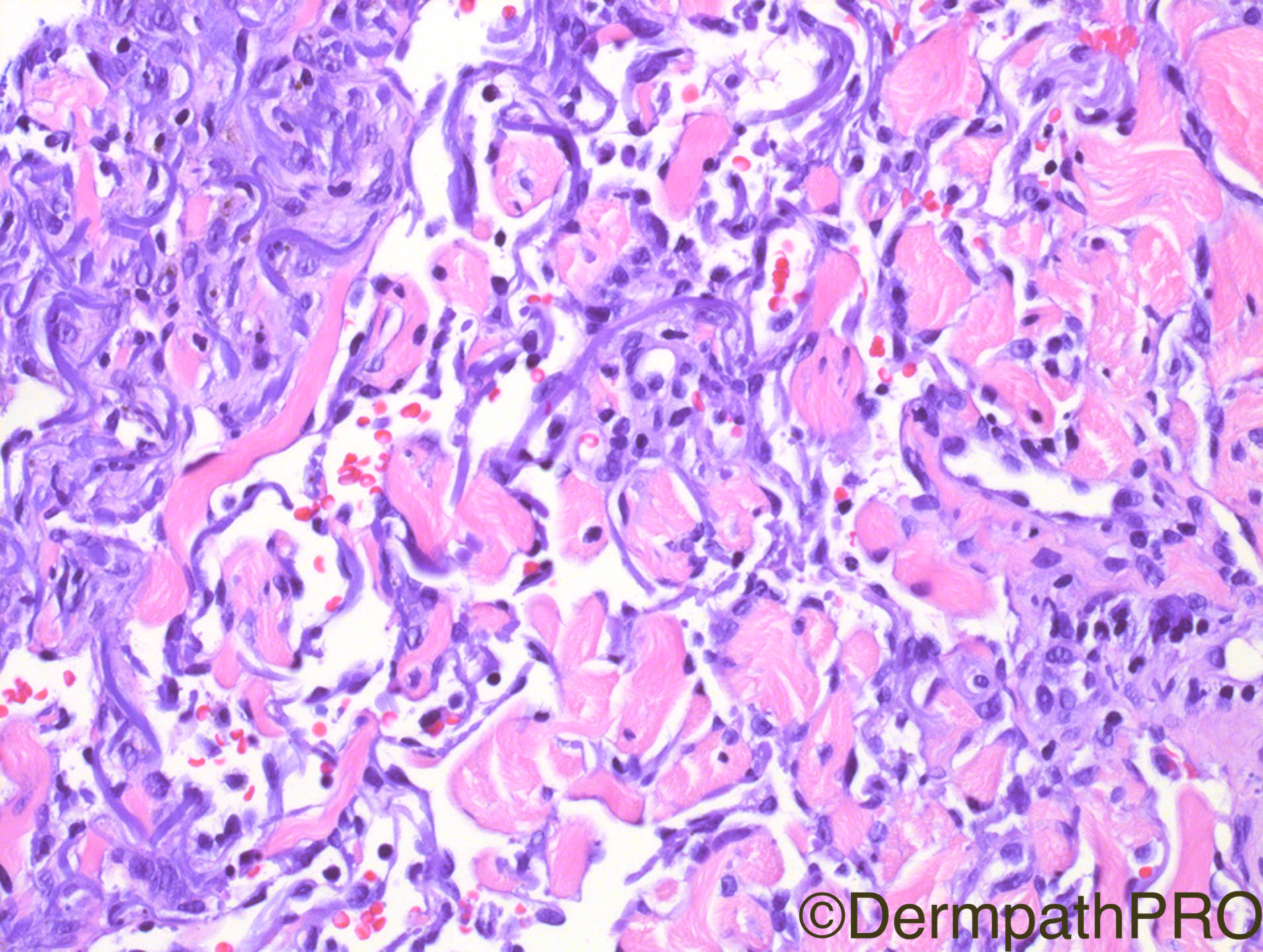
Clinical History: 65 year old male with erythematous scalp lesion

Case Posted by Dr Hafeez Diwan


  Report Record

User Feedback


Admin_Dermpath

Posted

Dr Hafeez Diwan rounds of what has been a great month for DermpathPRO.

Cheers, Geoff Cross - DermpathPRO Projects

Share this comment


Link to comment
share_externally

urmilapandey

Posted

necrosis, some endothelial atypia, can't see any pre-existing vascular space where this proliferation is occuring, hence am thinking on the lines of an angiosarcoma

Share this comment


Link to comment
share_externally

Robledo F. Rocha

Posted

Taking into account the patient’s age and the lesion’s site, I’d tend to construe those irregularly branching thin-walled vessels with dissecting growth pattern as an angiosarcoma.

Share this comment


Link to comment
share_externally

vincenzo polizzi

Posted

Agree with above, but I would like know about EMA- CD31-D2-40, to rule out an odd ectopic meningothelial hamartomatous lesion in adult ( never seen before! )

Share this comment


Link to comment
share_externally

vincenzo polizzi

Posted

Is there a psammoma body in last fig bottom right??? 

Share this comment


Link to comment
share_externally

Raul Perret

Posted

I have heard of a case that dr. Diwan had once but never actually saw the case. It was about a patient that had findings compatible with angiosarcoma but concomitantly showed large areas of necrosis and bacterial colonies. Apparently he suggested to treat the patient for the infection before confirming the dx of angiosarc. Apparently the treatment for the infection made also dissapear the vascular proliferation. If it is this case, I find it extremely challenging

Share this comment


Link to comment
share_externally

Dr. Mona Abdel-Halim

Posted

20 minutes ago, Raul Perret said:

I have heard of a case that dr. Diwan had once but never actually saw the case. It was about a patient that had findings compatible with angiosarcoma but concomitantly showed large areas of necrosis and bacterial colonies. Apparently he suggested to treat the patient for the infection before confirming the dx of angiosarc. Apparently the treatment for the infection made also dissapear the vascular proliferation. If it is this case, I find it extremely challenging

Wao :-)

Share this comment


Link to comment
share_externally

Dr. Richard Carr

Posted

Angiosarcoma is a treacherous diagnosis!  I would recommend looking at the patient but one has to consider the possibility on this histology. I find immunos can be quite helpful.  Reactive vascular proliferations usually have endothelial staining for endothelial markes and rings of smooth muscle actin staining around the vessels (pericytes and smooth muscle cells), the latter not seen in angiosarcomas. Raul's post is most interesting!

Share this comment


Link to comment
share_externally

Raul Perret

Posted

I was searching where did I heard that and actually is in Dr. Diwan's blog link 

Share this comment


Link to comment
share_externally

Thank Dr. Diwan for this interesting case. To me this is angiosarcoma until proven otherwise. Can't wait to see the dx. Cheers!

Share this comment


Link to comment
share_externally

Nitin Khirwadkar

Posted

Raul, thanks for referring to the blog. Just had a look at what Dr Diwan said about that case, and it looks that this is the case! 

Without the reference, would have probably favoured/consistent with an angiosarcoma . Right clinical setting, age! However, I have never seen angiosarcomas with so much necrosis or bacterial debris.

Share this comment


Link to comment
share_externally

Arti Bakshi

Posted

Wow! ....i'm sure i had read that blog at some point but didnt connect it with this case... Raul's memory is clearly photographic!

Share this comment


Link to comment
share_externally

Dr. Hafeez Diwan

Posted

Angiosarcoma, superinfected with Enterobacter.  This was a large, erythematous lesion on the scalp of this patient.

Share this comment


Link to comment
share_externally


Join the conversation

You can post now and register later. If you have an account, sign in now to post with your account.

Guest
Add a comment...

×   Pasted as rich text.   Paste as plain text instead

  Only 75 emoji are allowed.

×   Your link has been automatically embedded.   Display as a link instead

×   Your previous content has been restored.   Clear editor

×   You cannot paste images directly. Upload or insert images from URL.

Loading...

×
×
  • Create New...