Jump to content
In this section we have spot diagnoses posted on a daily basis since June 2010, now over 4000! You can review the archived cases and read the suggested diagnoses by users and the final comment by the contributors.
Case are uploaded each week day by 10 am UK time with the correct diagnosis will generally be posted at 8 pm UK time. Why not view the most recent spot diagnosis and proffer a diagnosis?

Case Number : Case 1637 - 4 October Posted By: Guest

Please read the clinical history and view the images by clicking on them before you proffer your diagnosis.
Submitted Date :
   (0 reviews)

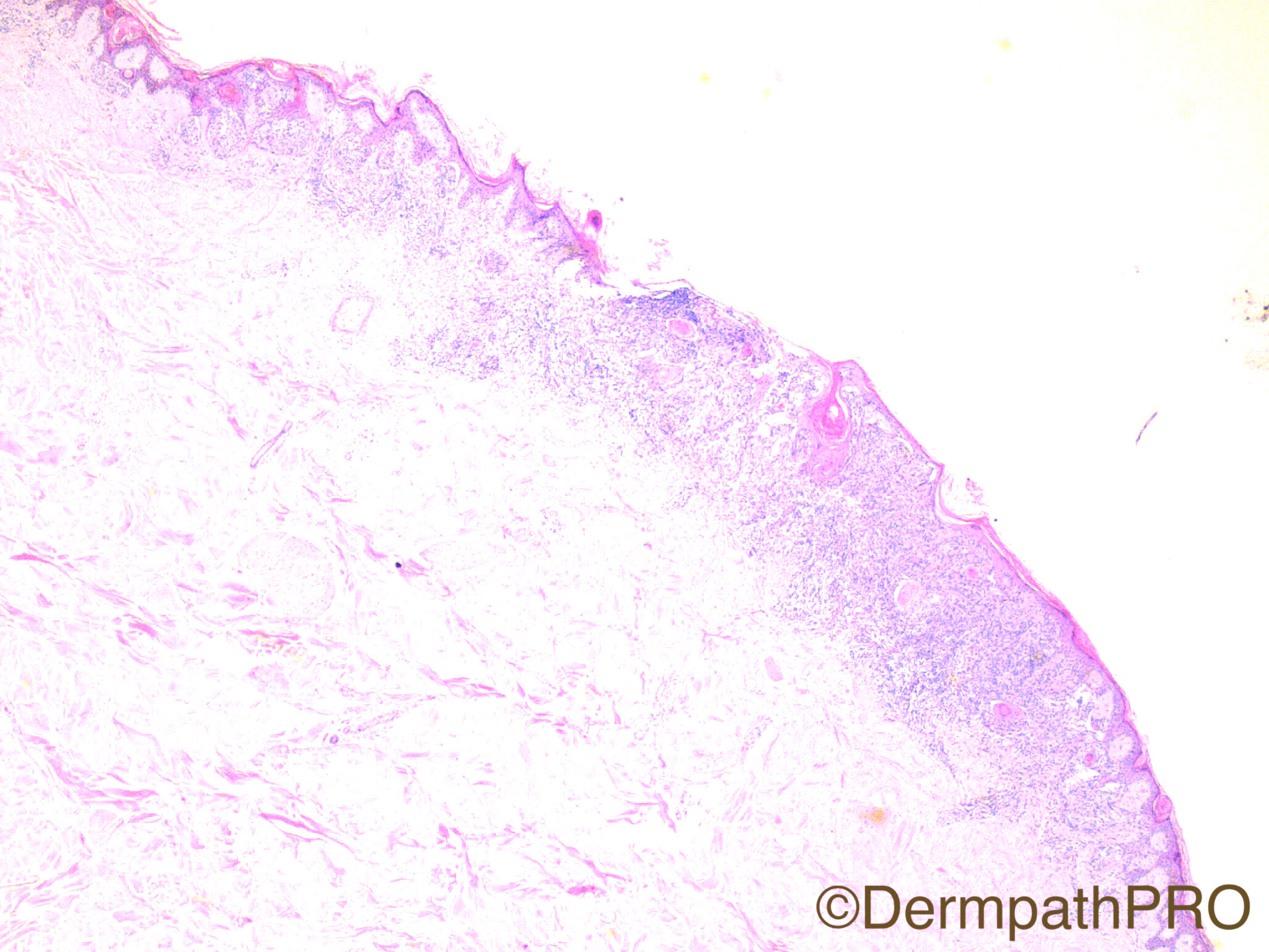
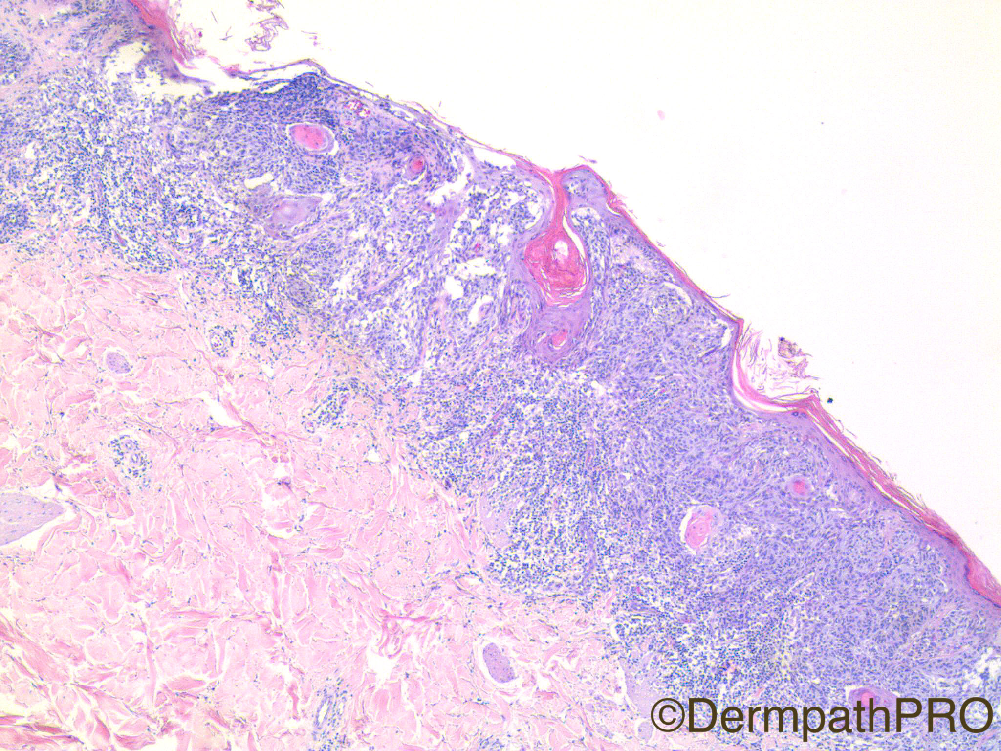
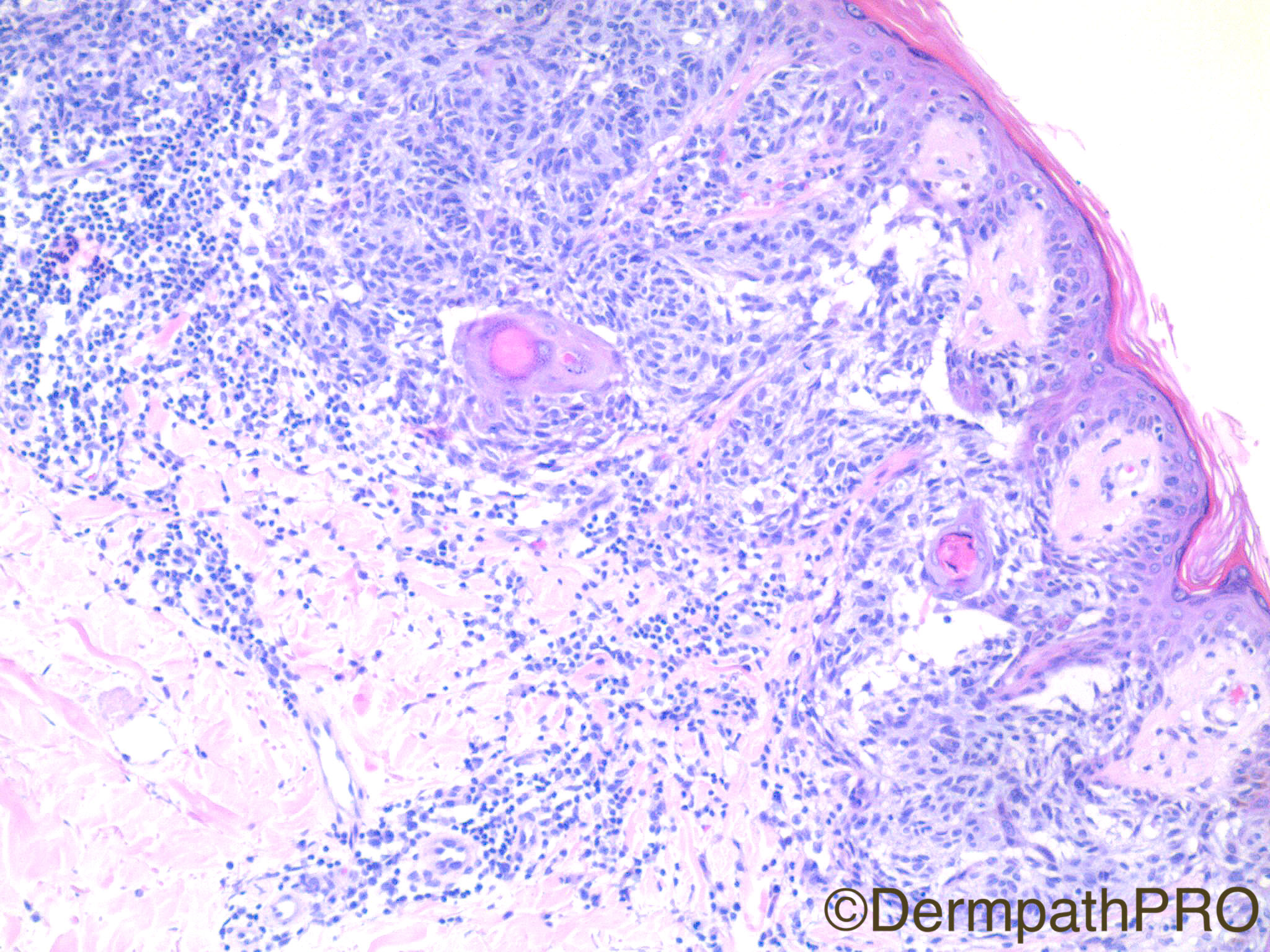
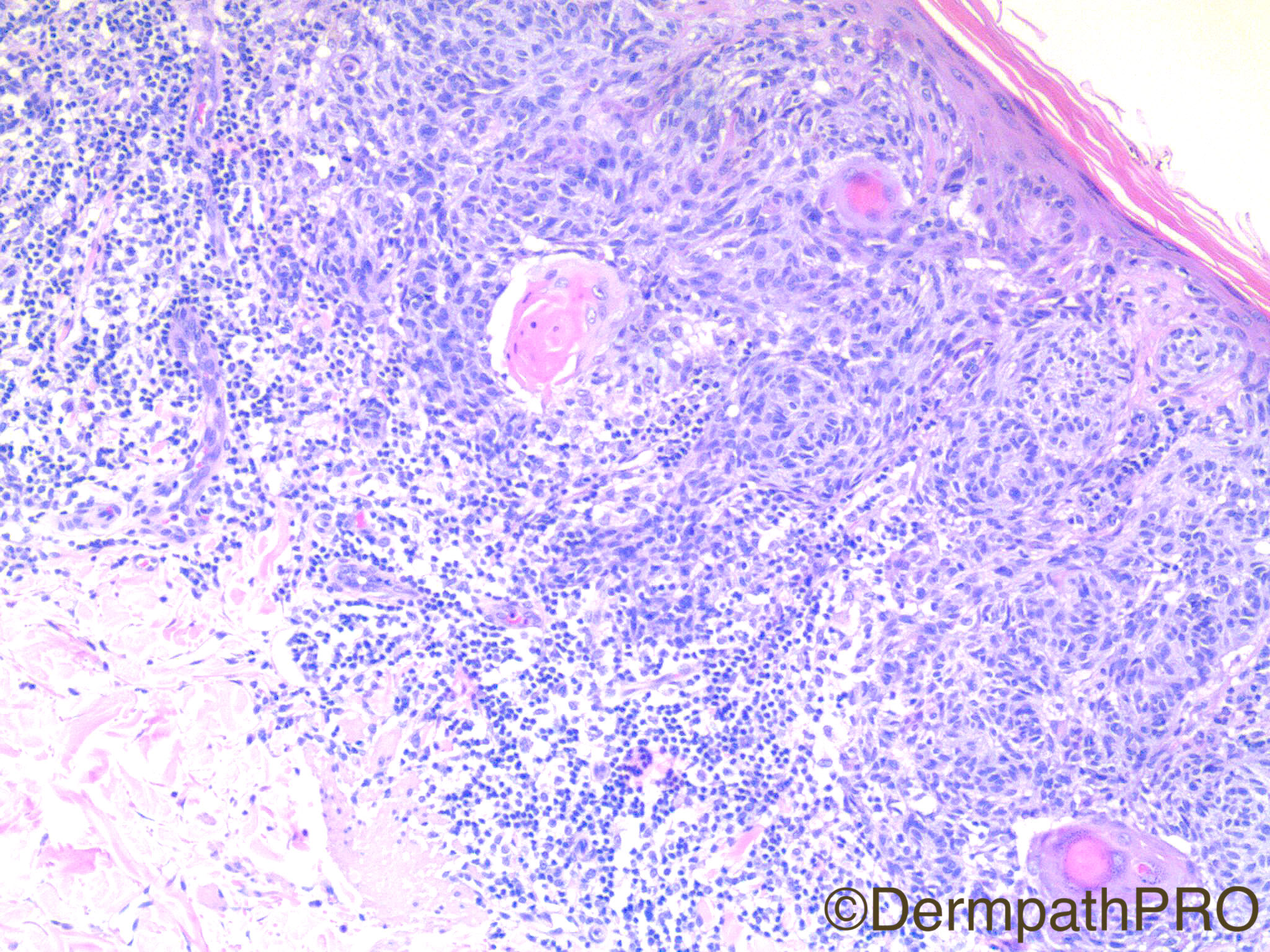
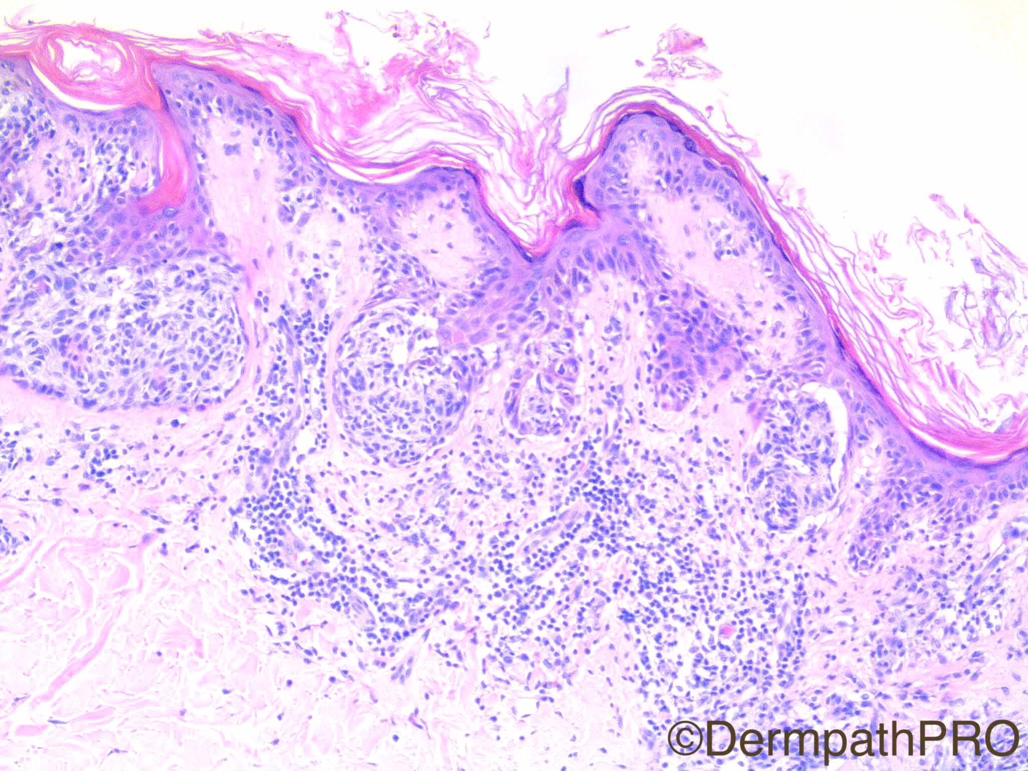
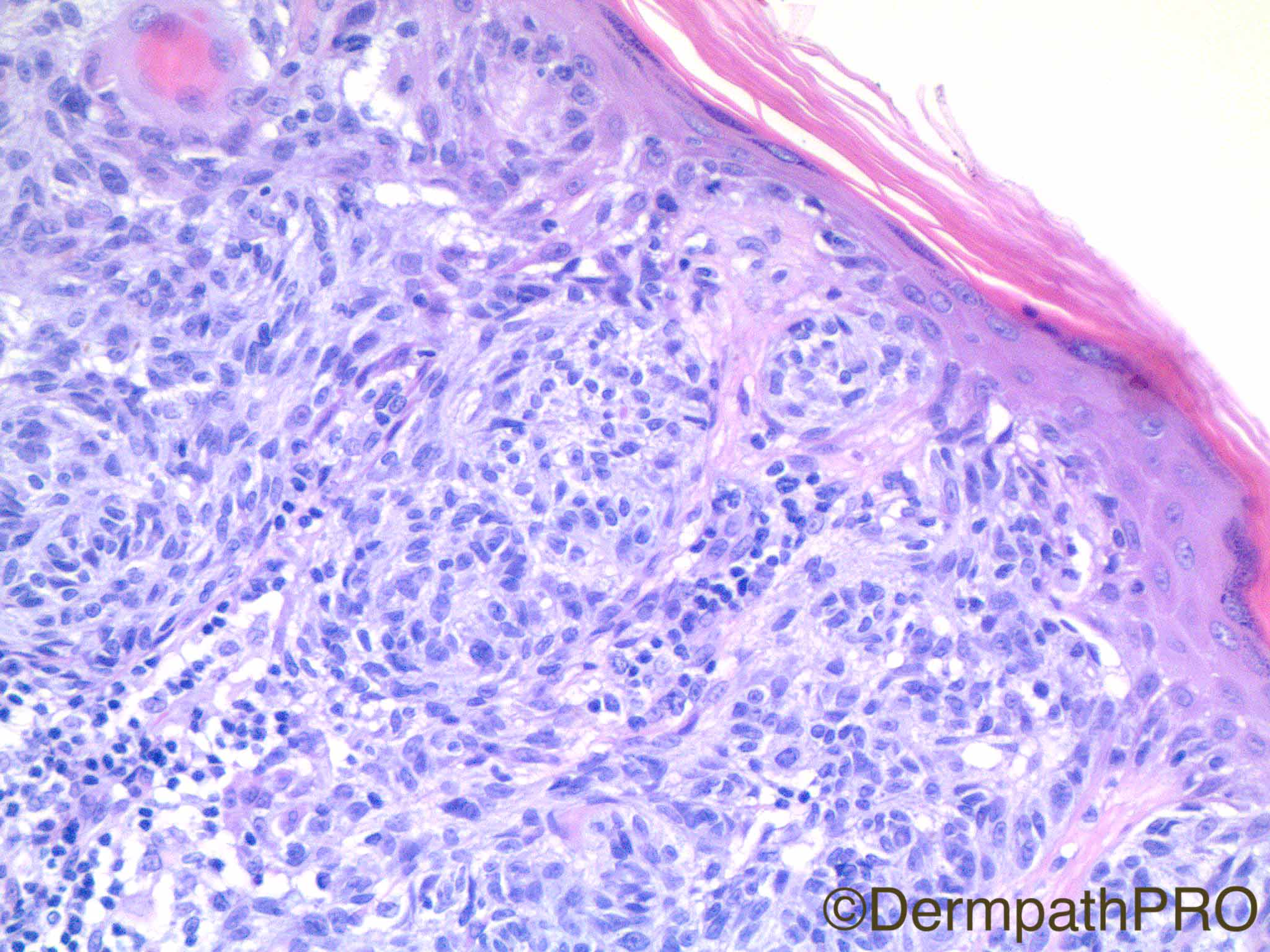
57 year old woman with 8 mm light/dark brown plaque with asymmetry and border notching on back.

Case Posted by Dr Uma Sundram

  Report Record

User Feedback


Admin_Dermpath

Posted

Another great case from Dr Uma Sundram.


Don't forget to check out Dr Walter Mooi case from the Manchester Dermatopathology Case which is the current Case Video of the Week, well worth the visit.


Cheers, Geoff Cross

AV and Data Limited - DermPathPro Projects

Share this comment


Link to comment
share_externally

Raul Perret

Posted

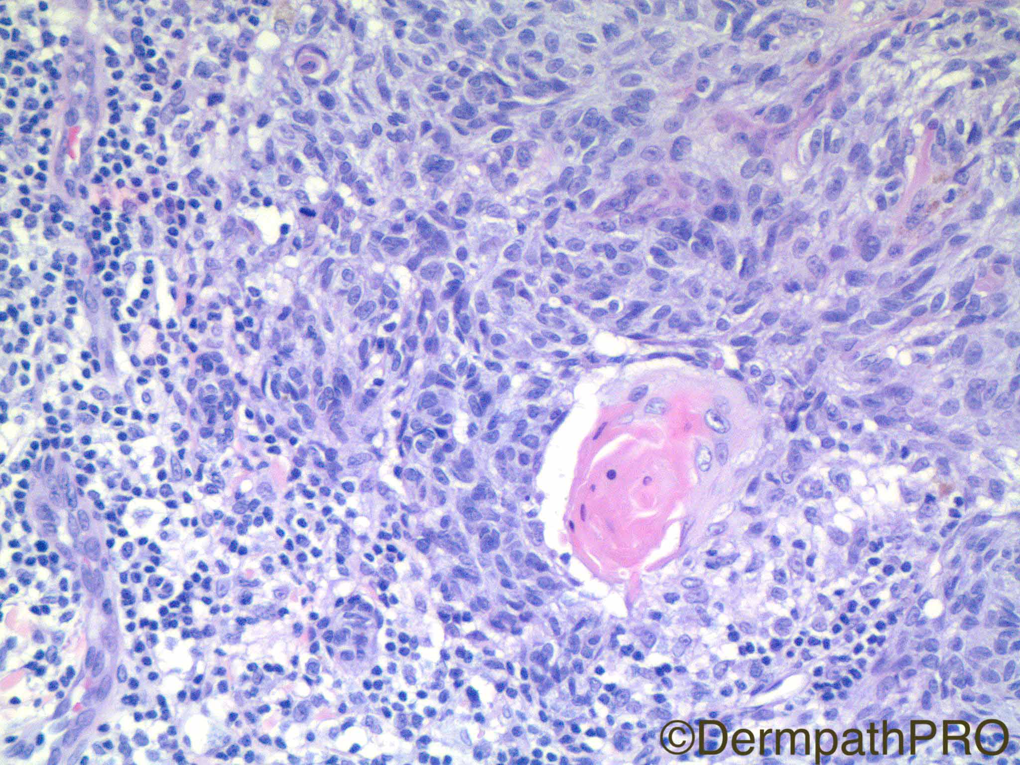
This is a thin lesion that has heterogeneous features. We have an area better seen in pic 3 that looks like a junctional nevus that from my point of view has at most mild dysplasia (should see the slide to better assess). Then the nests become larger and expansile towards the center and they coalesce acquiring a complex architectural organization, we can see some areas of retraction artifact surrounding some of the nests. The melanocytes are mildly to moderately atypical and surround adnexae, there is a single non-atypical mitosis in the last picture and a florid lymphocytic reaction is also evident. The clinical setting is worrisome. I would perform a nuclear melanocytic marker in order to better asses the breslow. But my diagnosis would be MELTUMP favouring benign behaviour and suggest follow up, It would not bother me to perform Fish or CGH in this case.

Share this comment


Link to comment
share_externally

vincenzo polizzi

Posted

Agree with above comments, but favor melanoma, with some halo effect suggesting a partial regression.

Share this comment


Link to comment
share_externally

Nitin Khirwadkar

Posted

Agree with most of the comments made above. I am worried with the deep mitosis in the last figure, brisk lymphoid infiltrate and the confluence of nests. p16 may help. I would recommend complete excision, favouring a melanoma.

Share this comment


Link to comment
share_externally

Alice Roberts

Posted

After getting more clinical history, ie duration, recent change. I'd do FISH and/or MyPath gene expression studies. I'm worried about melanoma. More of an in situ component would be helpful in that diagnosis but I'd do deeper cuts also.

Share this comment


Link to comment
share_externally

I can see an atypical lesion with "asymmetry and border notching" clinically and large nested melanocytes with small but atypical nuclei and few or no pagetoid spread on histopathology. In this person (57 yo) I think this is an "in situ" nevoid melanoma. This is a good case to be supported by FISH or CGH.

Share this comment


Link to comment
share_externally

Alice Roberts

Posted

I do favor melanoma though... Epidermal consumption/ ulceration.

Share this comment


Link to comment
share_externally

Dr. Richard Carr

Posted

Difficult but based on clinical and what we see I favour a relatively thin SSMM arising in the setting of a mildly dysplastic naevus.

Share this comment


Link to comment
share_externally

Dr. Mona Abdel-Halim

Posted

Melanoma arising within a dysplastic nevus

Share this comment


Link to comment
share_externally

Uma Sundram

Posted

Extremely difficult case and great to see the excellent discussion! We too favored a melanoma arising within a dysplastic nevus and MyPath was positive, in the melanoma range. The deep mitosis, sheet like growth pattern and epidermal consumption were all concerning features for us.

Share this comment


Link to comment
share_externally


Join the conversation

You can post now and register later. If you have an account, sign in now to post with your account.

Guest
Add a comment...

×   Pasted as rich text.   Paste as plain text instead

  Only 75 emoji are allowed.

×   Your link has been automatically embedded.   Display as a link instead

×   Your previous content has been restored.   Clear editor

×   You cannot paste images directly. Upload or insert images from URL.

Loading...

×
×
  • Create New...