Jump to content
In this section we have spot diagnoses posted on a daily basis since June 2010, now over 4000! You can review the archived cases and read the suggested diagnoses by users and the final comment by the contributors.
Case are uploaded each week day by 10 am UK time with the correct diagnosis will generally be posted at 8 pm UK time. Why not view the most recent spot diagnosis and proffer a diagnosis?

Case Number : Case 1644 - 13 October Posted By: Guest

Please read the clinical history and view the images by clicking on them before you proffer your diagnosis.
Submitted Date :
   (0 reviews)

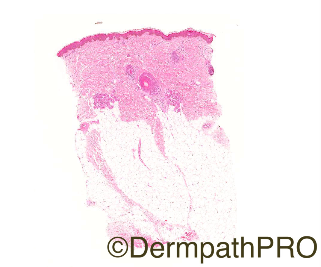
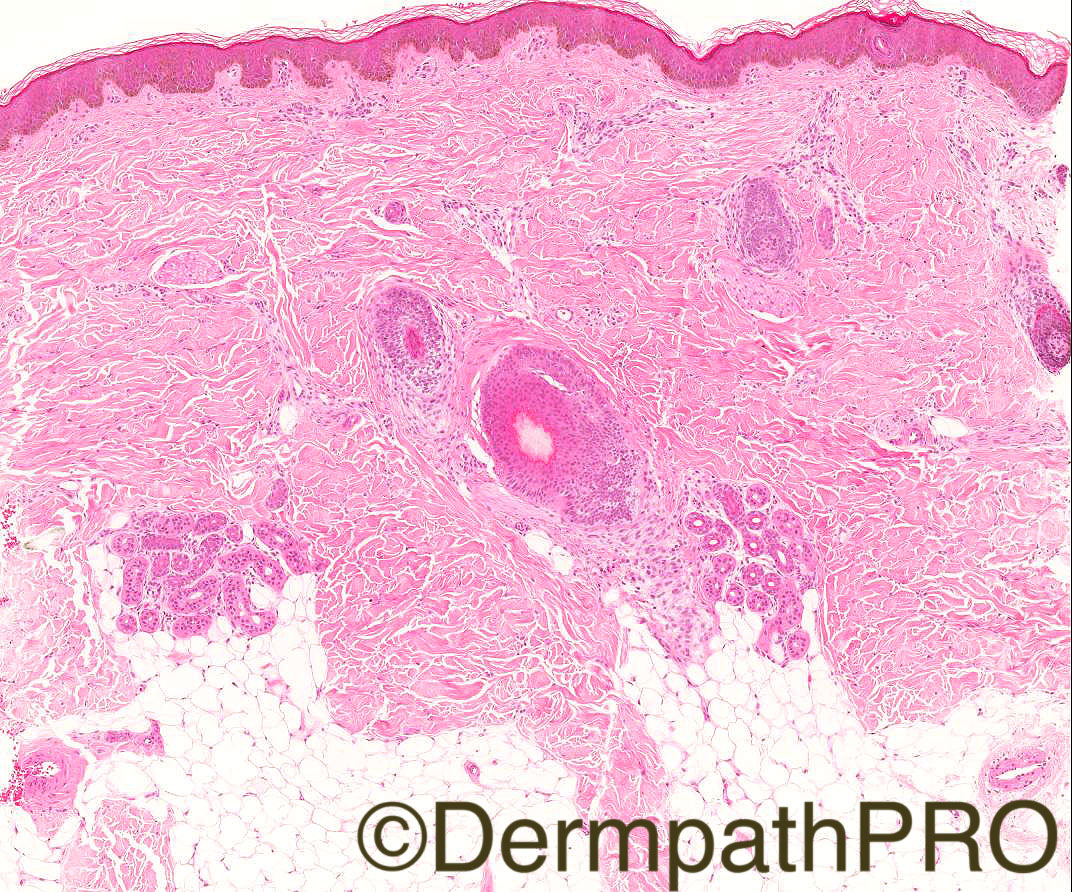
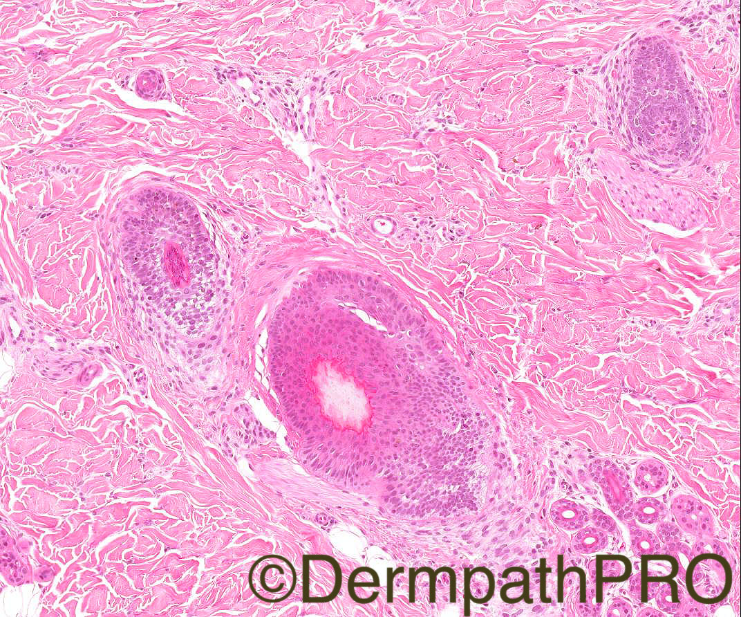
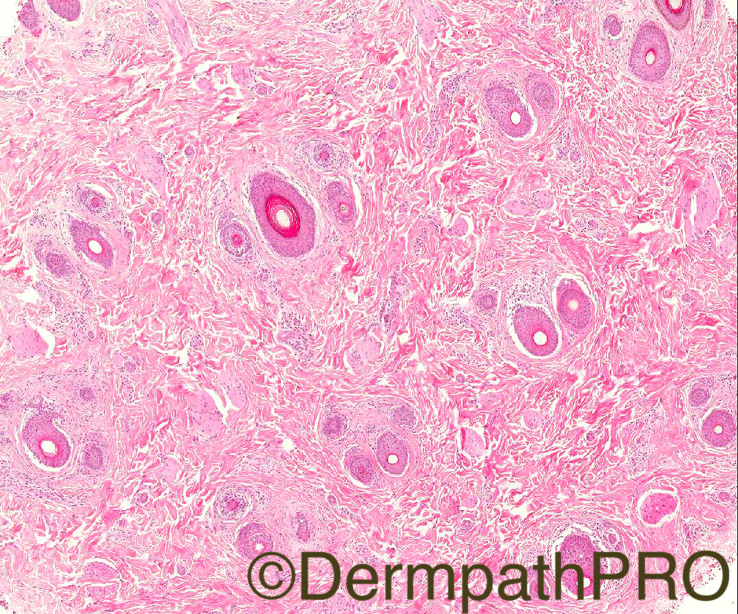
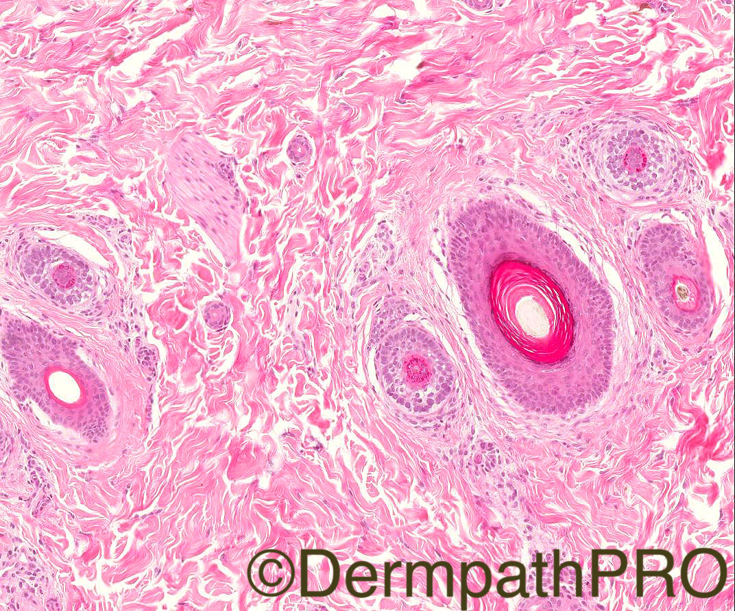
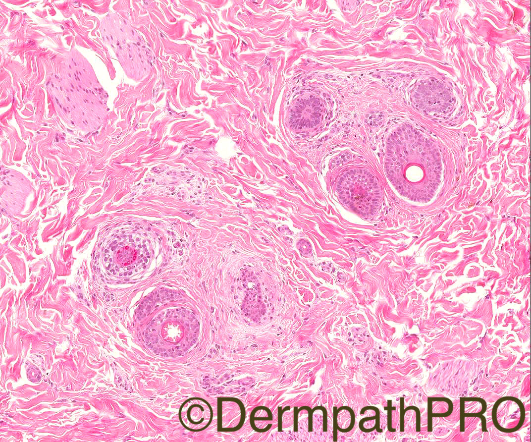
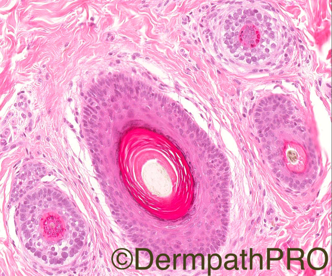
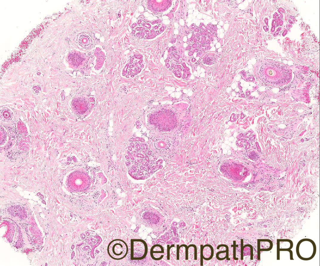
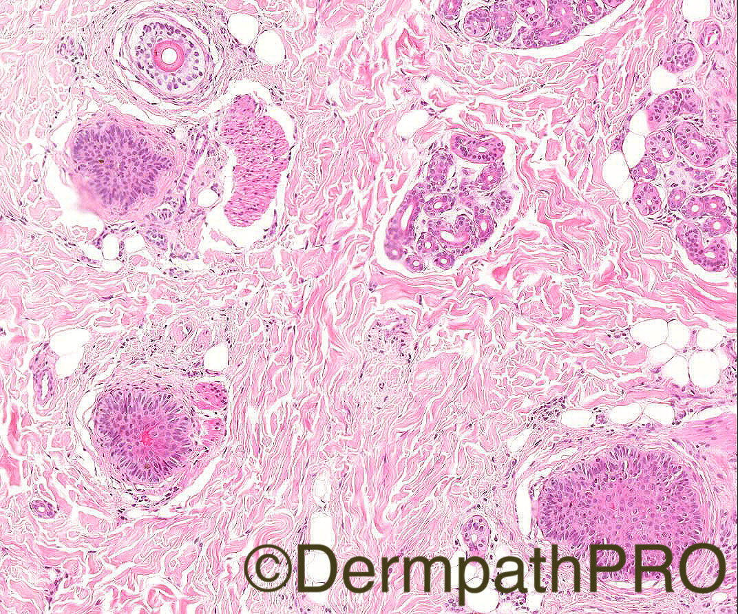
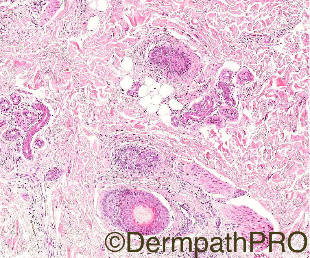
‘2 year old girl with area of reduced hair growth in  parietal scalp, 2 biopsies from scalp’. Images 1,2, 3- vertical sections. Images 4,5,6,7- horizontal section at level of isthmus. Images 8, 9, 10- horizontal sections at dermo-subcutaneous junction
 
Case Posted by Dr Arti Backshi


  Report Record

User Feedback


Admin_Dermpath

Posted

Another beautiful set of images from Dr Arti Backshi

Share this comment


Link to comment
share_externally

Arti Bakshi

Posted

Are some of you still having problems logging in/adding comments/enlarging images? If so, I can pass this on to the administrator. 

Share this comment


Link to comment
share_externally

Raul Perret

Posted

I see a predominance of catagen and telogen hairs, lack of inflammatory infiltrate, abnormal cornification of IRS and lots of clefts. I think this is a case of loose anagen hair syndrome, the main differential diagnosis is AA that is more frequent in the pediatric population


Share this comment


Link to comment
share_externally

Raul Perret

Posted

reviewing the case, It bothers me a bit the high amount of catagen and telogen. Should correlate with trichogram but I favor LAHS over AA

Share this comment


Link to comment
share_externally

Dr. Mona Abdel-Halim

Posted

The most striking feature is the excess catagen and telogen together with minimal inflammation. Can be seen in longstanding AA but at such stages, compensatory hypertrophy of seb lobules is usually seen which we do not have here. There is a follicle with excess keratin. There are clefts around hair follicles. I do not c pigment casts but still will raise the possibility of trichotillomania. 

Share this comment


Link to comment
share_externally

Raul Perret

Posted (edited)

I have commited a mistake when saying abnormal cornification of IRS. It is just abnormal cornification as Mona mentioned, we cannot actually see the IRS in that follicle. Trichotillomania without pigment casts or hemorrhage sounds weird... I think that the only pathology that can show us all the features we see here is AA in a late phase. I have very little experience with alopecia but I would not discard LAHS   

Edited by Raul Perret

Share this comment


Link to comment
share_externally

Raul Perret

Posted (edited)

oh dear it is kind of annoying not being able to edit the text of comments anymore. Where we read I mean "us" hope I am not getting banned of the web :D

Edited by Raul Perret

Share this comment


Link to comment
share_externally

Admin_Dermpath

Posted

Raul,

I will have a word with the site guru and see if this can be changed.

Cheers, Geoff

Share this comment


Link to comment
share_externally

Admin_Dermpath

Posted

Raul etc,

I have changed settings so that you can edit your postings.

You are currently a limited member and soon you may not be able to post, have you considered membership?

Cheers, Geoff

Share this comment


Link to comment
share_externally

Arti Bakshi

Posted

Alopecia is a difficult area! Some of my trainees who visit this site have suggested that more description of the images would help them to understand the discussion better. So I am going over some of the main morphological findings.

As Raul and Mona rightly picked up, there is excess of telogen and catagen follicle (particularly seen in last 3 images). Normal count is 10-15%, in this case 40-50% of follicles were in telogen/catagen phase. There is another prominent finding seen in image 3, 4 and 5. Remember this is a horizontal section at level of isthmus/infundibulum, so normal anagen terminal follicles at this level should all look somewhat like the central follicle in image 7. The 3 other follicles in that image are clearly abnormal for this level. Any suggestions what these represent??

Also, Mona rightly pointed towards absence of sebaceous hyperplasia. Infact, there are hardly any sebaceous glands seen at all. Any thoughts why this may be?

Raul, loose anagen syndrome would show clefts between the inner and outer root sheath (or sometimes within inner root sheath), the clefts seen in these images are mainly surrounding the outer root sheath and stroma. Some fragmentation of IRS is a common artifact of processing (noted in lowest follicle in image 6). Also, almost all follicles would be anagen in LAHS, which is not the case here. (If it helps, LAHS was not a clinical consideration either).

Will leave it for another day. More comments welcome!!

Share this comment


Link to comment
share_externally

Dr. Mona Abdel-Halim

Posted

The other three follicles in image 7 are vellus or vellus like (miniaturized follicles). So we also have increased vellus to terminal hair ratio which I missed first and this is not seen in trichotillomania but favors longstanding AA especially with the clinical presentation in an infant. 

The absent sebaceous glands usually points to a scarring process and is also seen in psoriatic alopecia, both causes are not present here so since this girl is a 2 year old, can one explain this absence of seb glands by an age related state of sebaceous gland atrophy?


Share this comment


Link to comment
share_externally

Arash Daryakar

Posted

i think of Congenital Triangular Alopecia .

Share this comment


Link to comment
share_externally

Dr. Mona Abdel-Halim

Posted

42 minutes ago, Arash said:

i think of Congenital Triangular Alopecia .

I thought of it when I found the increased vellus hairs but will it explain the increased catagen and telogen? 

Share this comment


Link to comment
share_externally

vincenzo polizzi

Posted (edited)

I have two questions: is sebaceous glands loss an usual finding in scarring alopecia? So we couldn't find this in non scarring alopecia...

is there any mild peri follicular scar or am I wrong?

Edited by vincenzo polizzi

Share this comment


Link to comment
share_externally

Raul Perret

Posted

7 minutes ago, vincenzo polizzi said:

I have a question: is sebaceous glands loss an usual finding in scarring alopecia? 

Yes vincenzo is something seen usually. But we all agree that this case is not scarring alopecia as scars are absent and perifollicular fibrosis. As mona mentioned sebaceous hypoplasia/aplasia can be seen in psoriatic alopecia. I did not find a reference relating age and density of sebaceous glands though. Hormonal levels can play a role and apparently from the history this does not seem to be syndromic. 

Share this comment


Link to comment
share_externally

Arash Daryakar

Posted

6 hours ago, Dr. Mona Abdel Halim said:

I thought of it when I found the increased vellus hairs but will it explain the increased catagen and telogen? 

you are right. cant fit everything in this case. i think we should wait for Arti to better clarification.

Share this comment


Link to comment
share_externally

Arti Bakshi

Posted

Thanks for all your excellent comments!

This is a case of chronic alopecia areata.  Well done to Mona and Raul for considering the diagnosis inspite of absent peribulbar inflammation. Mona's second comment wraps up the case beautifully!

The key findings are marked shift into telogen phase and prominent miniaturisation. (The terminal:vellus ratio was 1:1 in this case ; normal is 7:1). This combination should always make one think of chronic AA. The main differentials of high telogen counts ie  telogen effluvium and traction alopecia do not show miniaturisation. Conversely the other main cause of miniaturisation ie androgenic alopecia will not show such high telogen counts (although borderline increase of 15-20% not unusual). Temporal traingular alopecia has a typical distribution and again one would not expect telogen shift in this entity. Remember, that chronic AA often lacks peribulbar inflammation, so undue reliance on this one feature can lead to underdiagnosis. AA was the clinical impression too in this case.

 For the beginners in alopecia pathology, vellus/vellus like follicles have their bulbs in upper or mid reticular dermis (seen in images 1 and 2) and on horizontal sections are recognised by follicles with inner root sheath thickness greater than or equal to the hair shaft diameter. (image 7).

Share this comment


Link to comment
share_externally

Arti Bakshi

Posted

With regards to Vincenzo's question...absence of sebaceous glands is a usual feature of scarring alopecia and one that I find quite useful. But there are caveats...psoriatic alopecia being one them. I was puzzled by paucity of sebaceous glands in this case, which is clearly not a scarring process clinically or histologically. I reached the same conclusion as Mona that the sebaceous atrophy is due to the age of the child. Kazakov's textbook (of adnexal tm) discusses the evolution of sebaceous glands rather well. (pg 333). ...'In the newborn the sebaceous glands are fully developed but atrophy in the ensuing months due to drop in maternal androgens to become a mantle (an immature sebaceous gland) They again become hyperplastic at puberty'. In some of the levels in this case, one could see atrophic mantles surrounding hair follicles.

Do apologise for the excessively long posts, but I do find alopecia pathology rather fascinating!!

Share this comment


Link to comment
share_externally

Arash Daryakar

Posted

Thanks dear Arti for your perfect explanation.

please consider to post more alopecia cases. i think we need more !

So ,you mean because of prominent increase of Telogen/catagen hair we should think of chronic AA...

In fact in AA we have more Telogen/catagen hairs than conditions like Telogen effluvium.

Share this comment


Link to comment
share_externally

Arti Bakshi

Posted

Yes, completely agree, Arash. Chronic telogen effluvium usially shows milder excess of telogen counts and counts in excess of 40-50% would be unusual for this entity, whereas this is the norm in chronic AA. In typical cases, the clinical would be fairly distinctive (diffuse thinning in TE vs patch of hair loss in AA) . The problem comes in the (less common ) diffuse forms of alopecia areata.....but that is a discussion for another day!

Share this comment


Link to comment
share_externally


Join the conversation

You can post now and register later. If you have an account, sign in now to post with your account.

Guest
Add a comment...

×   Pasted as rich text.   Paste as plain text instead

  Only 75 emoji are allowed.

×   Your link has been automatically embedded.   Display as a link instead

×   Your previous content has been restored.   Clear editor

×   You cannot paste images directly. Upload or insert images from URL.

Loading...

×
×
  • Create New...