Jump to content
In this section we have spot diagnoses posted on a daily basis since June 2010, now over 4000! You can review the archived cases and read the suggested diagnoses by users and the final comment by the contributors.
Case are uploaded each week day by 10 am UK time with the correct diagnosis will generally be posted at 8 pm UK time. Why not view the most recent spot diagnosis and proffer a diagnosis?

Case Number : Case 1645 - 14 October Posted By: Guest

Please read the clinical history and view the images by clicking on them before you proffer your diagnosis.
Submitted Date :
   (0 reviews)

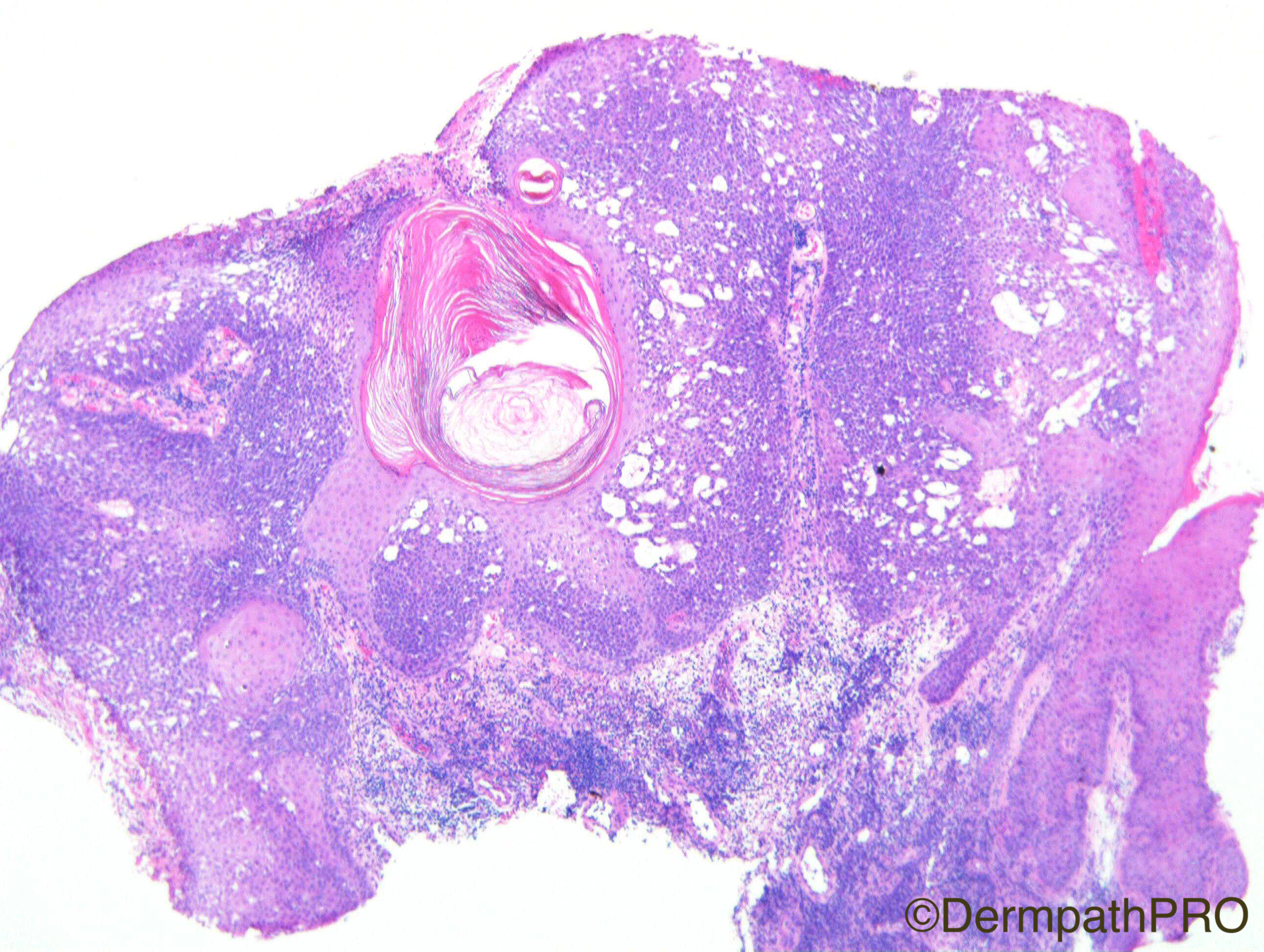
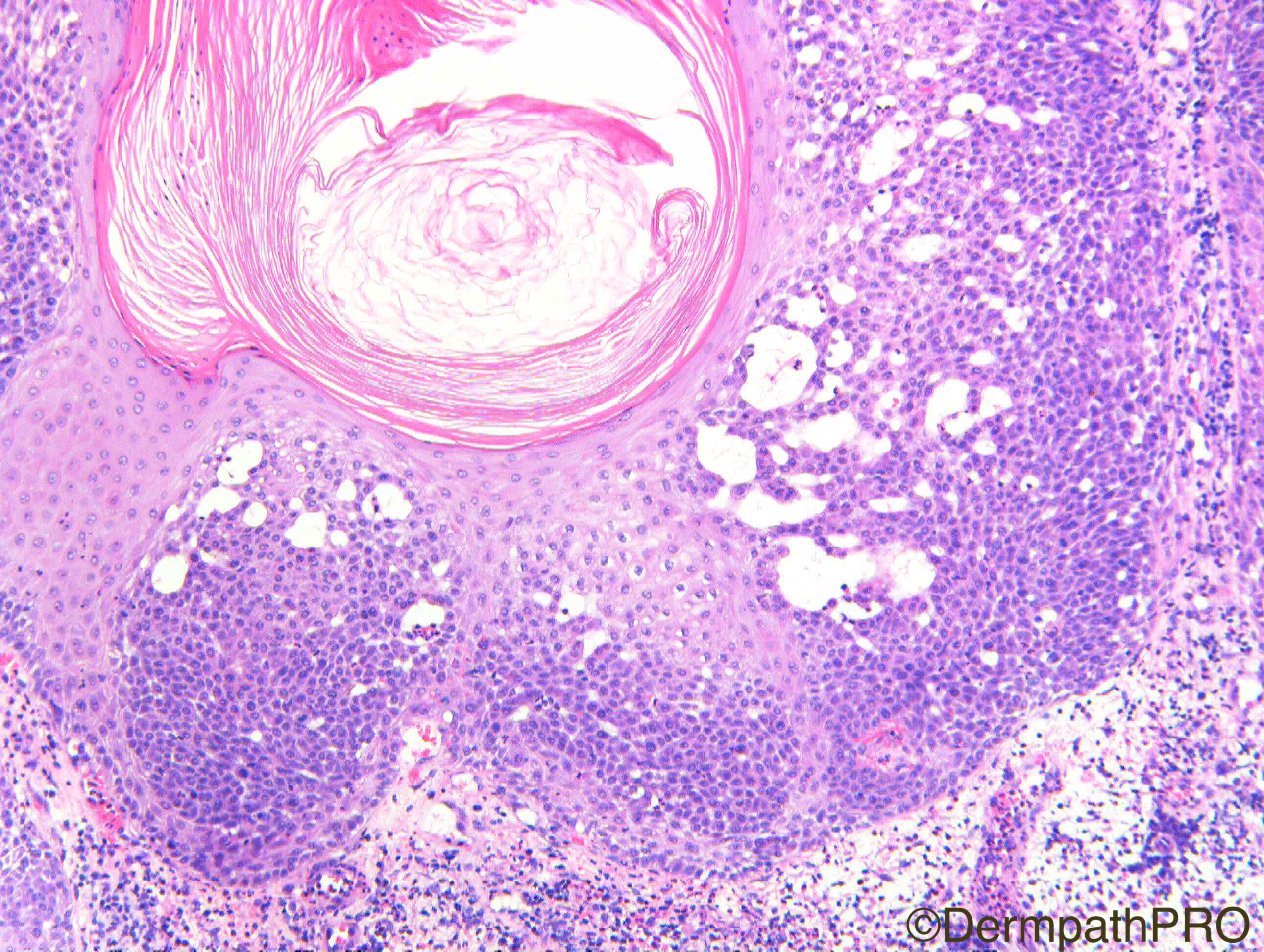
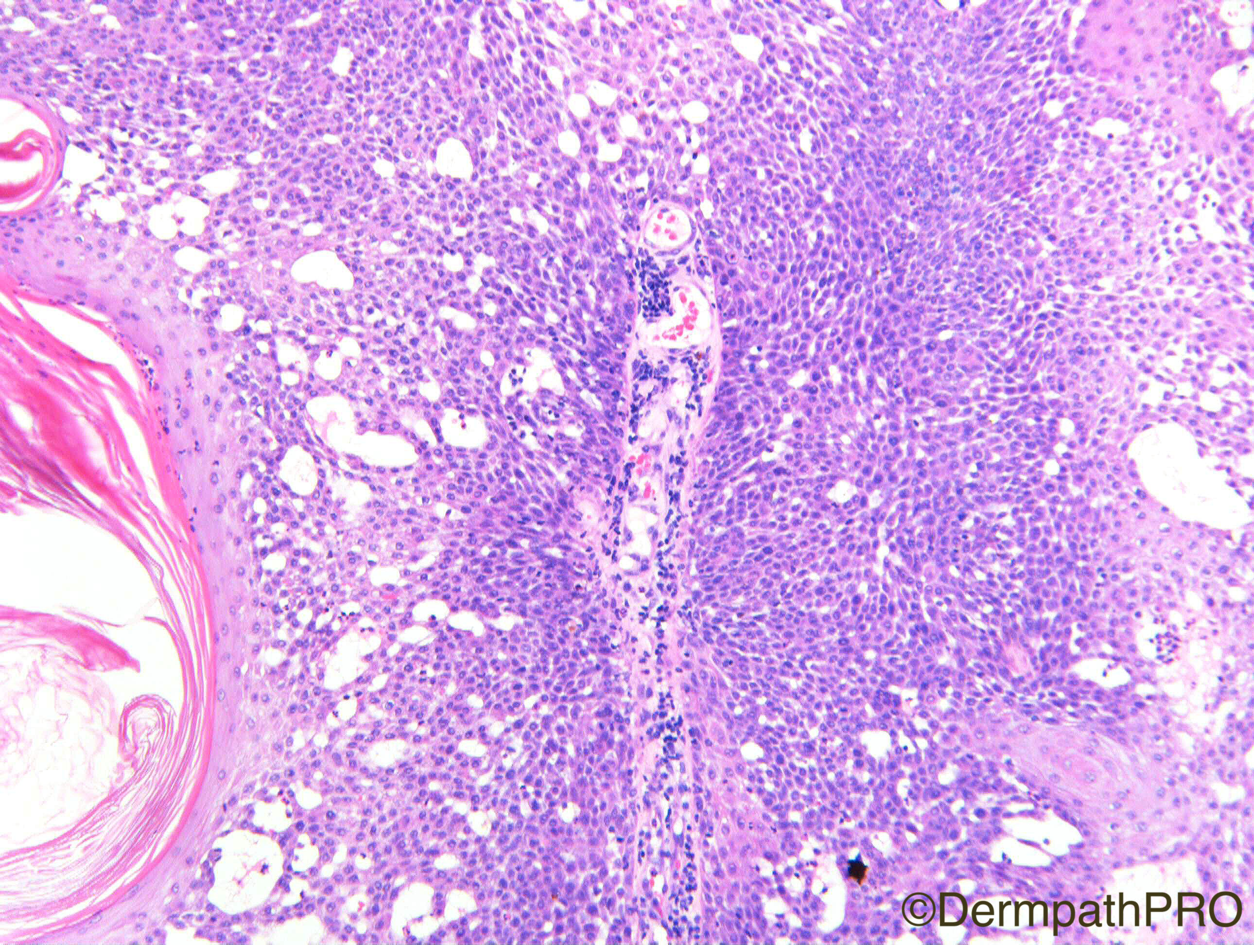
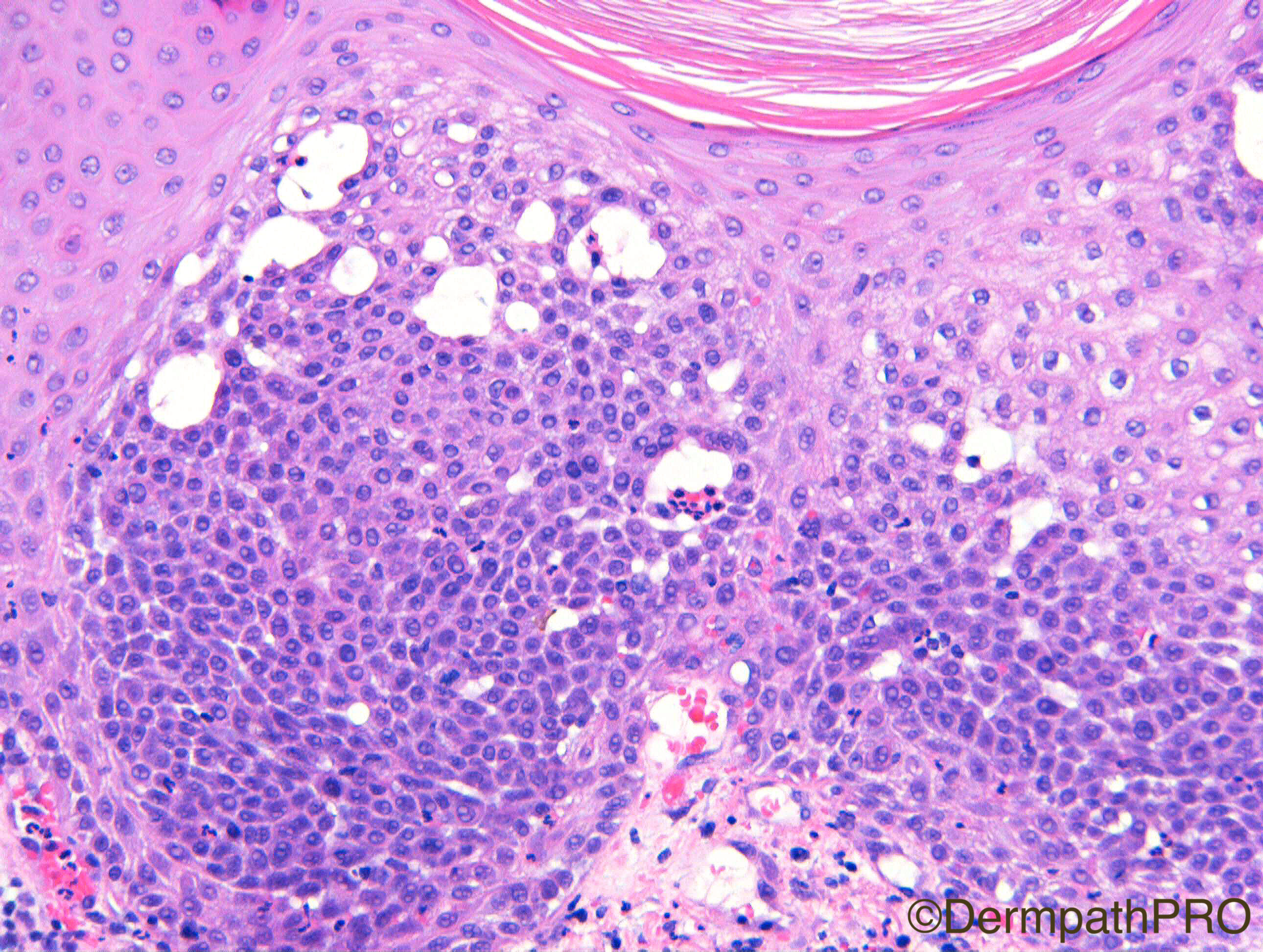
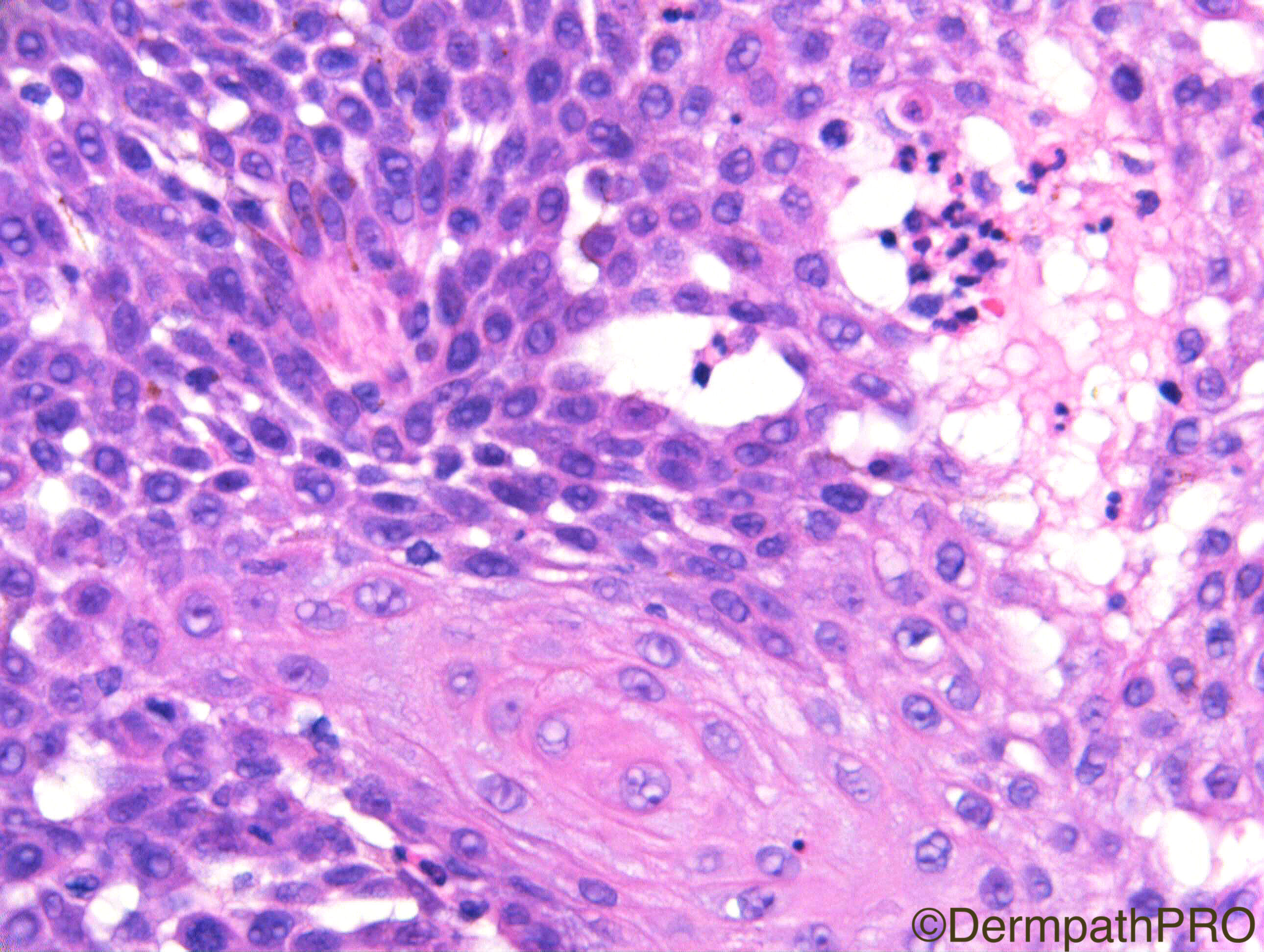
M85. 20 x 20mm eroded friable plaque arising in old scar. ?SCC

Case Posted by Dr Richard A Carr
Edited by Admin_Dermpath


  Report Record

User Feedback


Admin_Dermpath

Posted

Rounding off the week with a cracking case from Dr Richard A Carr, more to be posted at 6pm 

Cheers, Geoff

Share this comment


Link to comment
share_externally

Nitin Khirwadkar

Posted

Agree, follicular SCC. Nice mucin? The scar is secondary to a previous excision of? Kobnerisation?

Share this comment


Link to comment
share_externally

Arti Bakshi

Posted

I was thinking more along the lines of inverted follicular keratosis, which can also show epithelial mucin.

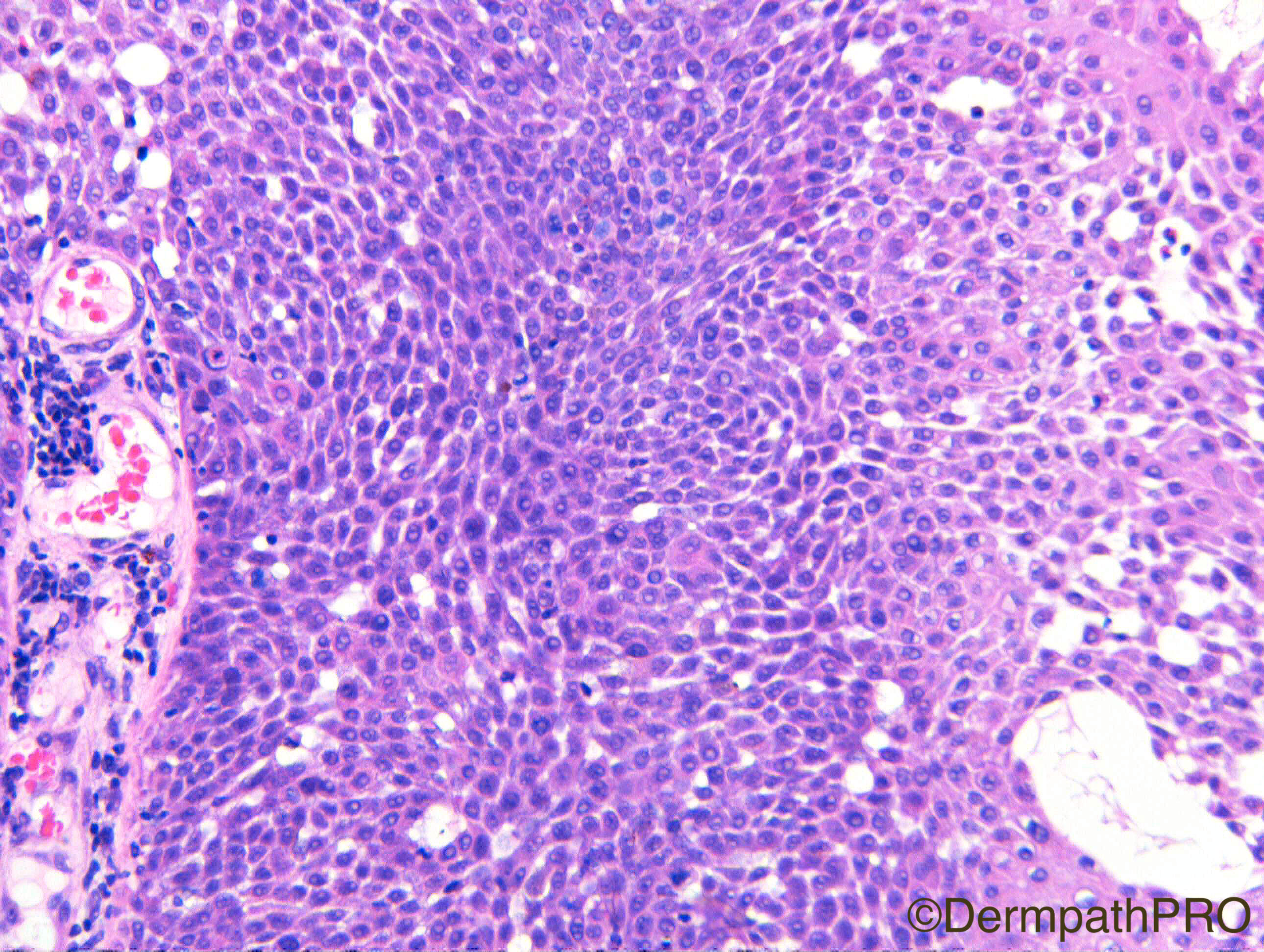
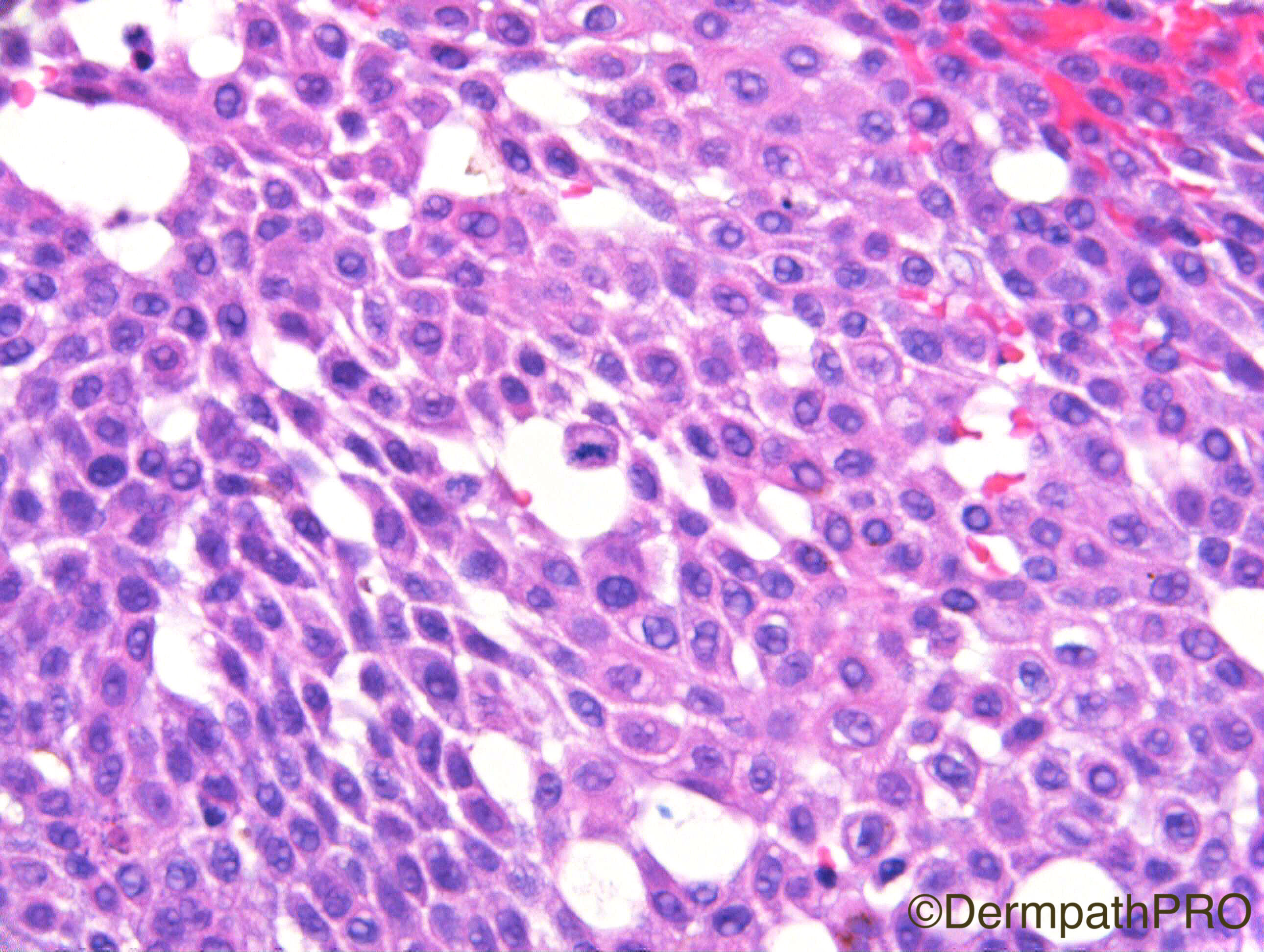
There is prominent basal cell proliferation and increased mitoses would be expected in such areas. Not convinced that there is significant atypia for follicular scc.

Share this comment


Link to comment
share_externally

Raul Perret

Posted (edited)

1 hour ago, arti bakshi said:

I was thinking more along the lines of inverted follicular keratosis, which can also show epithelial mucin.

There is prominent basal cell proliferation and increased mitoses would be expected in such areas. Not convinced that there is significant atypia for follicular scc.

Agree it was the impression i had too. Irritated sebk with mucin and a pseudoglandular morphology. Last picture showing abrupt transition from basaloid and mature keratinocytes is quite suggestive

Edited by Raul Perret

Share this comment


Link to comment
share_externally

Dr. Mona Abdel-Halim

Posted

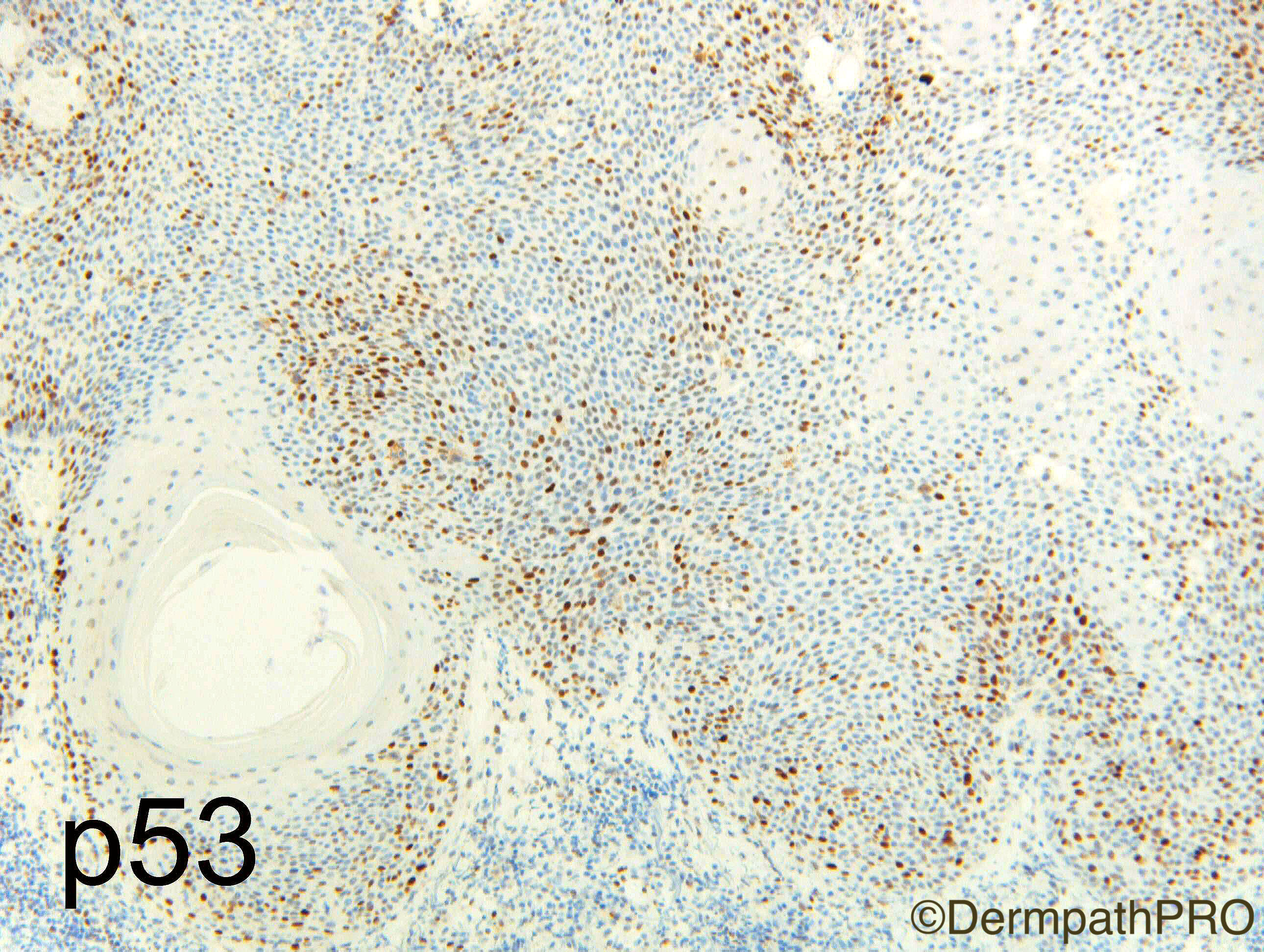
Arti and Raul are right as regards the lack of significant atypia. The clinical is worrisome though. Eroded plaque on a long standing scar. Mildly pleomorphic fSCC can be very difficult to differentiate from IFK. I thibk P53 will help. 

Share this comment


Link to comment
share_externally

Raul Perret

Posted

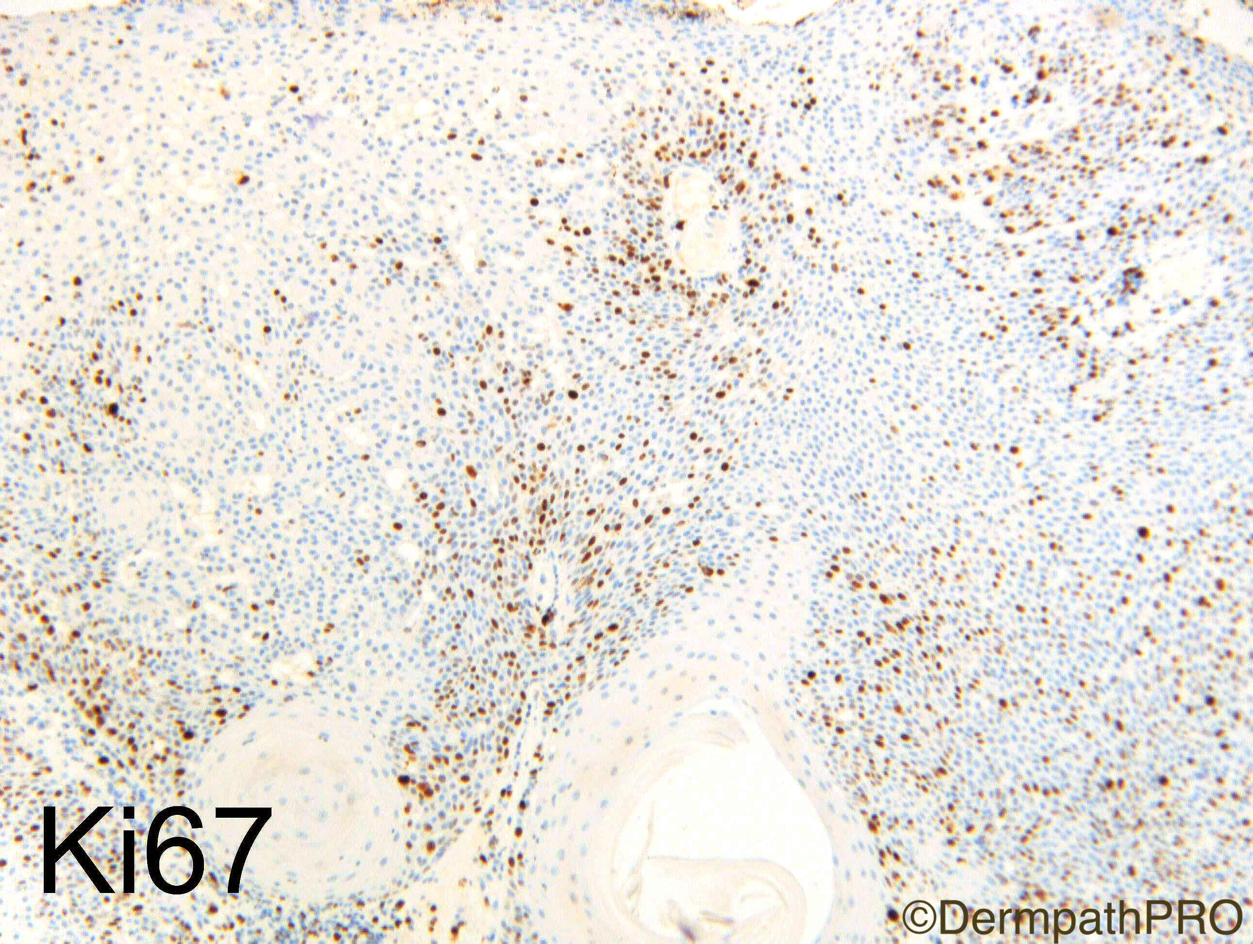
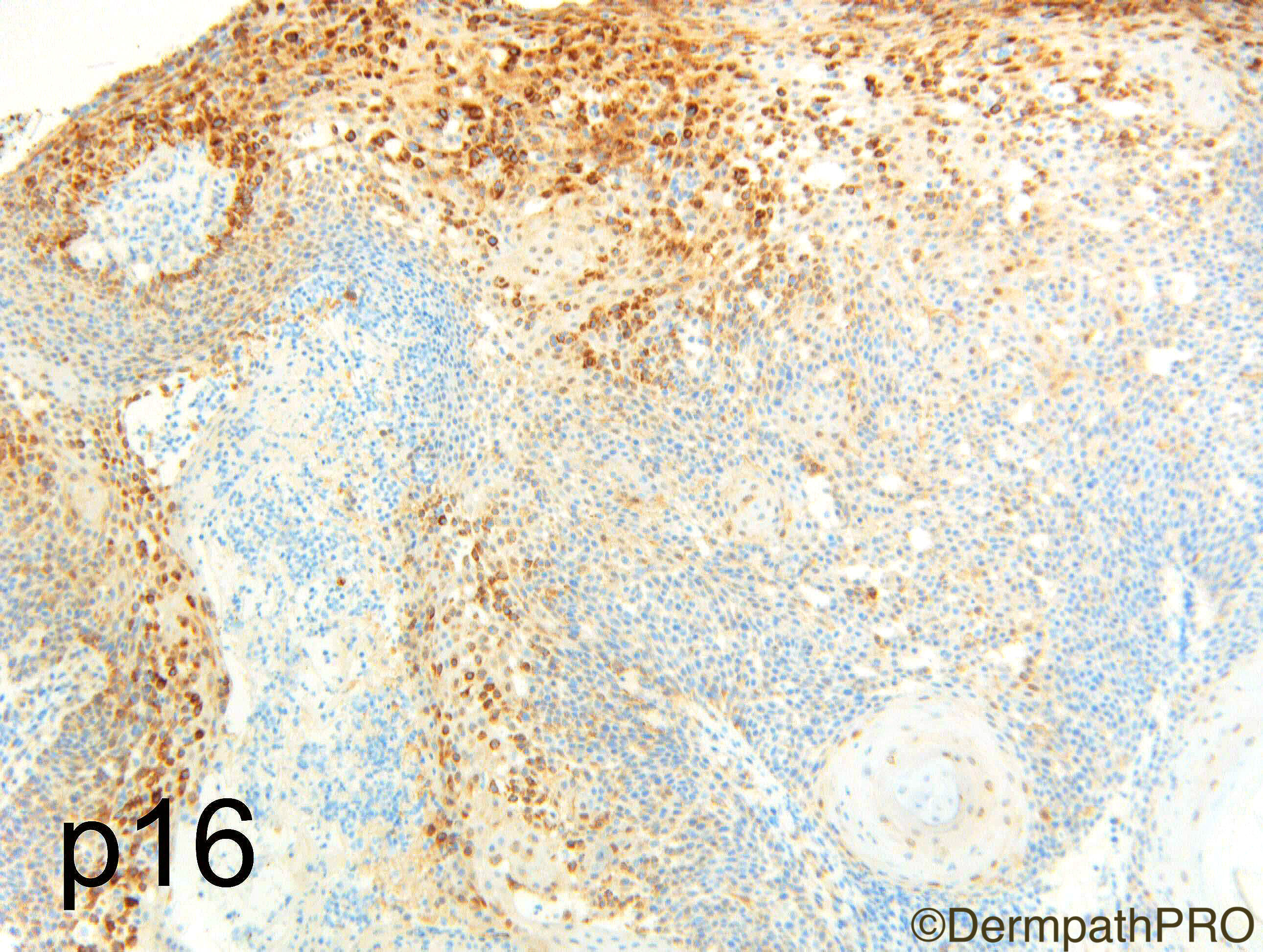
The pattern of the immunos is that of a benign lesion so the diagnosis of sebk stands

Share this comment


Link to comment
share_externally

Dr. Mona Abdel-Halim

Posted

Yes, agreed

immunos benign.

IFK although very unusual clinically ..


Share this comment


Link to comment
share_externally

Nitin Khirwadkar

Posted

Agree with IFK. IHC definitely benign. Did think about it, but favoured a FSCC.

Share this comment


Link to comment
share_externally

vincenzo polizzi

Posted

I agree with IFK, but something doesn't fit perfectly: the basaloid component looks like intraepidermal poroma/hydroacanthoma simplex..(.this recalls the reasonings about the eccrine nature of SK). However for diagnostic purpose I agree with above.

Share this comment


Link to comment
share_externally

Dr. Richard Carr

Posted

I had to eat humble pie on this case as I was ready to sign it out as a basaloid bowen's/SCC in situ when I was held back by a visiting dermpath trainee from the Netherlands.  Why is this not irritated seborrhoeic keratosis?  My low power spot diagnosis (basaloid, acantholytic, miotitic) had to be challenged and on further scrutiny - no atypical mitotic figures, relative uniformity of cells. So IHC requested that supported a diagnosis of irritated seborrhoeic keratosis / IFK.  I agree the history is concerning and I commented in the report that I have seen such mitotically active / clonal cases develop frank dysplasia. I currently believe seborrhoeic keratosis / IFK are infundibular keratoses (i.e. follicular tumours) hence can be exceedingly close to follicular SCC in situ (note they don't occur on non-hair bearing skin). Well done Arti / Raul and in particular Kimberly Van Oord!

Share this comment


Link to comment
share_externally

Dr. Richard Carr

Posted

Re: Vincenzo's point.  I have seen clonal lesions that I called SEBK develop over time in to porocarcinomas (at least two cases) so perhaps there is more to it than my first comment but we have to remember SEBK are very common lesions and perhaps are the innocent bystander and subtle clonal bowen's (epithelioma of Borst Jadhassohn) are very close to malignant hidroacanthoma morphologically.

Share this comment


Link to comment
share_externally


Join the conversation

You can post now and register later. If you have an account, sign in now to post with your account.

Guest
Add a comment...

×   Pasted as rich text.   Paste as plain text instead

  Only 75 emoji are allowed.

×   Your link has been automatically embedded.   Display as a link instead

×   Your previous content has been restored.   Clear editor

×   You cannot paste images directly. Upload or insert images from URL.

Loading...

×
×
  • Create New...