Jump to content
In this section we have spot diagnoses posted on a daily basis since June 2010, now over 4000! You can review the archived cases and read the suggested diagnoses by users and the final comment by the contributors.
Case are uploaded each week day by 10 am UK time with the correct diagnosis will generally be posted at 8 pm UK time. Why not view the most recent spot diagnosis and proffer a diagnosis?

Case Number : Case 1646 - 17 October Posted By: Guest

Please read the clinical history and view the images by clicking on them before you proffer your diagnosis.
Submitted Date :
   (0 reviews)

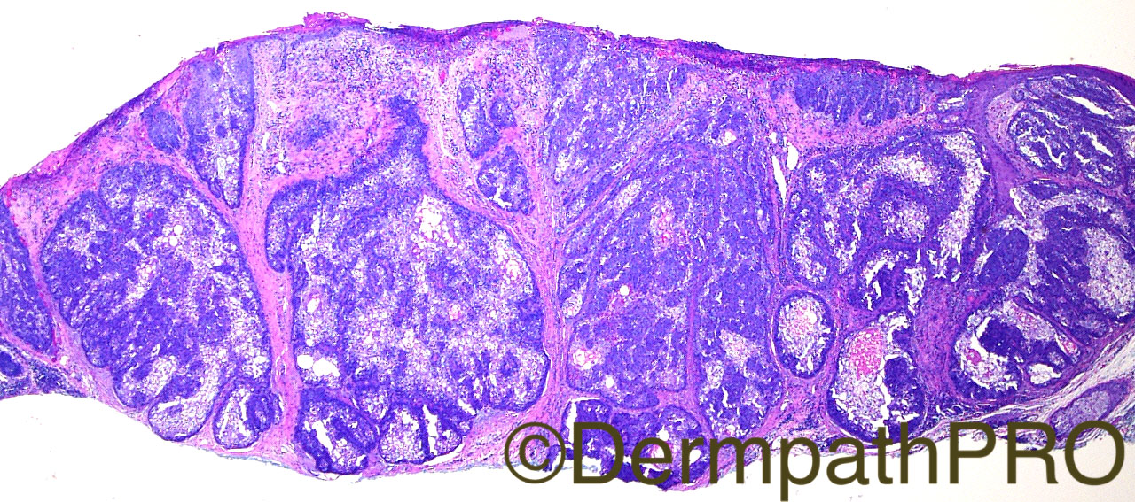
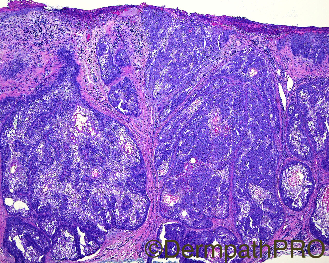
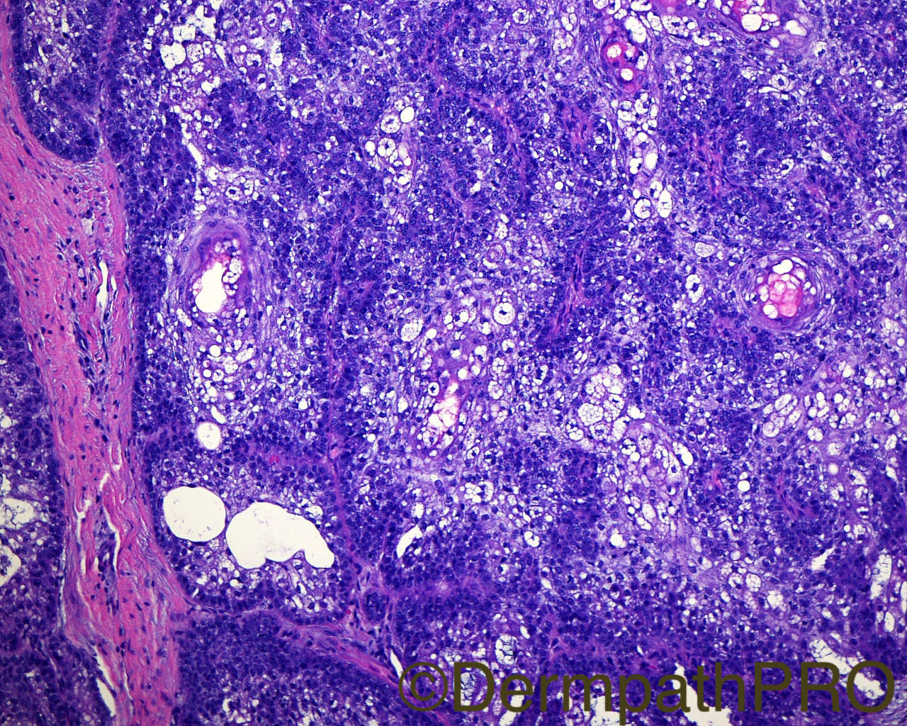
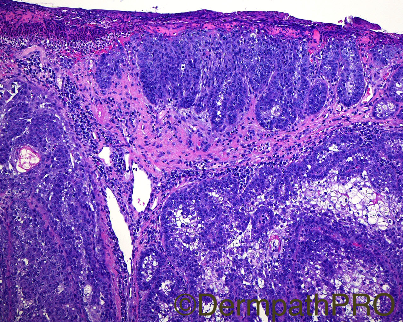
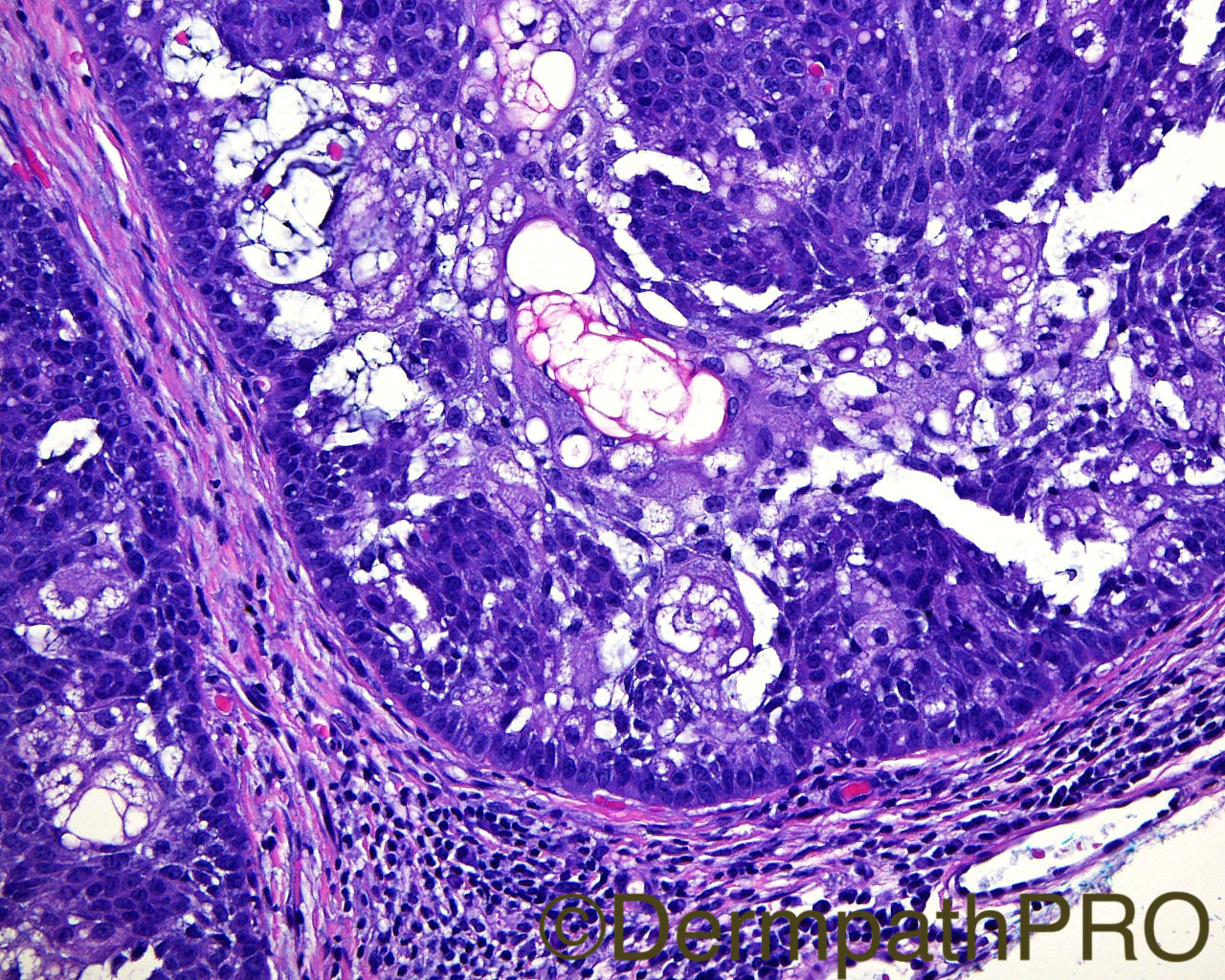
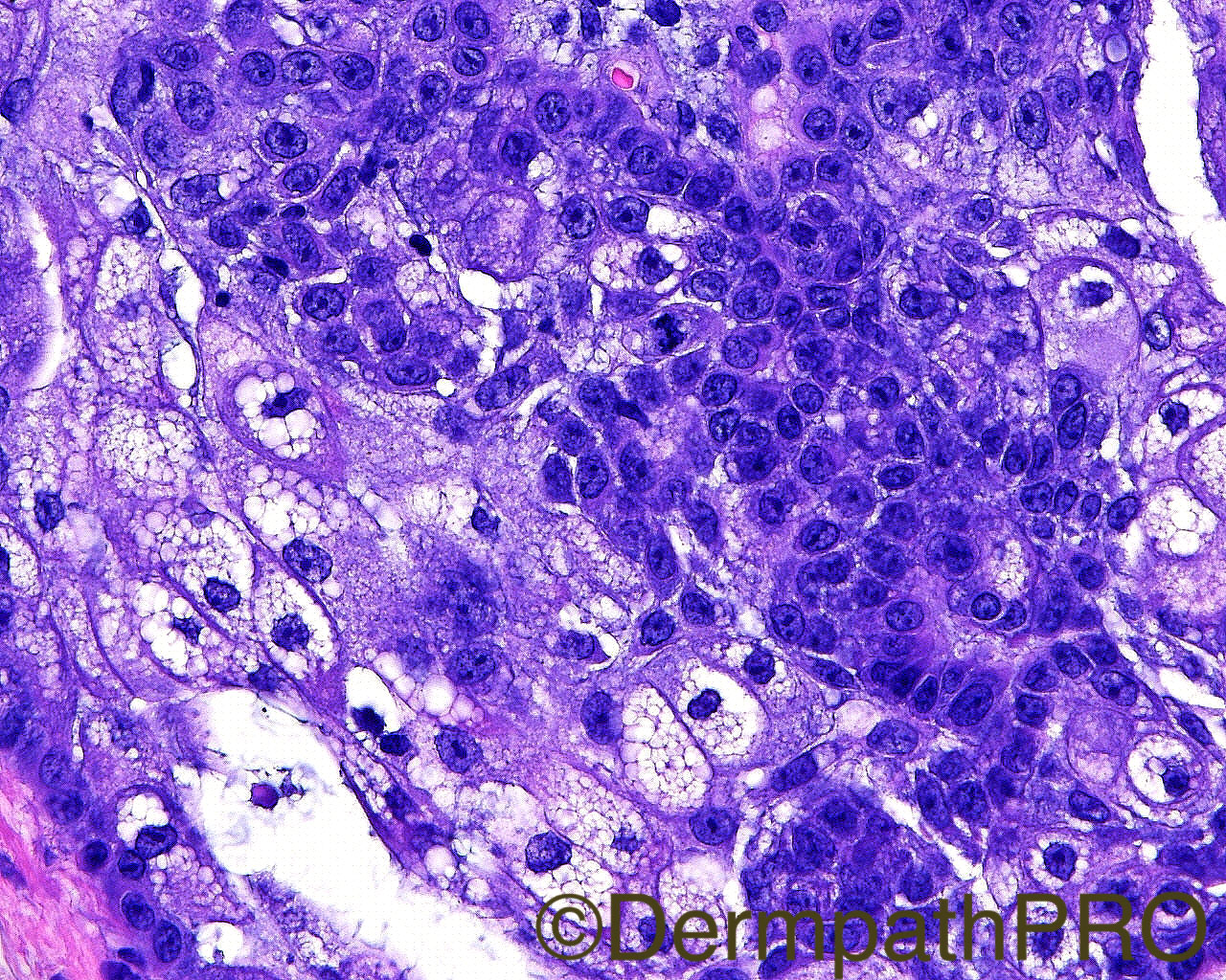
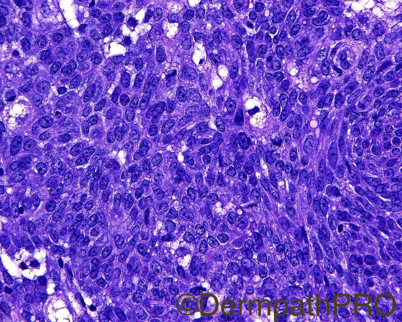
The patient is an 80-year-old man with a shave biopsy of a lesion on the left neck. Information provided by the referring dermatopathologist: "Clinically, it was basal cell carcinoma. This sebaceous neoplasm has features I suppose could be seen in the spectrum of sebaceoma. There is some increase in mitotic figures and some cellular atypia. Would this reach the threshold for sebaceous carcinoma to you?"

Case Posted by Dr Mark Hurt


  Report Record

User Feedback


Admin_Dermpath

Posted

Get your week off to a great start with this interesting Spot Diagnosis Case

Cheers, Geoff

Share this comment


Link to comment
share_externally

vincenzo polizzi

Posted

Difficult case. The more than mild sebaceous differentiation with poor germinative component fits better to adenoma... but too much atypia and mitosis and a bowenoid shoulder. So I favor low grade sebaceous carcinoma

Share this comment


Link to comment
share_externally

Dr. Mona Abdel-Halim

Posted

It is difficult!!! I am inclined more to call it sebaceous adenoma but if my case will double check with an expert first !!!!  I think the overall sebaceous component is more than the basaloid germinative component. About more than half of the each lobule is mature sebocytes. Very well circumscribed. Not that pleomorphic. Not infiltrative. Giant sebaceous adenomas can be mitotically active. Can mimic BCC clinically also. It is difficult though !! Muir Torre syndrome should be verified.

Share this comment


Link to comment
share_externally

Raul Perret

Posted (edited)

I thought there was a minimal predominance of immature cells but this is subjective i guess. Atypia is low as well as mitotic index. Tumor is quite lobular but has a marked horizontal extension, picture number 4 shows compromise of epidermis. From my perspective this is sebaceous carcinoma rather low grade. 

Edited by Raul Perret

Share this comment


Link to comment
share_externally

Arash Daryakar

Posted

This is an ulcerated multilobulated neoplasm focally attached to overlying epidermis with a sharp and pushing boundary to surrounding dermis . there are foci of mitoses but no obvious atypia.normal looking mitoses and ulceration can also be found in sebaceous adenoma.IHC study for ki67,p53 , Bcl-2 and p21 may help to make distinction from low grade sebaceous carcinoma.some intra and peritumoral lymphocytes may point to MTS(?).

overall i lean more toward to sebaceous adenoma.

waiting for Mark's precious idea!

Share this comment


Link to comment
share_externally

Nitin Khirwadkar

Posted

I would favour a low grade sebaceous carcinoma. Don't like the atypia. A Ki67 and p53 will be hopefully helpful. To rule out MTS.  Waiting to hear from Dr Hurt.

Share this comment


Link to comment
share_externally

Dr. Richard Carr

Posted

If this is a sebaceous carcinoma it is clearly in situ (lobulated pushing only borders). Analagous to our concept in follicular SCC in situ. I found image quality sub-optimal to assess the cytology with confidence but I suspect I would have let it go as an adenoma myself based on the architecture.  We should be aware of a difference in individual's approach in this somewhat contentious / challenging area but a mutant / null p53 would certainly pitch the scales.

Share this comment


Link to comment
share_externally


Join the conversation

You can post now and register later. If you have an account, sign in now to post with your account.

Guest
Add a comment...

×   Pasted as rich text.   Paste as plain text instead

  Only 75 emoji are allowed.

×   Your link has been automatically embedded.   Display as a link instead

×   Your previous content has been restored.   Clear editor

×   You cannot paste images directly. Upload or insert images from URL.

Loading...

×
×
  • Create New...