Jump to content
In this section we have spot diagnoses posted on a daily basis since June 2010, now over 4000! You can review the archived cases and read the suggested diagnoses by users and the final comment by the contributors.
Case are uploaded each week day by 10 am UK time with the correct diagnosis will generally be posted at 8 pm UK time. Why not view the most recent spot diagnosis and proffer a diagnosis?

Case Number : Case 1647 - 18 October Posted By: Guest

Please read the clinical history and view the images by clicking on them before you proffer your diagnosis.
Submitted Date :
   (0 reviews)

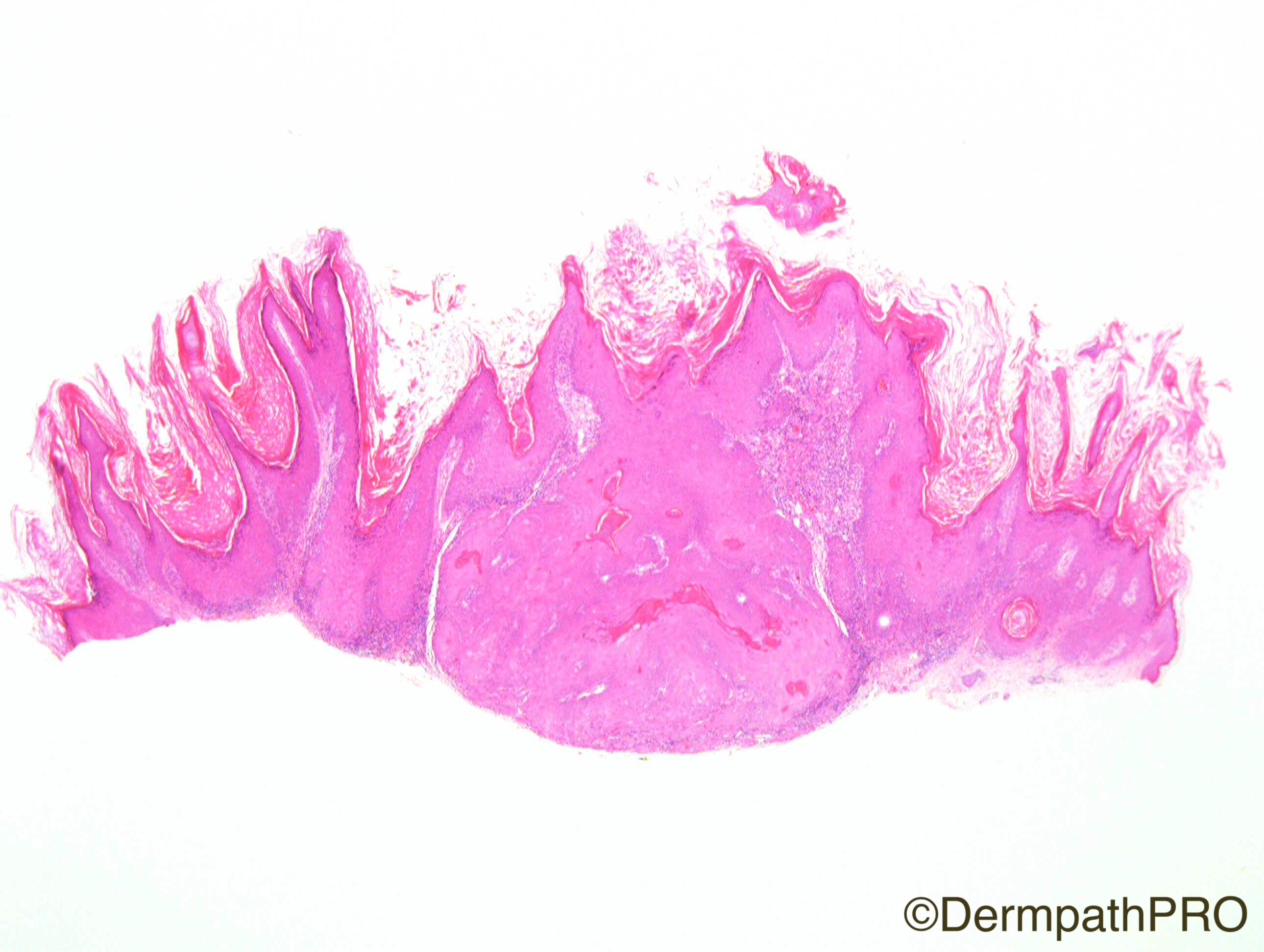
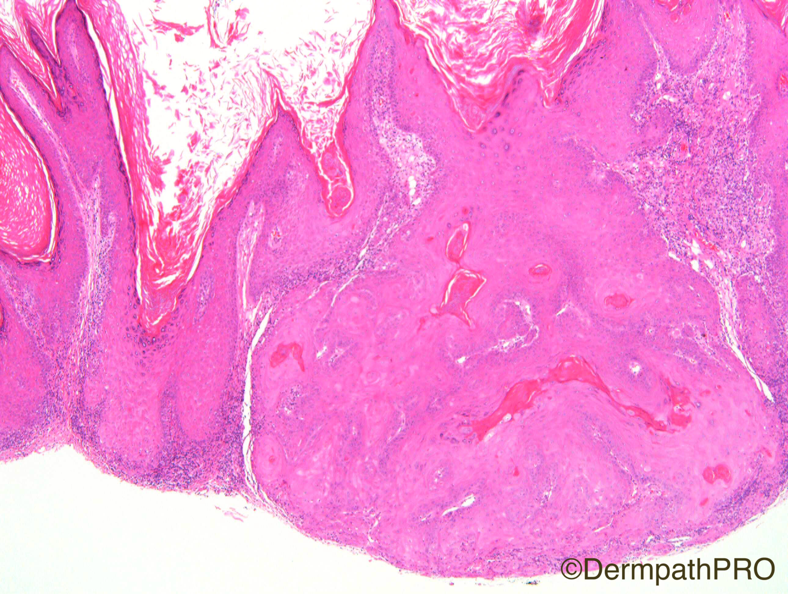
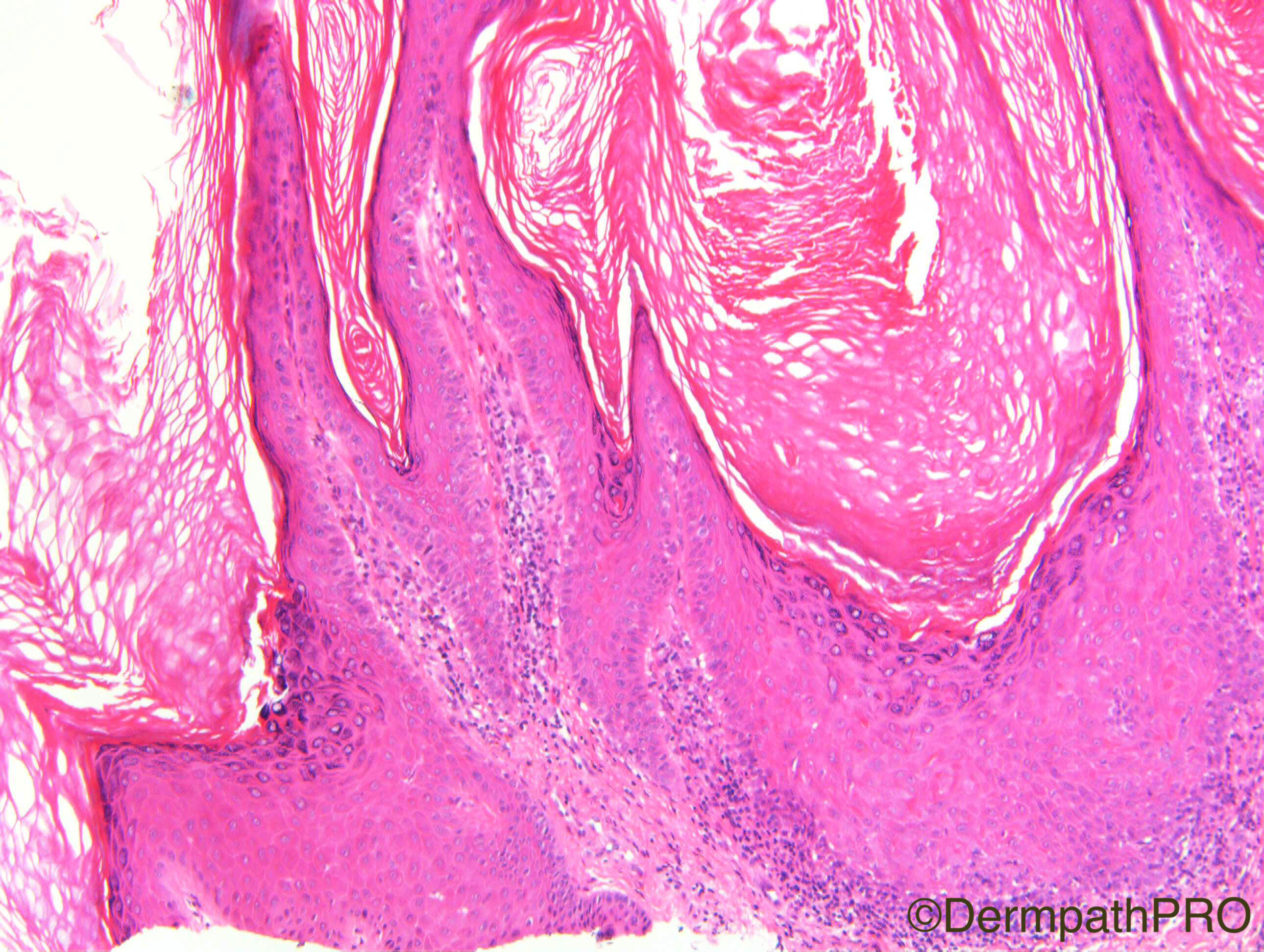
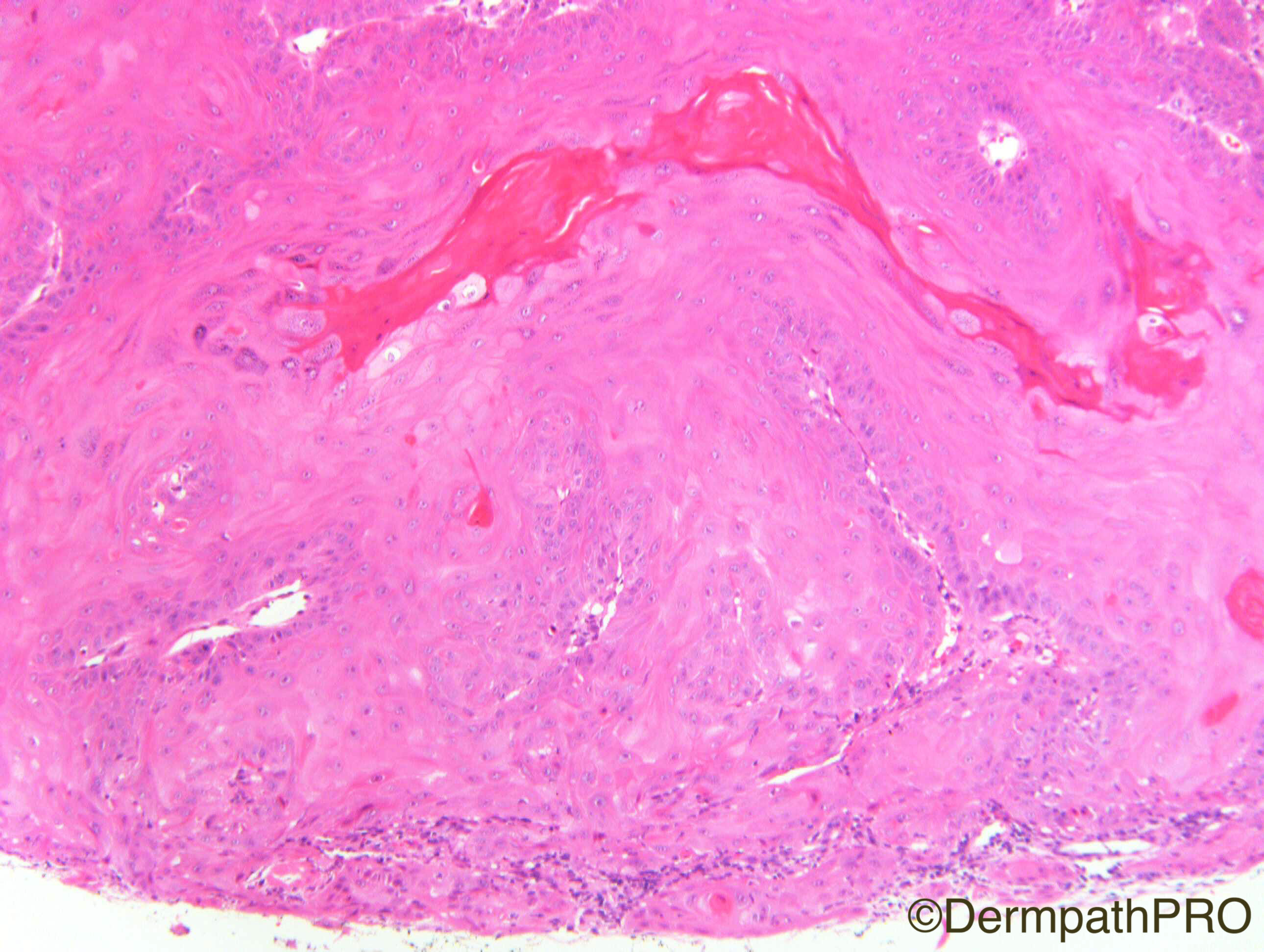
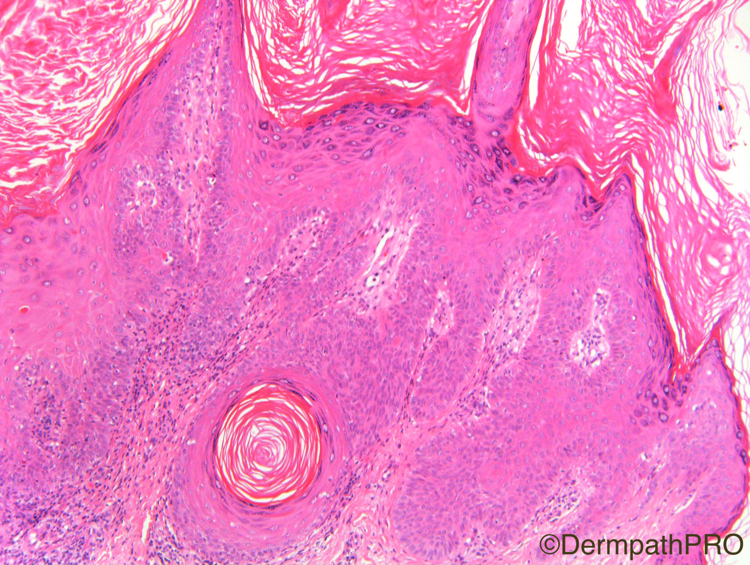
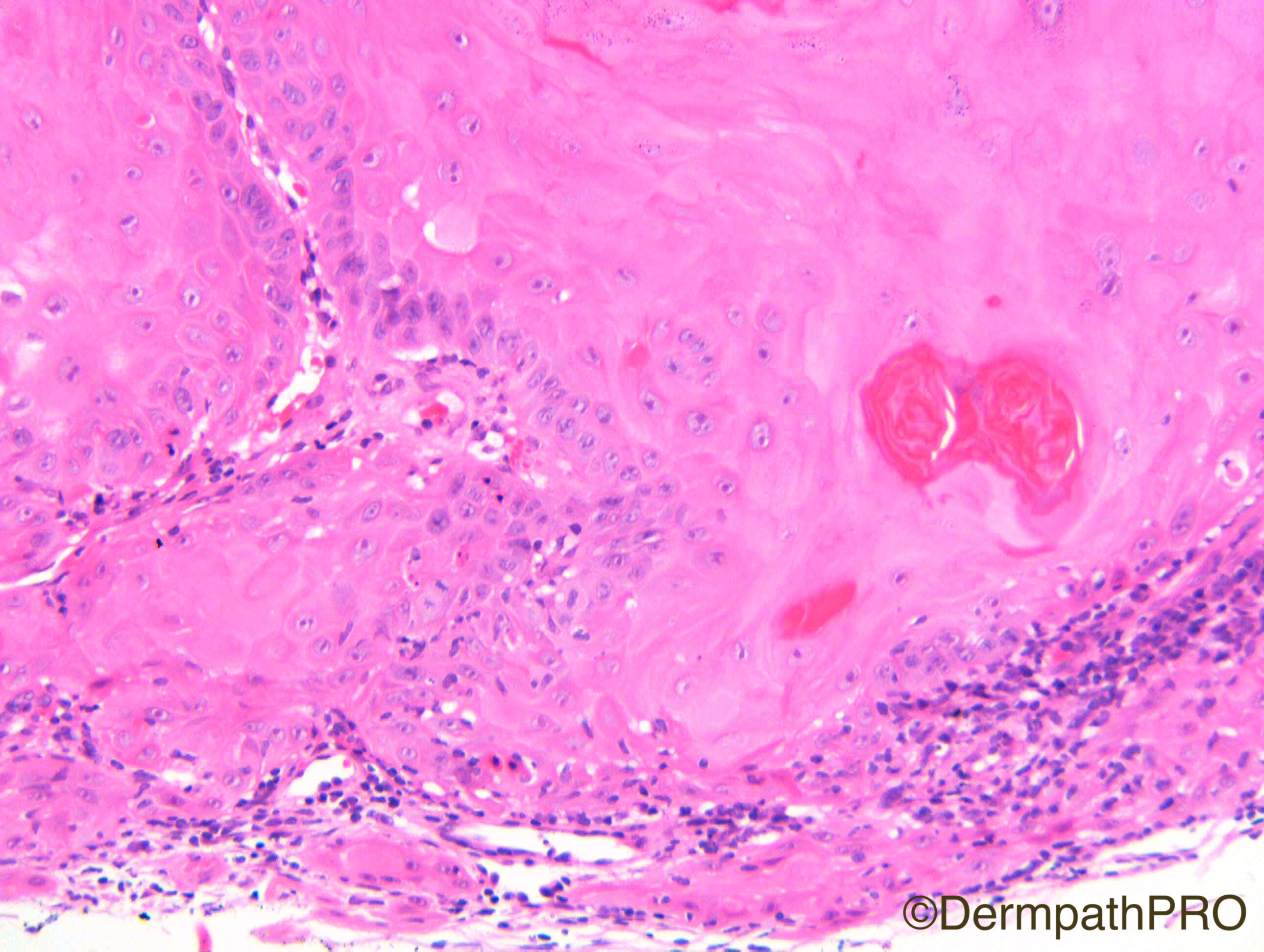
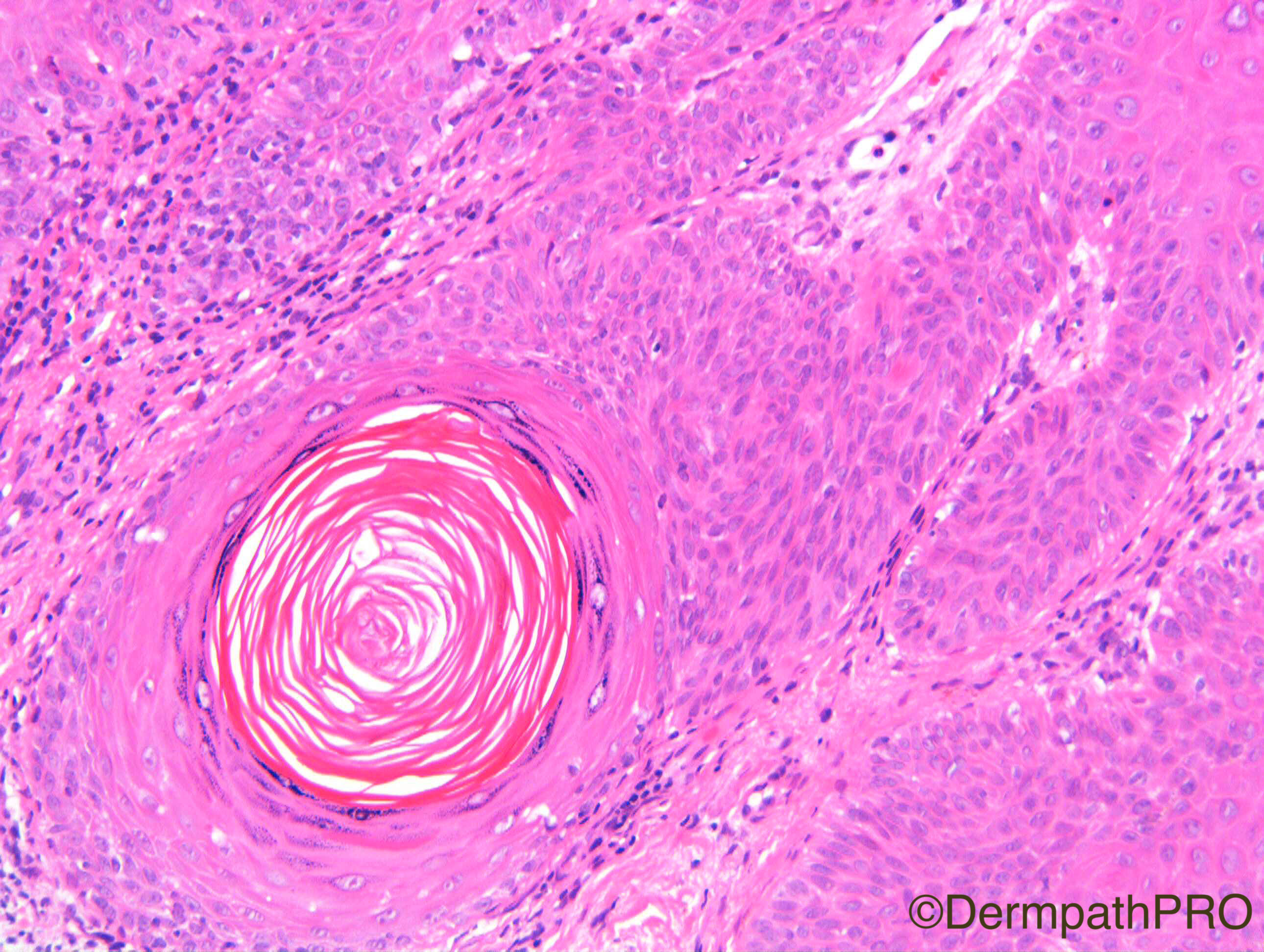
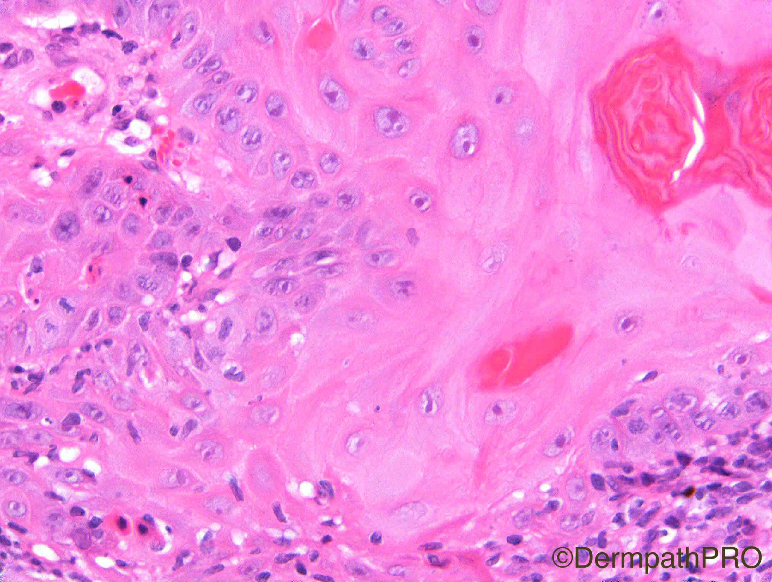
F80. Forearm lesion. Viral wart?

Case Posted by Dr Richard Carr
Edited by Admin_Dermpath


  Report Record

User Feedback


Admin_Dermpath

Posted

A special case from Dr Richard Carr to get your DermPath skills tested..

Share this comment


Link to comment
share_externally

Dr. Mona Abdel-Halim

Posted

We've seen this before Geoff !! 

Share this comment


Link to comment
share_externally

Admin_Dermpath

Posted

Sorry Mona, here is a different case which should be OK.


Cheers, Geoff

Share this comment


Link to comment
share_externally

Dr. Mona Abdel-Halim

Posted

KA

or wart with a keratoacanthomatous like growth pattern

Share this comment


Link to comment
share_externally

This is a difficult lesion. I can see some basaloid cells with infundibular cystic formation (figure 3) that is a part of a seborrheic keratosis for me, but in the center of this tumor there´s another growth that looks like a desmoplastic trichilemmoma. I would like to see other comments about this case.

Share this comment


Link to comment
share_externally

vincenzo polizzi

Posted

My first thought was Verrucous Carcinoma, but due the fact I wasn't thinking about KA...Now rule out KA is very difficult. However there is another option: a well differentiated, predominantly infundibular type of follicular scc, because of the SK-like feature well remarked by Igor...Obviously cpc is mandatory ( a lesion grown in 1 month for example would make me favor KA! ).

Share this comment


Link to comment
share_externally

Arash Daryakar

Posted

why not IFK?(again!)

Although, i think KA is also a good differential.Hard case.

Share this comment


Link to comment
share_externally

Raul Perret

Posted

Well I think also this is not an easy case and I expect a lot of variability even among good pathologists. From my point of view this problem is due to the overlapping histological features. We dont have all the criteria for considering this a verruca (lacks large keratohyalin granules, parakeratosis, koylocytes) or an actinic keratosis with mild dysplasia (lacks parakeratosis and solar elastosis), if you want to throw there also sebk its fine for me but I think the ''keratocyst'' is just due to incidence of cut and it is not a real basaloid proliferation for me. I incline more for actinic keratosis with mild dysplasia on these images. Considering the second lesion we see an squamous proliferation with trichilemmal differentiation/origin I think I could not discard for example an early trichilemmal horn. For me this lesion is benign and I would not like to call it a carcinoma

Share this comment


Link to comment
share_externally

Eman El-Nabarawy

Posted

My first thought was fSCC but really can't find atypical features enough for that diagnosis.I can see abrupt transformation of the basal cell layer to large pale eosinophilic glassy keratinocytes with characteristic keratinization of keratoacanthoma (fig 6&8). So I favor KA in a stage of regression.

Share this comment


Link to comment
share_externally

Nitin Khirwadkar

Posted

A tricky and difficult case indeed! Have read the comments above. The lesion does show some features of a verruca and seborrhoeic keratosis. However, what is striking is the bulbous mass of variably atypical squamous cells. There are dyskeratotic cells, but I couldn't see perforation of elastic fibres. Another, similar focus, probably early one is also noted. Would favour hypertrophic actinic keratosis. Couldn't convince myself of outer root sheath differentiation. Waiting for an answer.

Share this comment


Link to comment
share_externally

Dr. Richard Carr

Posted

I called this KA-like proliferation (rather than ordinary actinic KA) occurring within an inflamed seborrhoeic keratosis.  I agree this may be subjective and contentious however I have now seen this co-occurrence on many occasions and think it is probably greatly under-reported (I recall at least 3 cases in the last few months). If you take out the central KA-like proliferation you have, on balance, features diagnostic for a seborrhoeic keratosis (SEBK). If you take out the lateral SEBK you have all the typical features of a KA (elastic entrapment would be nice but not entirely necessary for the KA-like proliferation in SEBK diagnosis here).  I also report KA regularly in the co-existence of mildly dysplastic actinic keratosis (AK), this can be controversial and challenging, and I have to admit as we saw earlier this week AK and SEBK can co-exist or be morphologically challenging to distinguish frequently.  I often report AK and even bowenoid AK with SEBK-like architecture to acknowledge this problem. The reason I use KA-like proliferation (when I see this in the setting of SEBK) is that I don't think it is an SCC (symmetrical, abrupt transition from peripheral highly proliferative to highly differentiated pilar type - outer root sheath at the isthmus -, lack of acantholysis or follicular mucin) but a reactive proliferation perhaps that has taken one more step on the neoplastic journey. That said on one occasion many years ago I did report a subtle spindle cell SCC coming straight off the base of a SEBK and we already saw this week mentioned porocarcinomas arising with SEBK. I always advise discussion of KA in the MDM setting but in instances such as the one illustrated I think my advice would be watchful waiting as I have yet to see a case recur or undergo frank malignant transformation.

Perhaps I should briefly state that I personally regard KA as a distinct clinicopathological entity (I don't think I have ever stated in writing or public that I regard KA as a wholly benign lesion - in brief it's complicated and you have to pay attention to the small print!). In my concept KA may, all be it very rarely, metastasise (perhaps 4 cases in the world literature i.e. very much less than BCC or even dermatofibroma!), occasionally undergo malignant transformation (I think some authors who don't allow marked pleomorphism as a typical feature of early proliferative KA will greatly over-estimate malignant transformation) or be morphologically and clinicopathologically indistinguishable from follicular SCC (relatively often analogous to atypical Spitz group). I always recommend clinicopathological correlation and preferably MDM discussion for parital biopsies and use a risk stratification approach to the reports (including words such as "typical histological features of KA", "in keeping with KA", "favouring KA but possibility of SCC not excluded" etc. etc.  It is analogous to Spitz tumours and I prefer to say "don't know i.e. uncertain malignant potential" when I don't know rather than just favour SCC when I don't know (I appreciate in this setting clinicians will invariably opt for complete excision). In Spitz tumours I try to place it to favouring benign or malignant behaviour based on the particular prognostic parameters and the approach to KA is similar - in particular very few cases behaving badly but we must be very careful not to forget the highly aggressive crateriform (KA-like) follicular SCC (see our review in Diagnostic Histopathology).  We even dealt with the uncertainty relating to KA v's follicular SCC in some fashion in UK guidelines for pathologists reporting SCC.  It is my opinion that the previous lack of robust diagnostic criteria for both KA and follicular SCC (due mainly to complete lack of appreciation of the latter diagnosis) led to much of the confusion we now have when some pathologists report KA very rarely if ever (to be fair the past greats left us in a mess with practically no or contradictory diagnostic criteria) and others reporting it in up to 30% of KA/SCC cases (e.g. Dr Weedon). As you can see from the above I am in the Weedon camp reporting KA very frequently (even in small biopsies) at this point in time but would like to keep an open mind for the future especially if we can start to do some really good clinical research (in a prospective study of clinical cases) and use molecular techniques to aid risk stratification as we do in Spitz tumours. I often say it is amazing a human went to the moon in the 1960's but there was practically no decent research in to a common skin disease that we still cannot pigeon hole in to cancer or not over 50 years later!  Please accept my apologies and I will get down from the pedestal now! 

Share this comment


Link to comment
share_externally


Join the conversation

You can post now and register later. If you have an account, sign in now to post with your account.

Guest
Add a comment...

×   Pasted as rich text.   Paste as plain text instead

  Only 75 emoji are allowed.

×   Your link has been automatically embedded.   Display as a link instead

×   Your previous content has been restored.   Clear editor

×   You cannot paste images directly. Upload or insert images from URL.

Loading...

×
×
  • Create New...