Jump to content
In this section we have spot diagnoses posted on a daily basis since June 2010, now over 4000! You can review the archived cases and read the suggested diagnoses by users and the final comment by the contributors.
Case are uploaded each week day by 10 am UK time with the correct diagnosis will generally be posted at 8 pm UK time. Why not view the most recent spot diagnosis and proffer a diagnosis?

Case Number : Case 1655 - 28 October Posted By: Guest

Please read the clinical history and view the images by clicking on them before you proffer your diagnosis.
Submitted Date :
   (0 reviews)

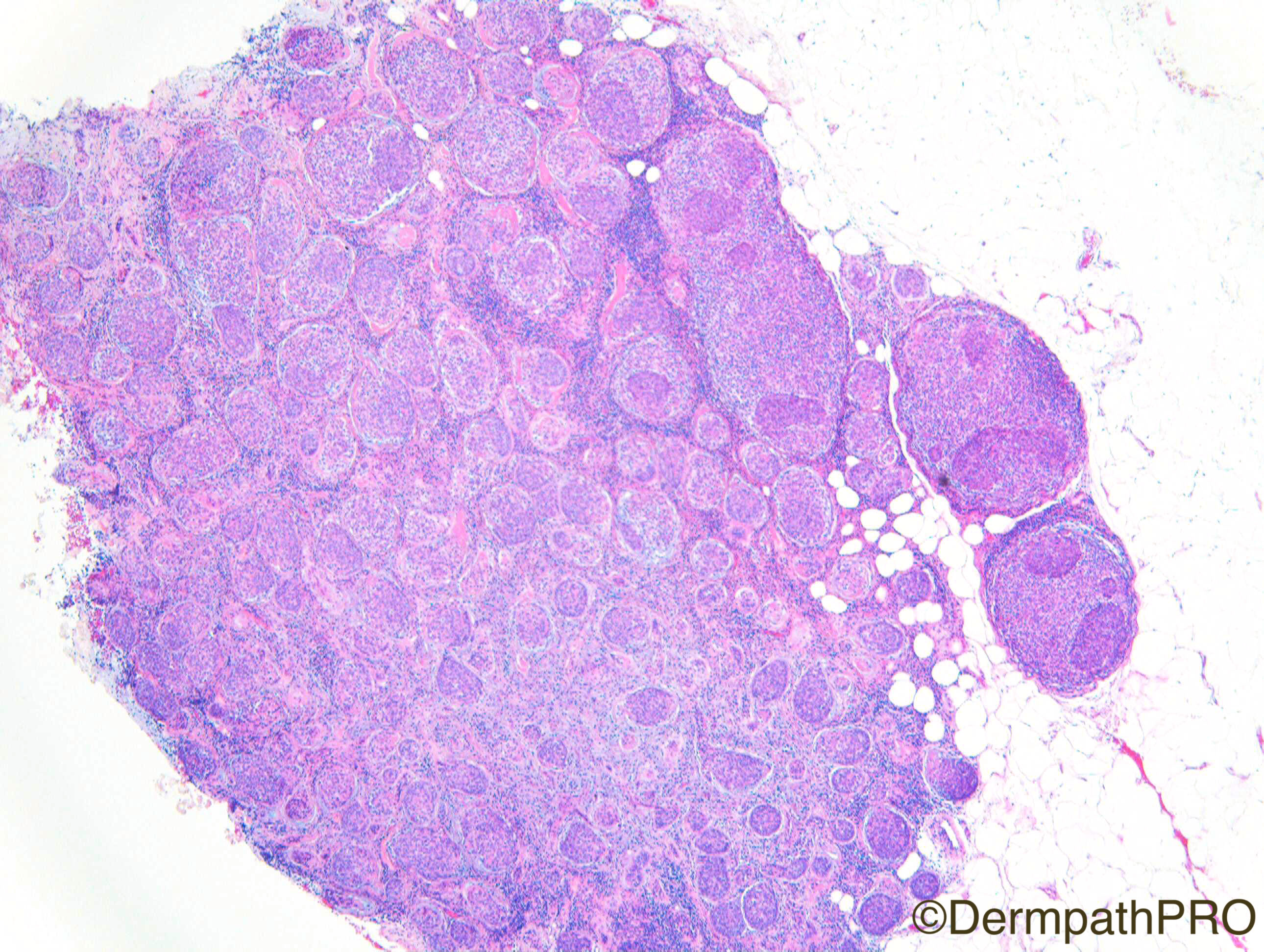
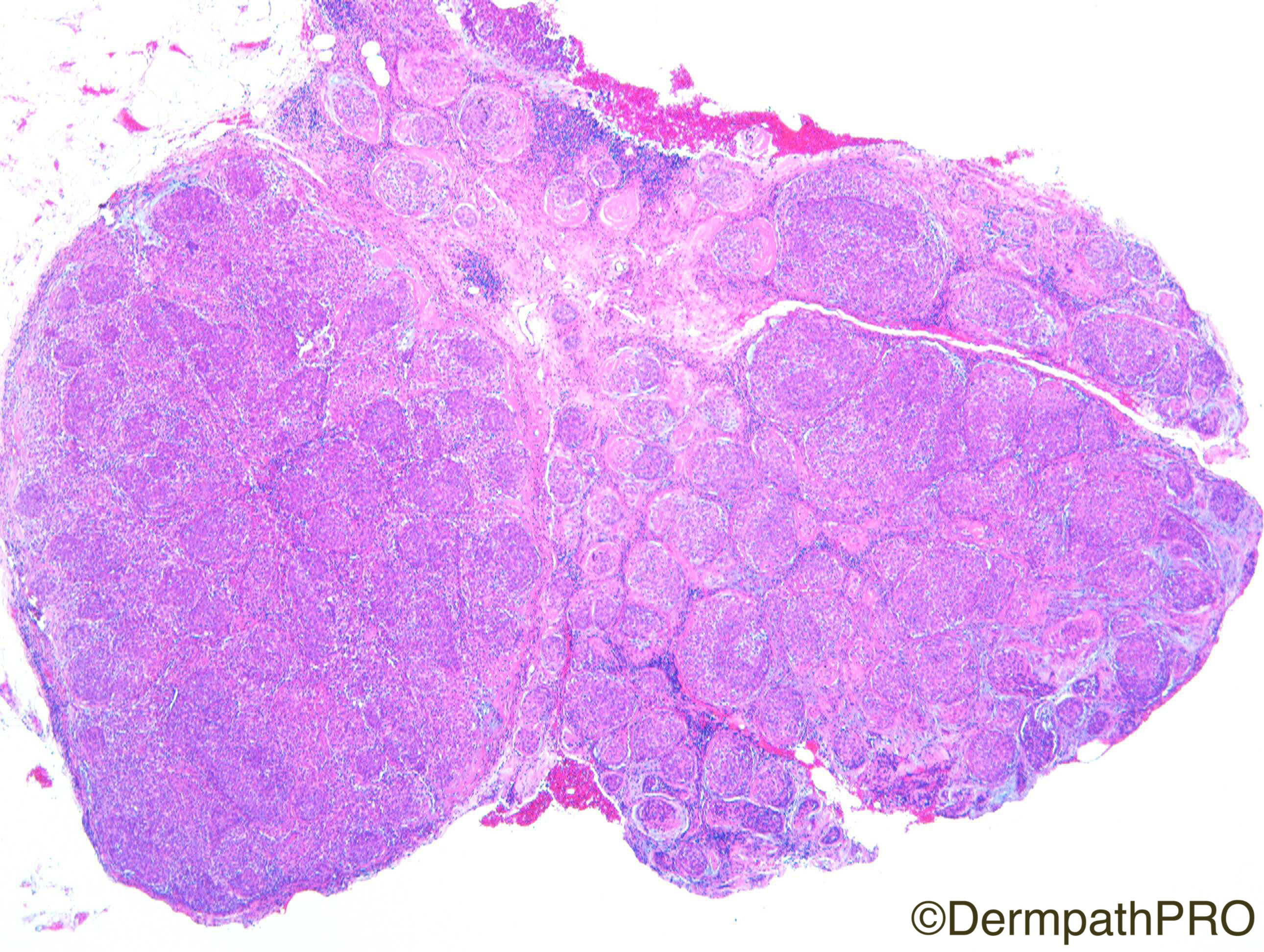
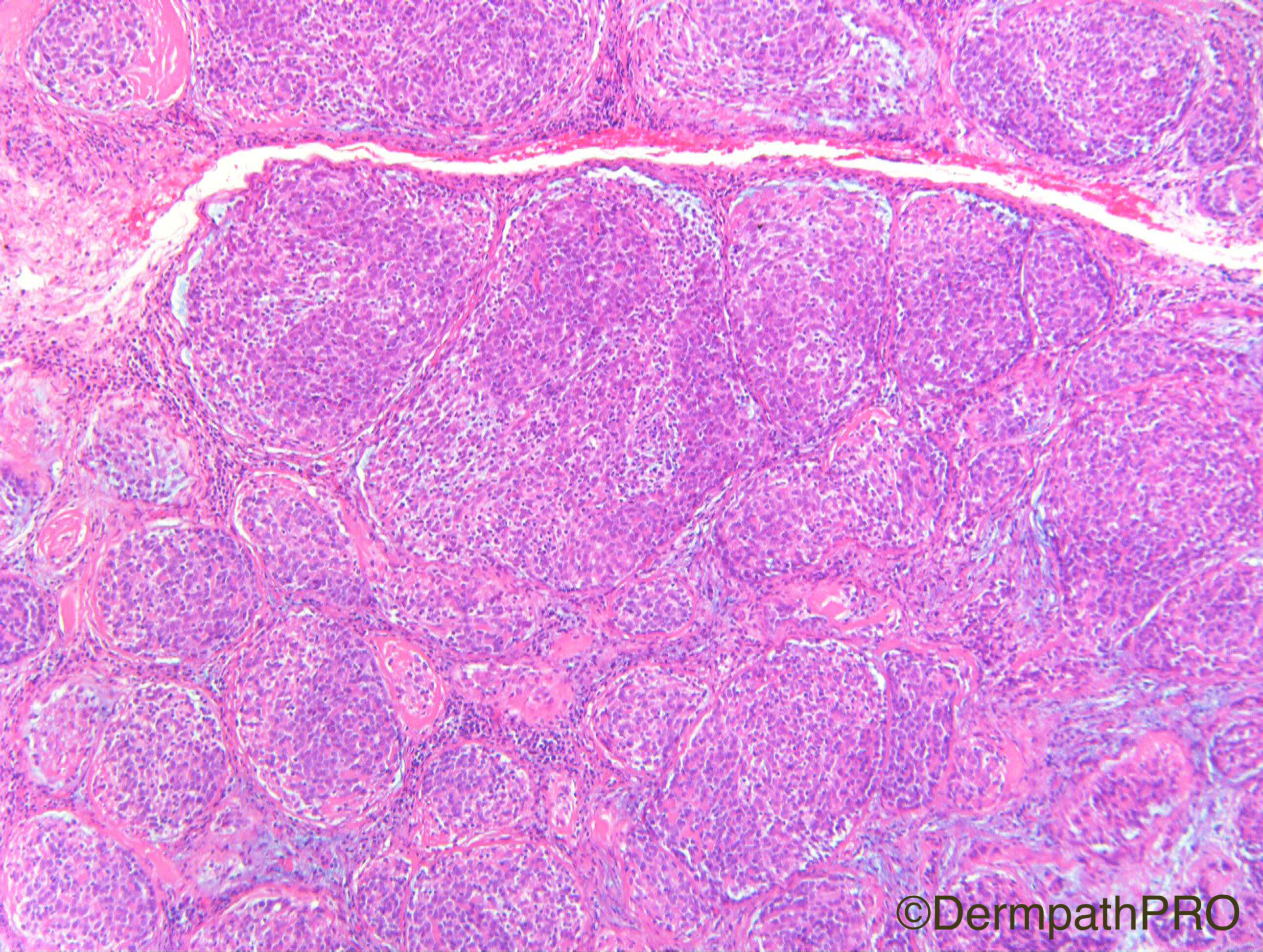
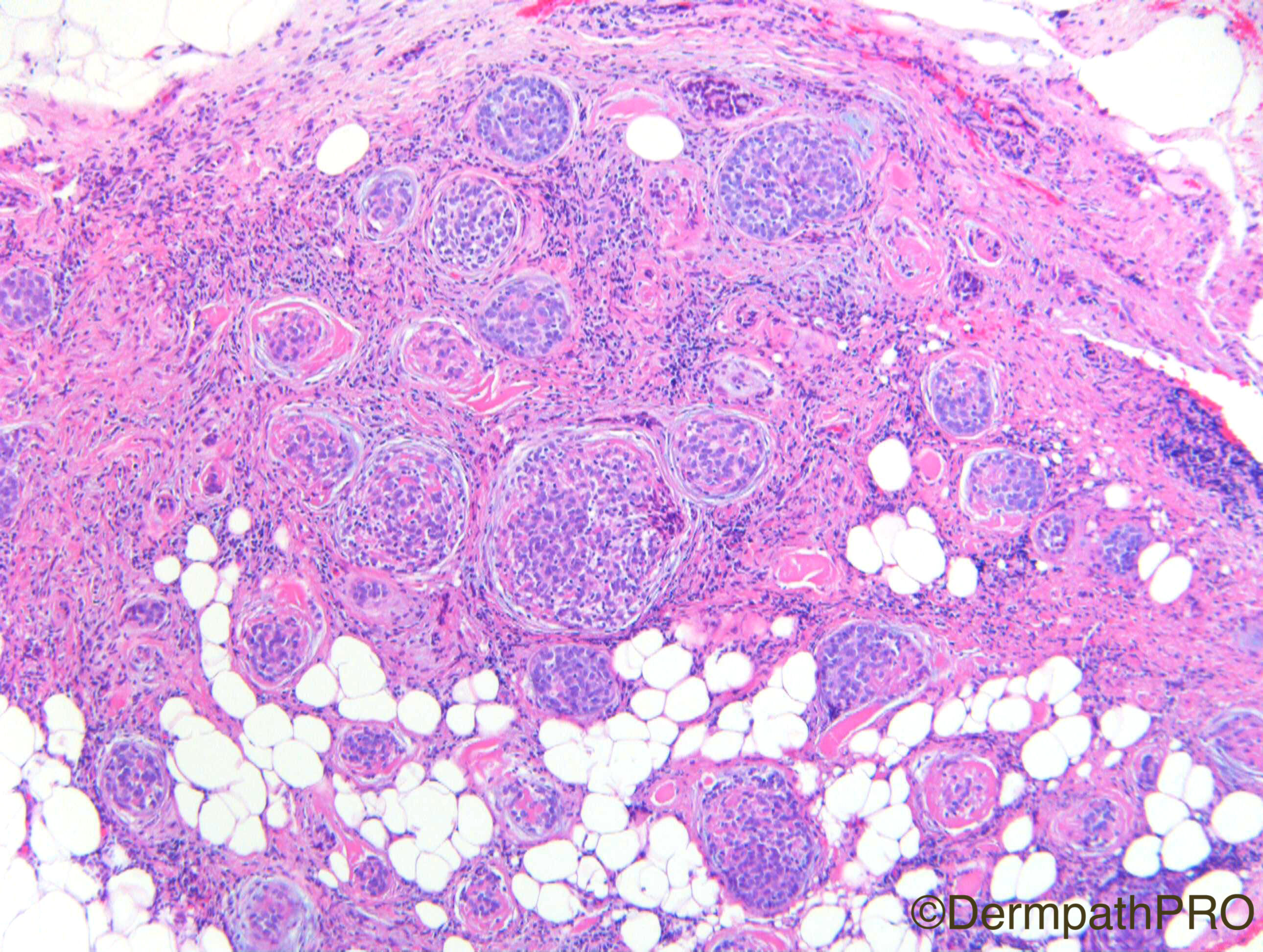
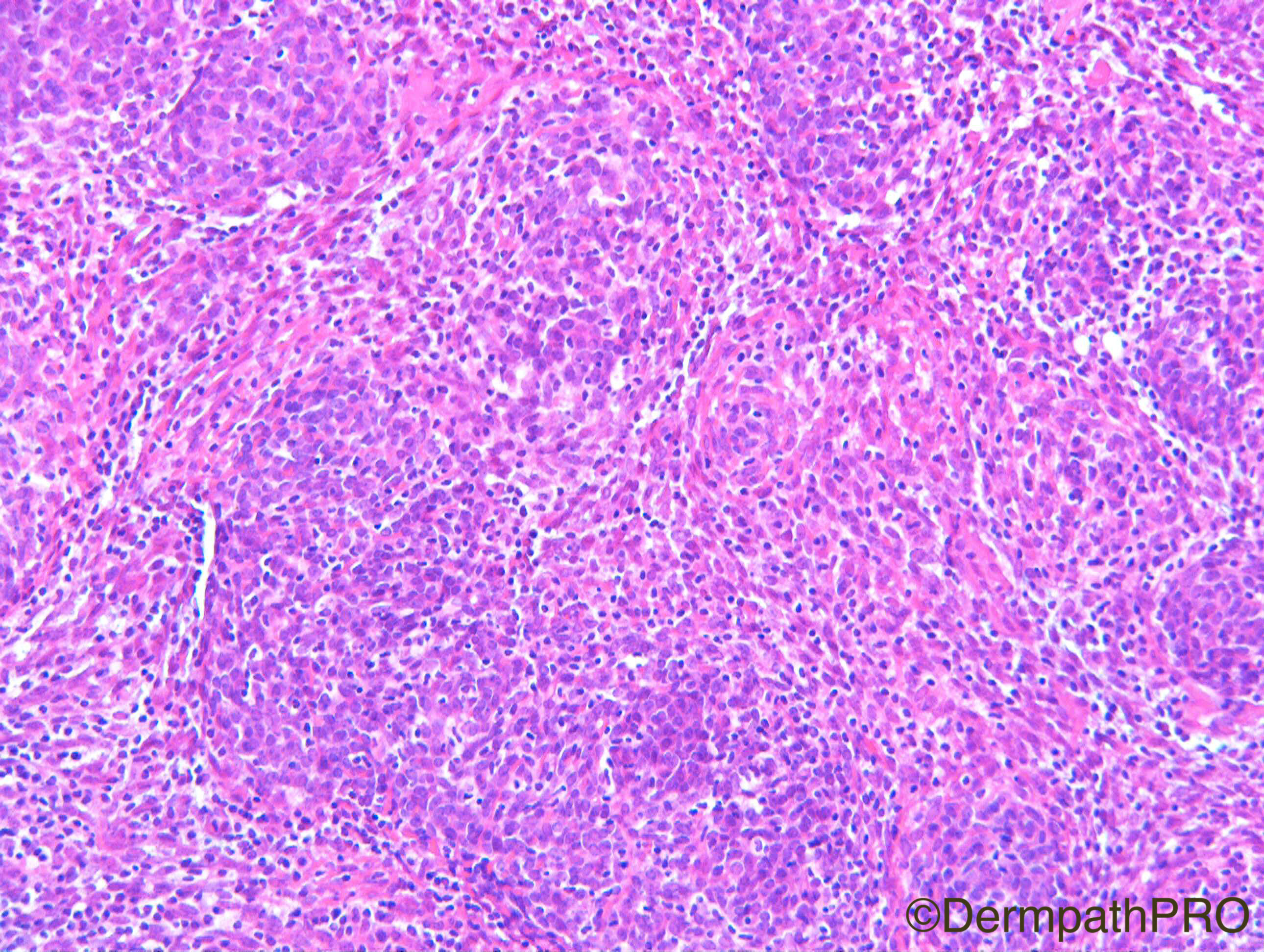
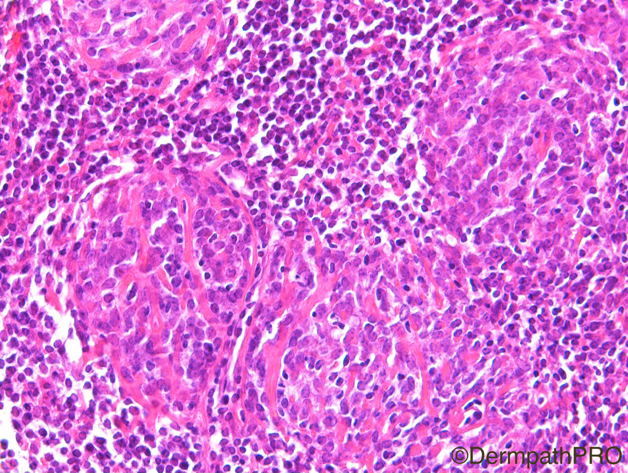
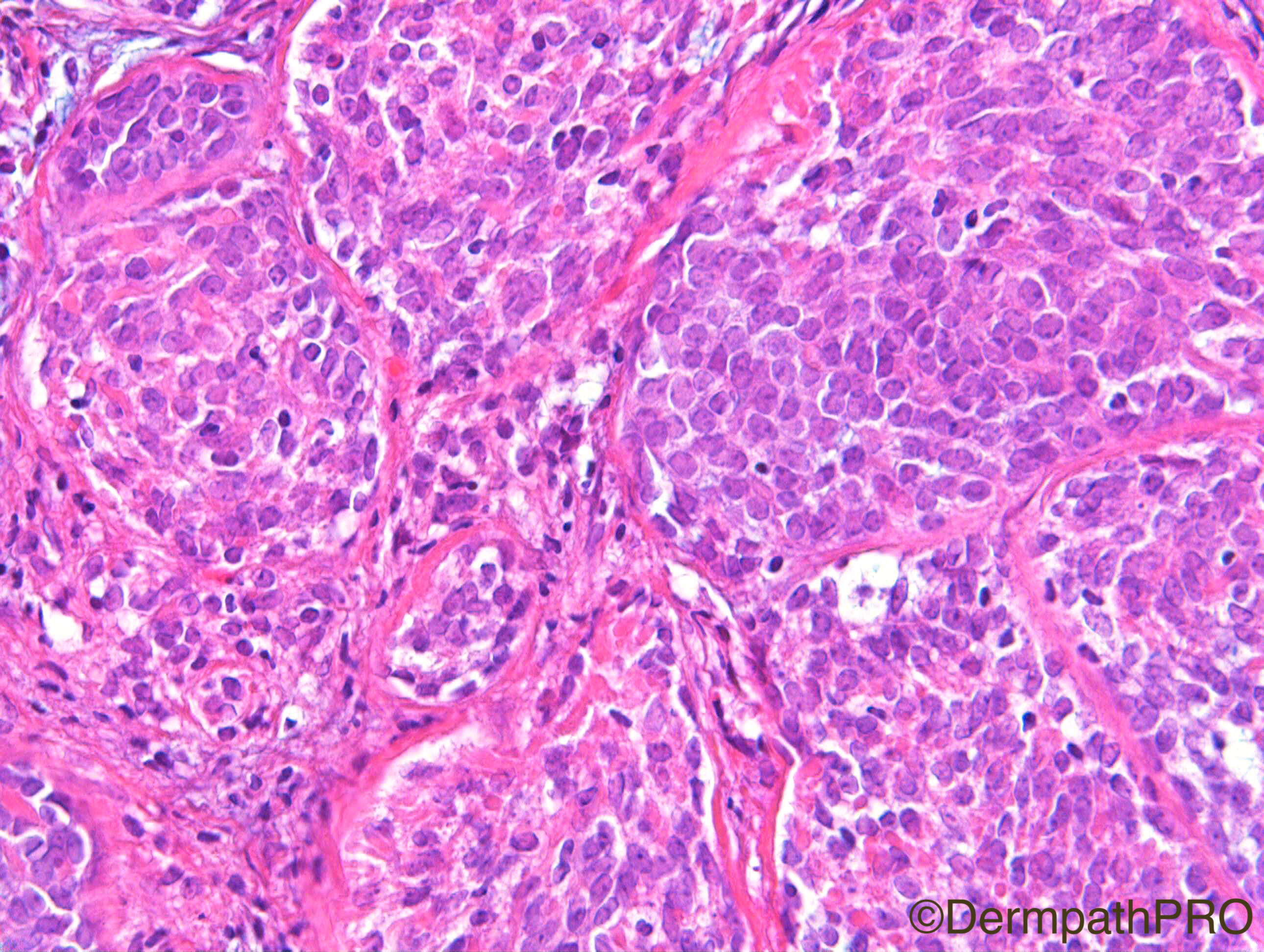
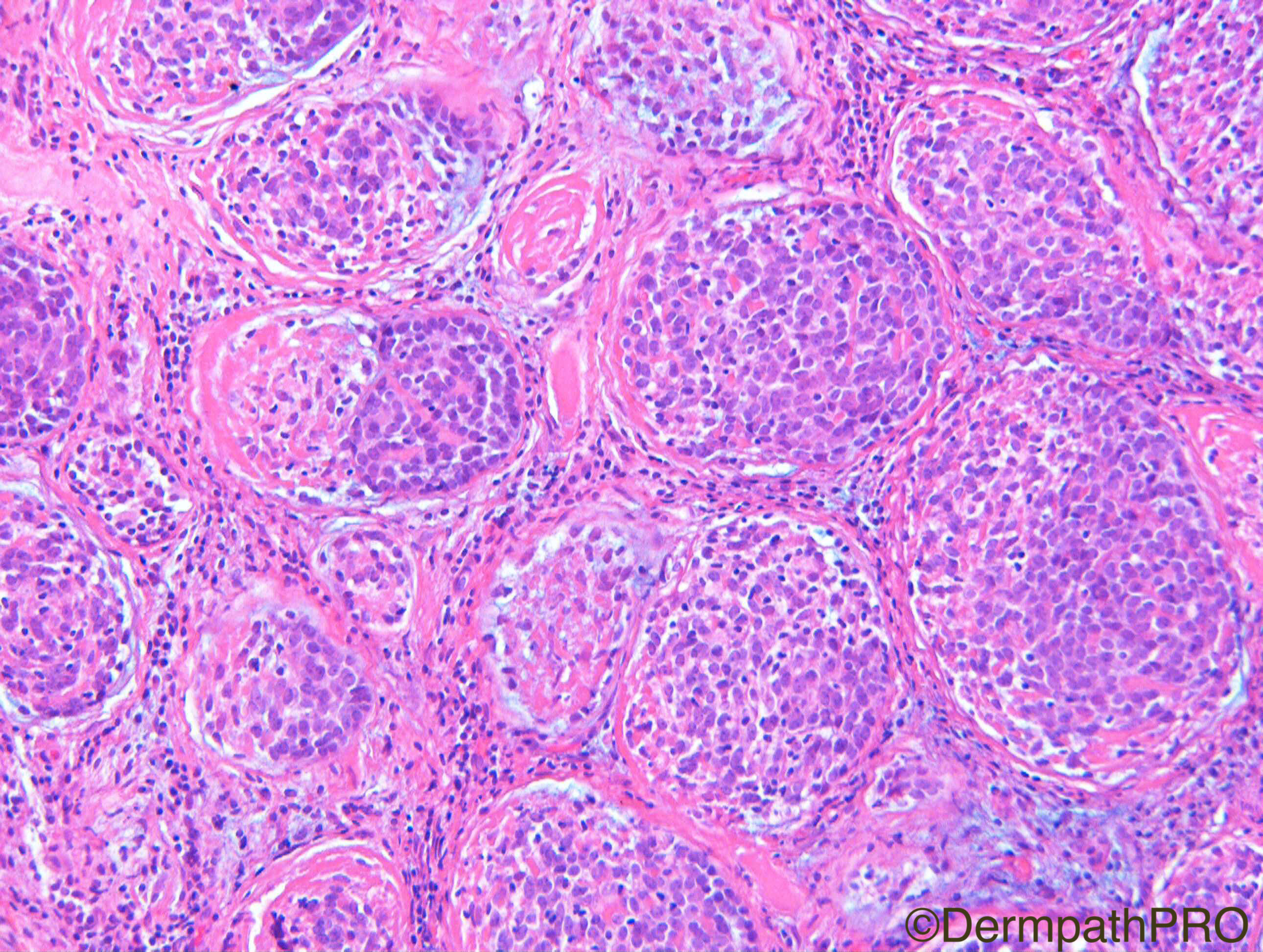
F30. Cystic lesion right scapula. c/o Dr Rand Hawari

Case Posted by Dr Richard A Carr

  Report Record

User Feedback


Admin_Dermpath

Posted

To close off the week we have a fine case from Dr Richard A Carr.

Share this comment


Link to comment
share_externally

vincenzo polizzi

Posted

A very unusual lymphadenoma

both for site and because looks much like trichoblastoma. 

Share this comment


Link to comment
share_externally

Nitin Khirwadkar

Posted

What about a lymphocyte rich spiradenoma? 

Share this comment


Link to comment
share_externally

Dr. Mona Abdel-Halim

Posted

Difficult case!! Some images r really like lymphocyte rich spiradenoma. Others made me think of lymphadenoma. May be it is an adnexal lesion with mixed features. Any possibility of Brooke Spiegler?? 

Share this comment


Link to comment
share_externally

Raul Perret

Posted

Sorry i disagree with my own comment. This is clearly not a plex fh tumor, should focus a bit more on the cases, 

Share this comment


Link to comment
share_externally

Raul Perret

Posted

I agree with the comments of the colleagues looks like a weird lymphadenoma and should discard BS.

Share this comment


Link to comment
share_externally

Robledo F. Rocha

Posted

I favor spiradenocylindroma. Tumor is located entirely in the subcutaneous adipose tissue and shows a multinodular growth pattern with hyaline material as droplets or narrow bands within epithelial aggregates. Prominence of lymphocytes in the stroma gives this tumor a thymoma resemblance.

Share this comment


Link to comment
share_externally

Arti Bakshi

Posted

Unusual case!....Agree with Nitin and Robledo. The nests of cells do not show peripheral palisading or central clear cells., typical of lymphadenoma. Hyaline material is also not a feature of trichoblastoma group of lesions.

So, would favour spiradenoma/cylindroma with prominent lymphocytes.

Share this comment


Link to comment
share_externally

Dr. Richard Carr

Posted

Nice discussion.  For the reasons given by Robledo (also very welcome back) and Arti this is a spiradenoma (paucilumnal again).  BerEP4 can highlight the ductal phenotype cells as small clusters in spiradenoma whereas in lymphadenoma there is usually a peripheral pattern of the staining in the basaloid cells. The basement membrane globules were the crucial clue in this instance to a spiradenoma (or spiradenocylindroma if you prefer).

Enjoy your weekends.

Share this comment


Link to comment
share_externally


Join the conversation

You can post now and register later. If you have an account, sign in now to post with your account.

Guest
Add a comment...

×   Pasted as rich text.   Paste as plain text instead

  Only 75 emoji are allowed.

×   Your link has been automatically embedded.   Display as a link instead

×   Your previous content has been restored.   Clear editor

×   You cannot paste images directly. Upload or insert images from URL.

Loading...

×
×
  • Create New...