Jump to content
In this section we have spot diagnoses posted on a daily basis since June 2010, now over 4000! You can review the archived cases and read the suggested diagnoses by users and the final comment by the contributors.
Case are uploaded each week day by 10 am UK time with the correct diagnosis will generally be posted at 8 pm UK time. Why not view the most recent spot diagnosis and proffer a diagnosis?

Case Number : Case 1656 - 31 October Posted By: Guest

Please read the clinical history and view the images by clicking on them before you proffer your diagnosis.
Submitted Date :
   (0 reviews)

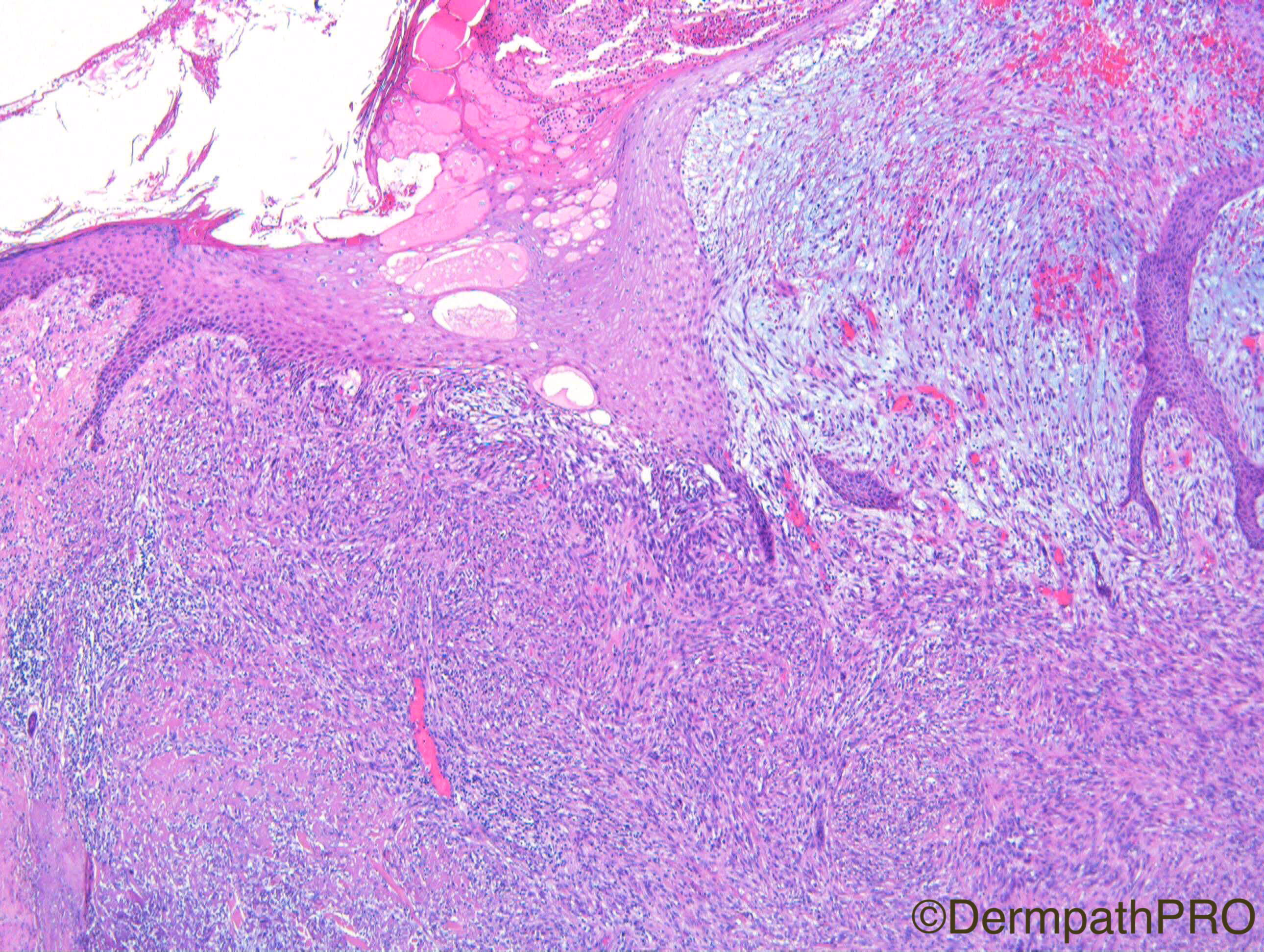
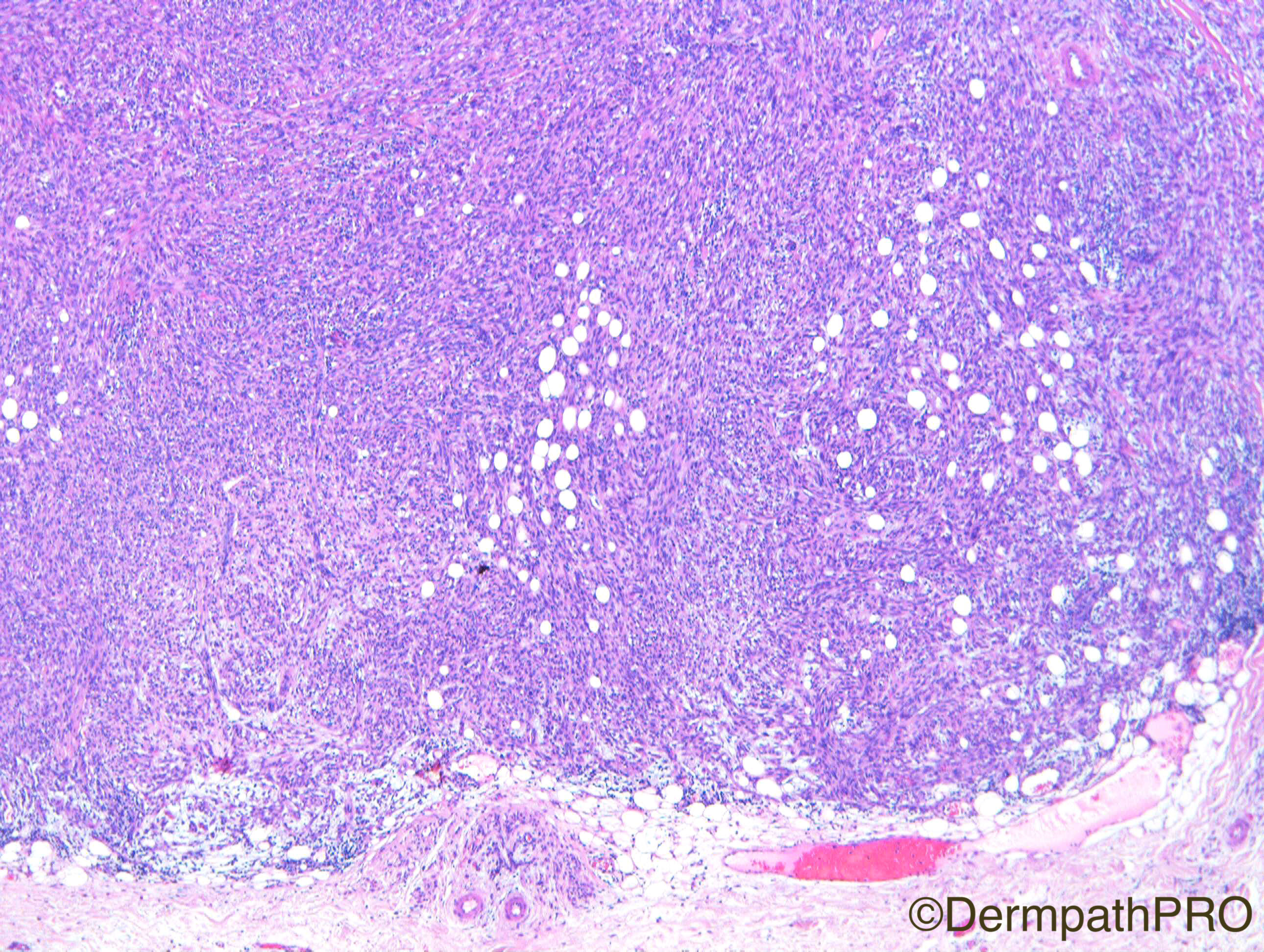
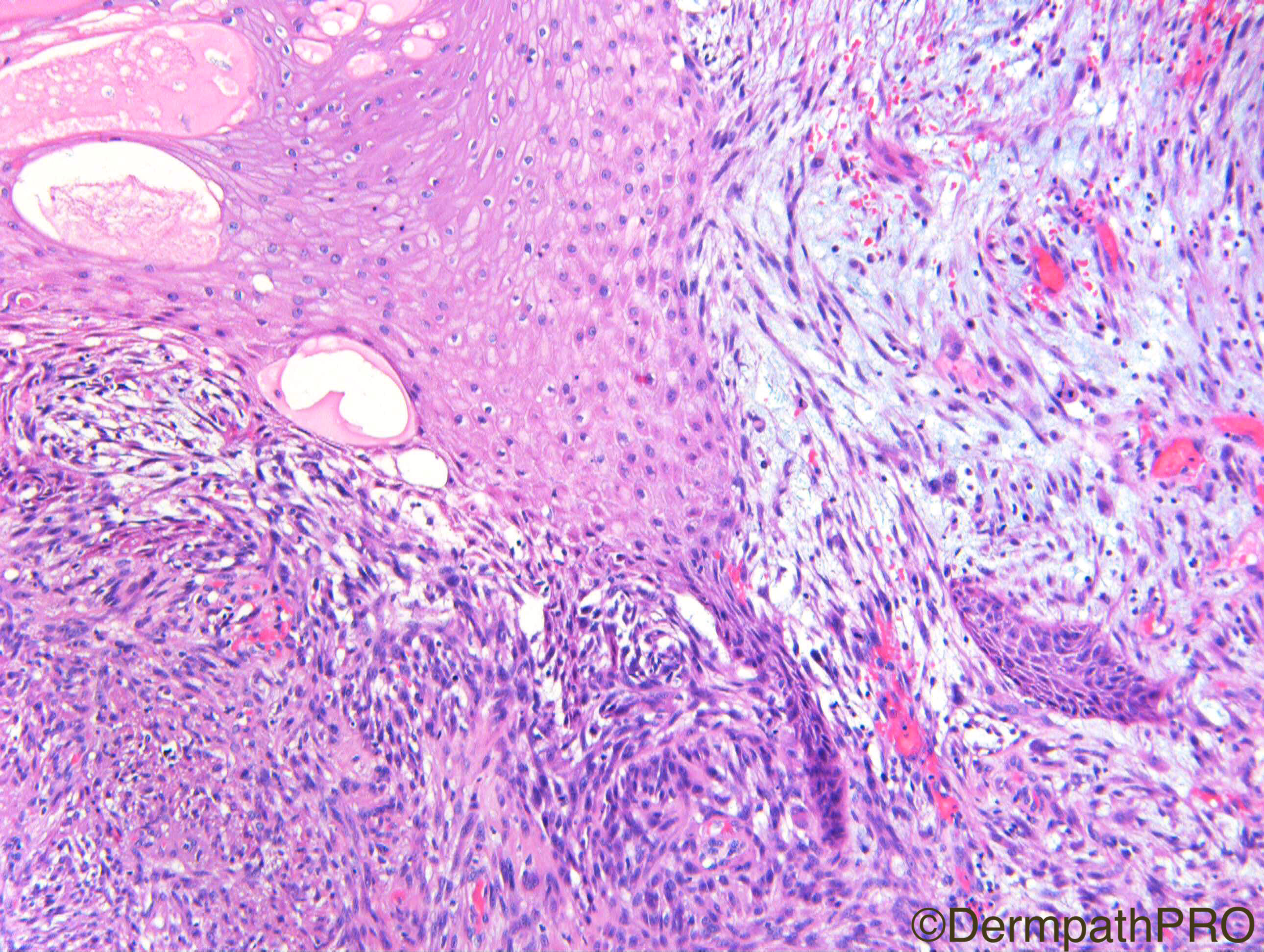
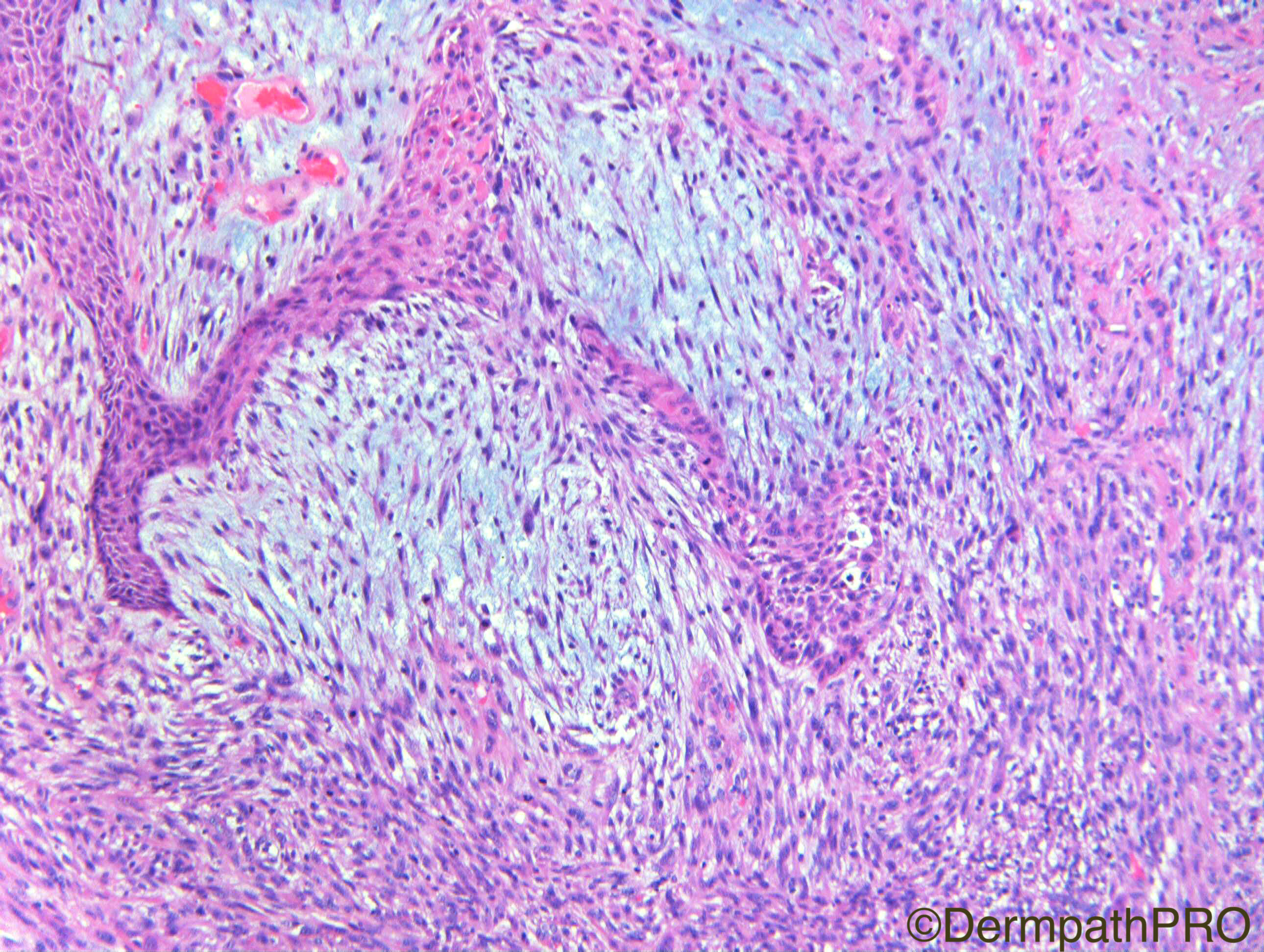
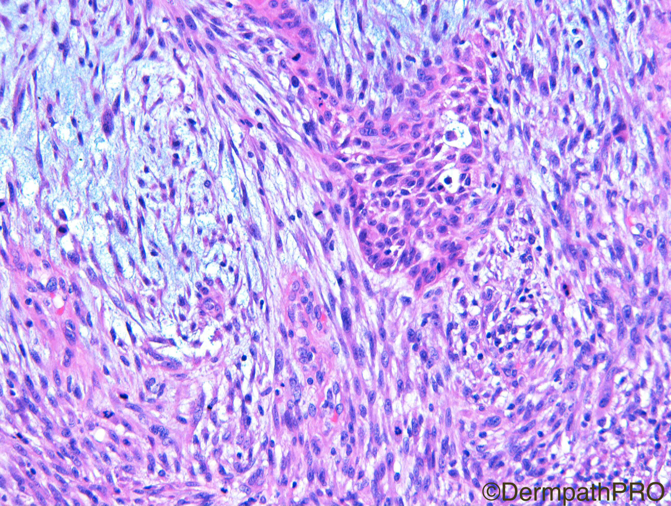
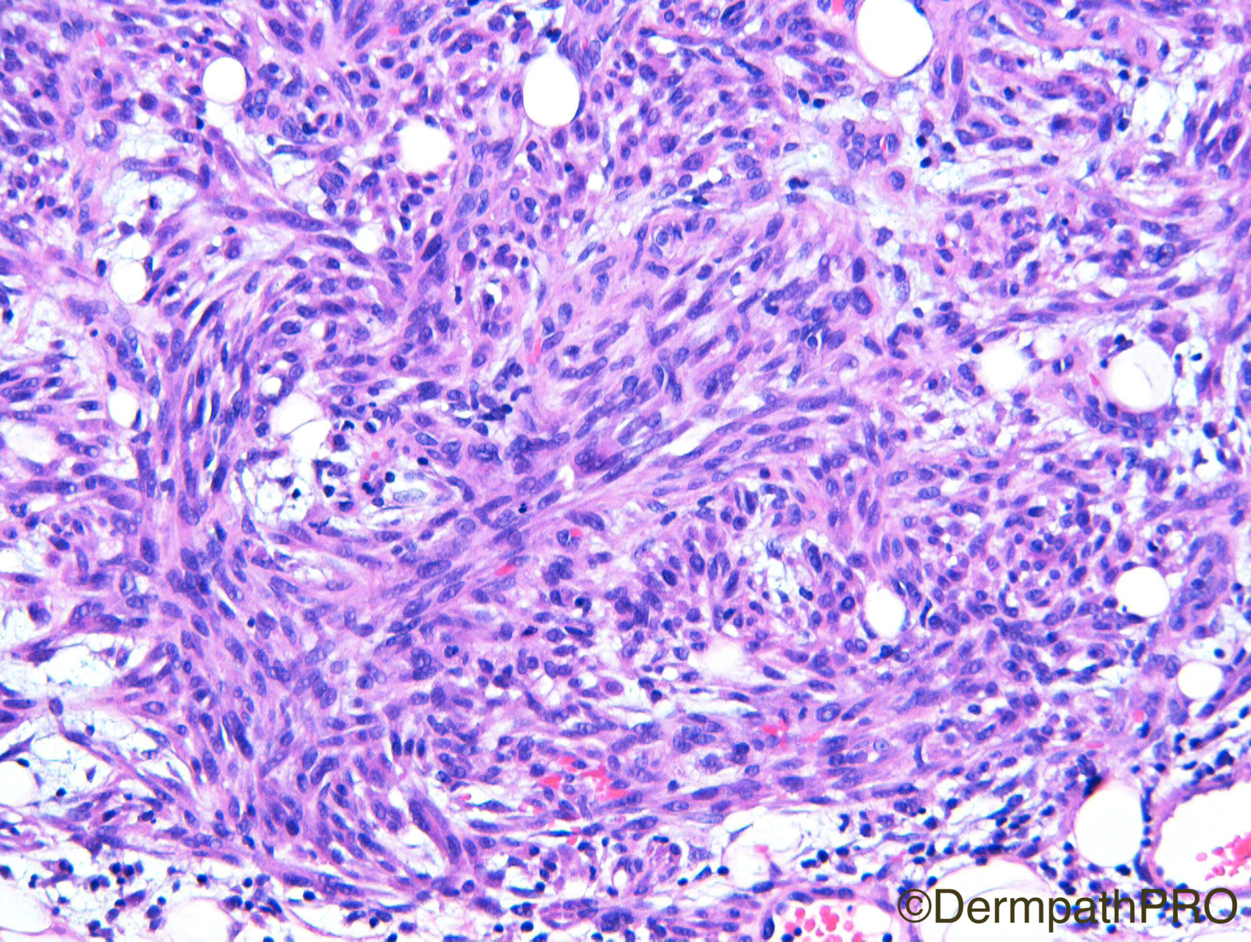
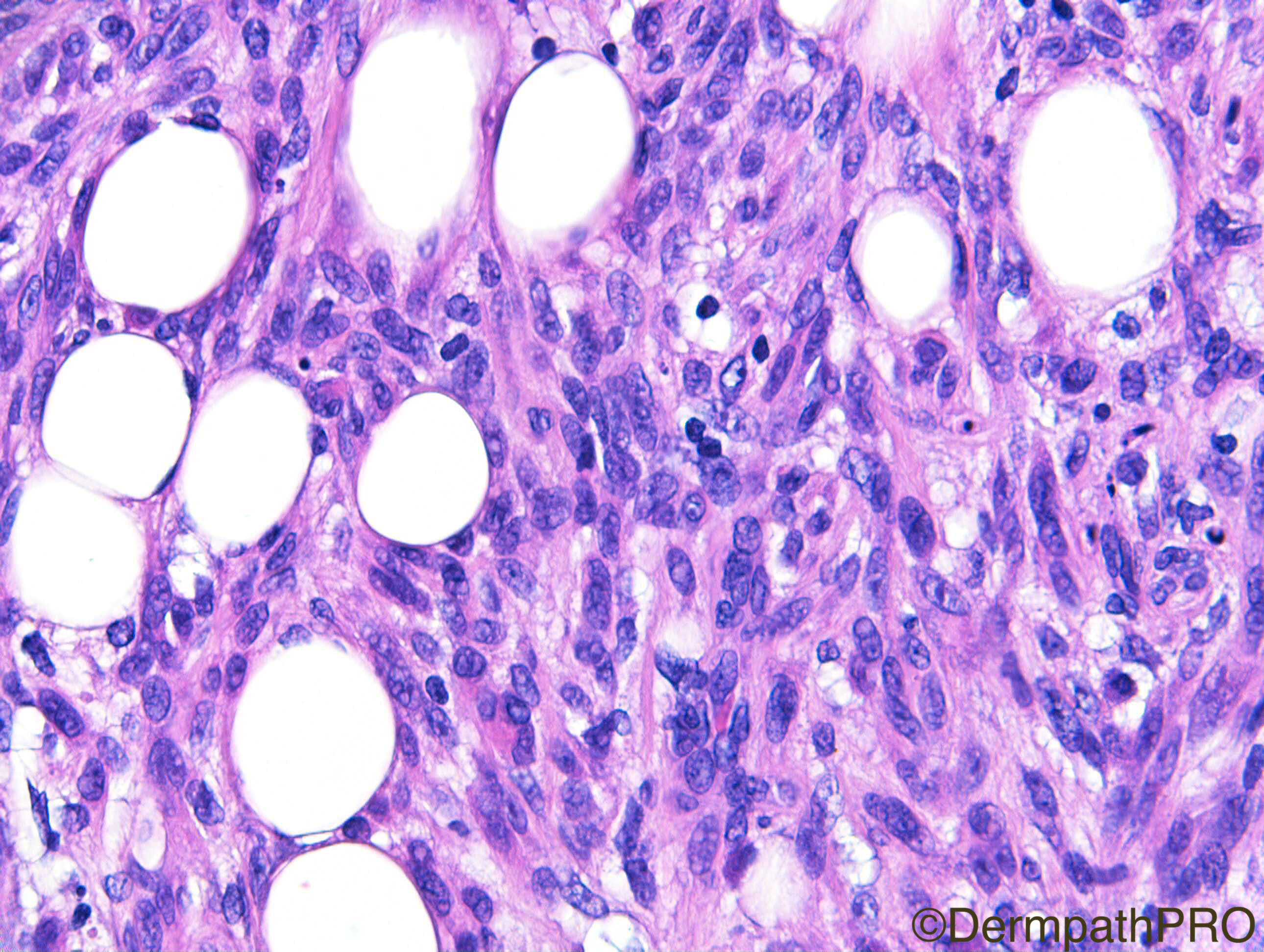
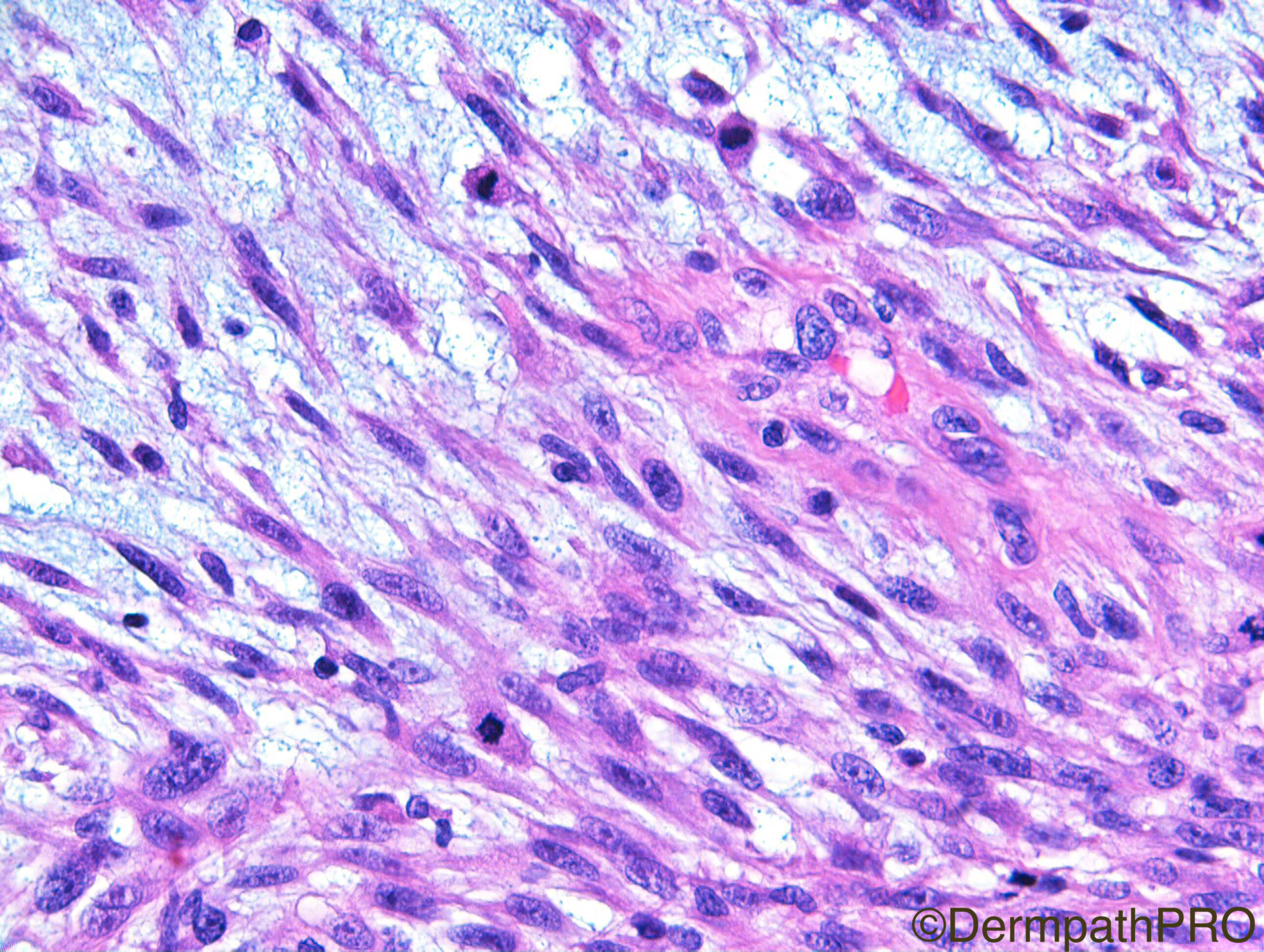
M75. Crusty bleeding nodules on scalp. Previous h/o SCC

Case Posted by Dr Richard Carr
Edited by Admin_Dermpath


  Report Record

User Feedback


Admin_Dermpath

Posted

Here is Richard's Hallowe'en treat for you all, just a little early as I am away in Paris until Monday evening. My lovely daughter Martha will be adding a number of images at 6pm on Monday afternoon.


Cheers, Geoff

Share this comment


Link to comment
share_externally

Dr. Mona Abdel-Halim

Posted

Sarcomatoid SCC is my first impression for IHC. 


Share this comment


Link to comment
share_externally

Raul Perret

Posted

agree sarcomatoid/spindle cell squamous cell carcinoma

Share this comment


Link to comment
share_externally

Robledo F. Rocha

Posted

Sarcomatoid squamous cell carcinoma is also my first thought. Neoplastic spindle cells seem to be released from the epidermis.

Share this comment


Link to comment
share_externally

vincenzo polizzi

Posted

Agree with spindle cell SCC, above all in fig 4/5. Fig 6 make me thing to a melanoma...but my spot diagnosis without IHC is SCC.

Share this comment


Link to comment
share_externally

Arash Daryakar

Posted

I think of spindle cell SCC,too. The neoplasm seems to attach to overlying epidermis.

Although Spindle melanoma is also in differential.

IHC study at least for cytokeratin,S100 ,melanA  are indicated.

Share this comment


Link to comment
share_externally

Eman El-Nabarawy

Posted

Spindle cell SCC for verification by immunos cytokeratin, S100, MelanA, desmin. 

Share this comment


Link to comment
share_externally

Arti Bakshi

Posted

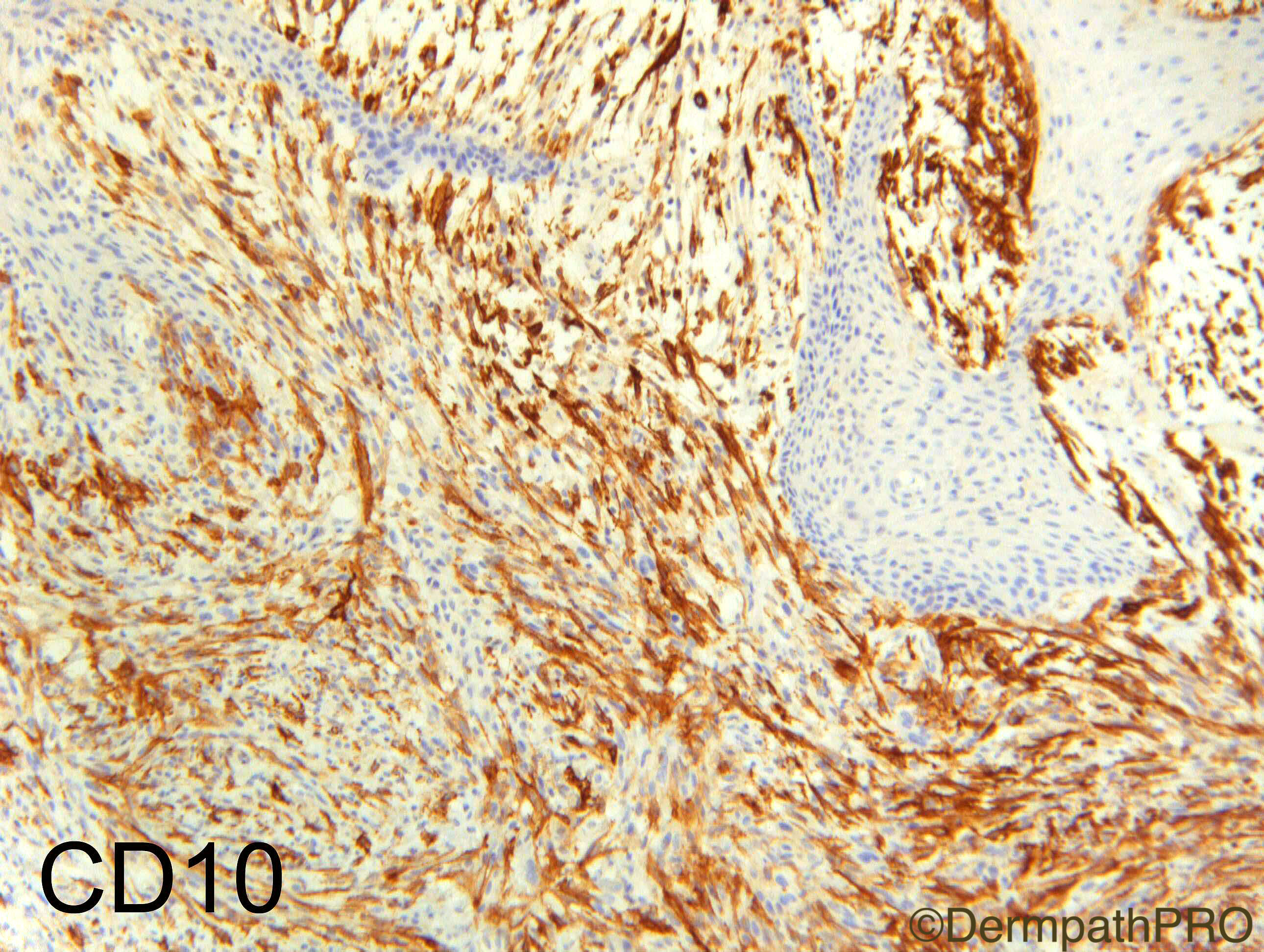
Yes, sarcomatoid SCC. Also shows the non specificity of CD10!

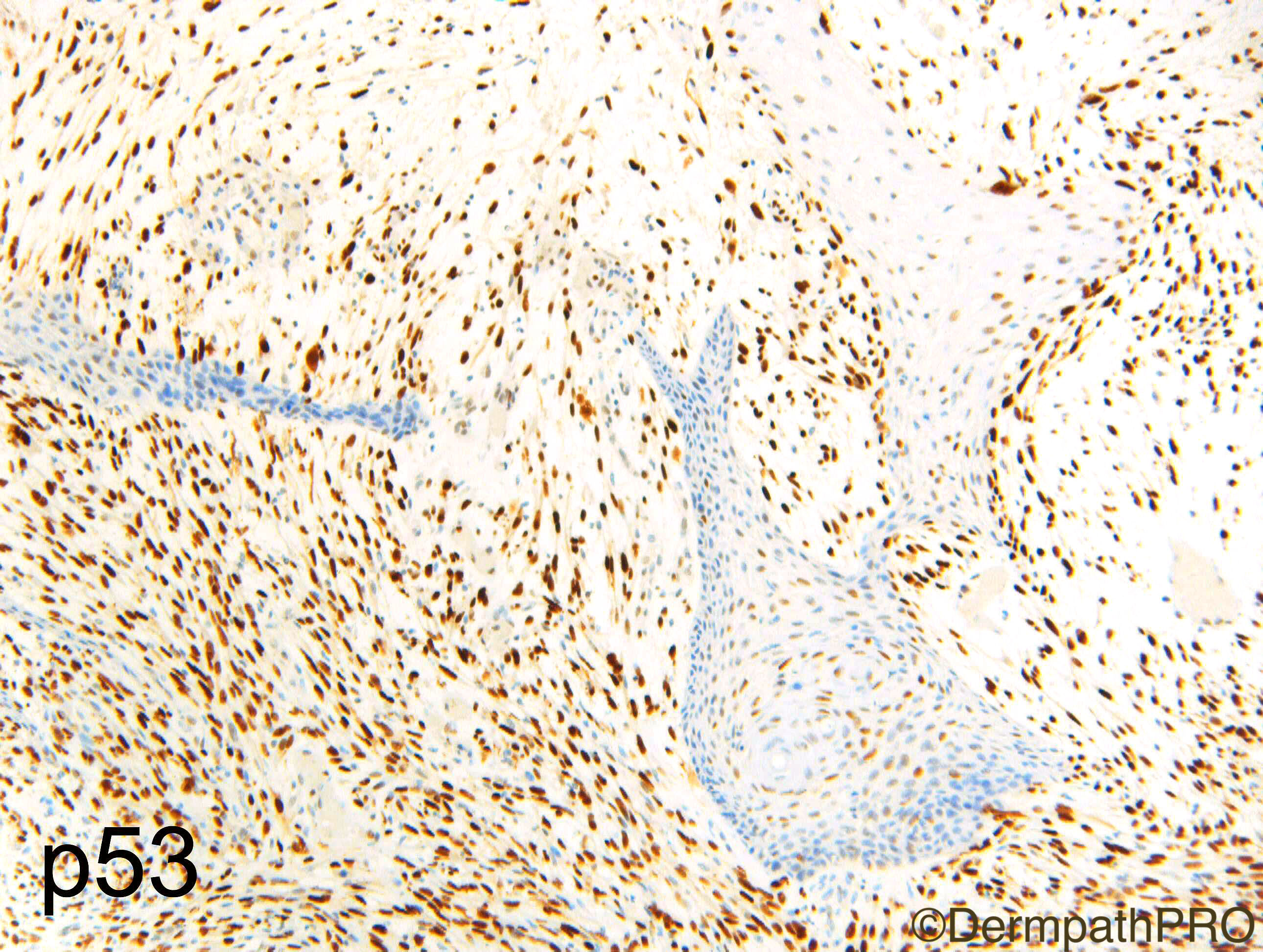
 Richard, how does a p53 help in this setting?

Share this comment


Link to comment
share_externally

Dr. Richard Carr

Posted

Yes well done all - very good!  This is sarcomatoid carcinoma (presumed a variant of cutaneous SCC).

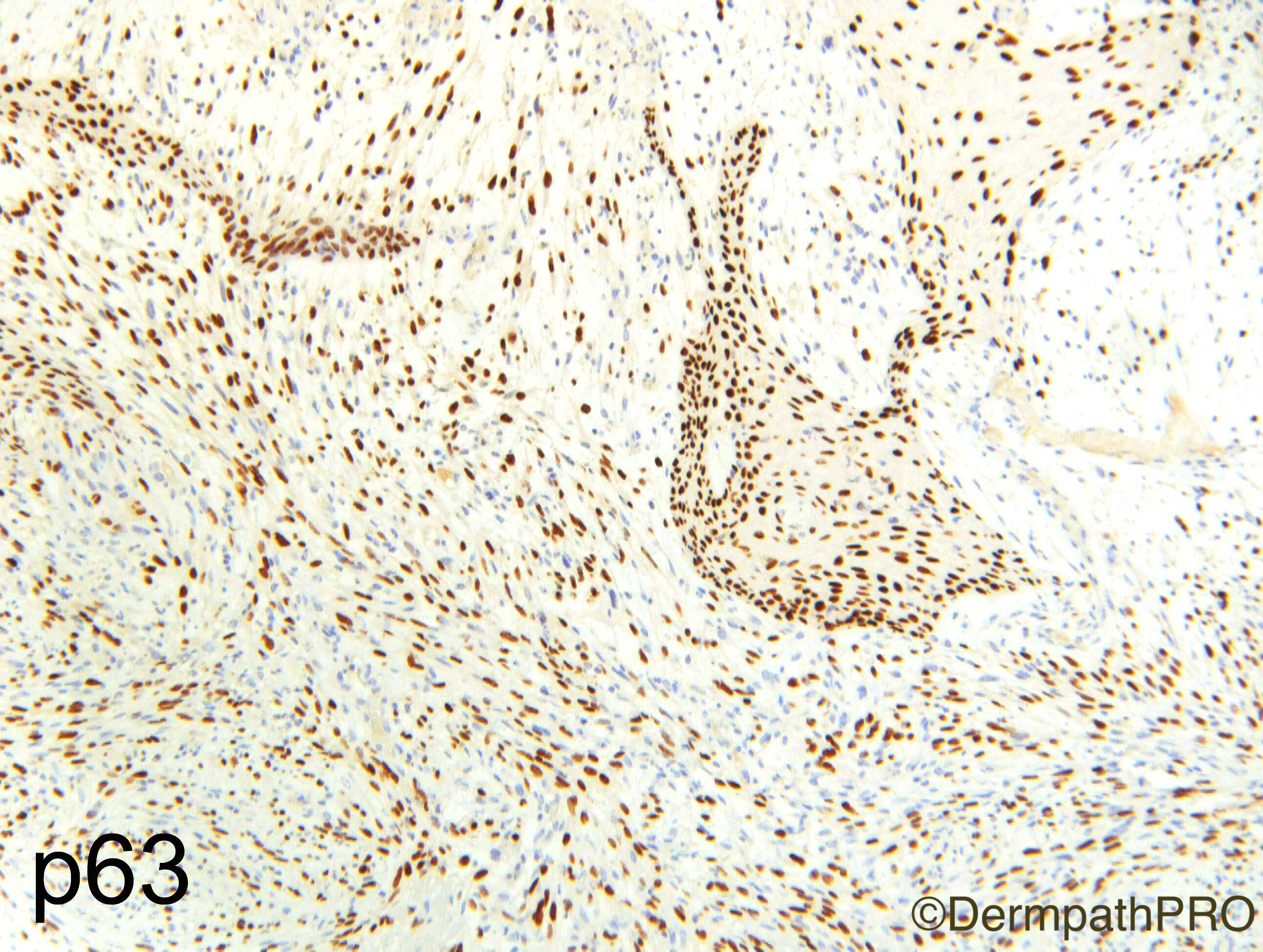
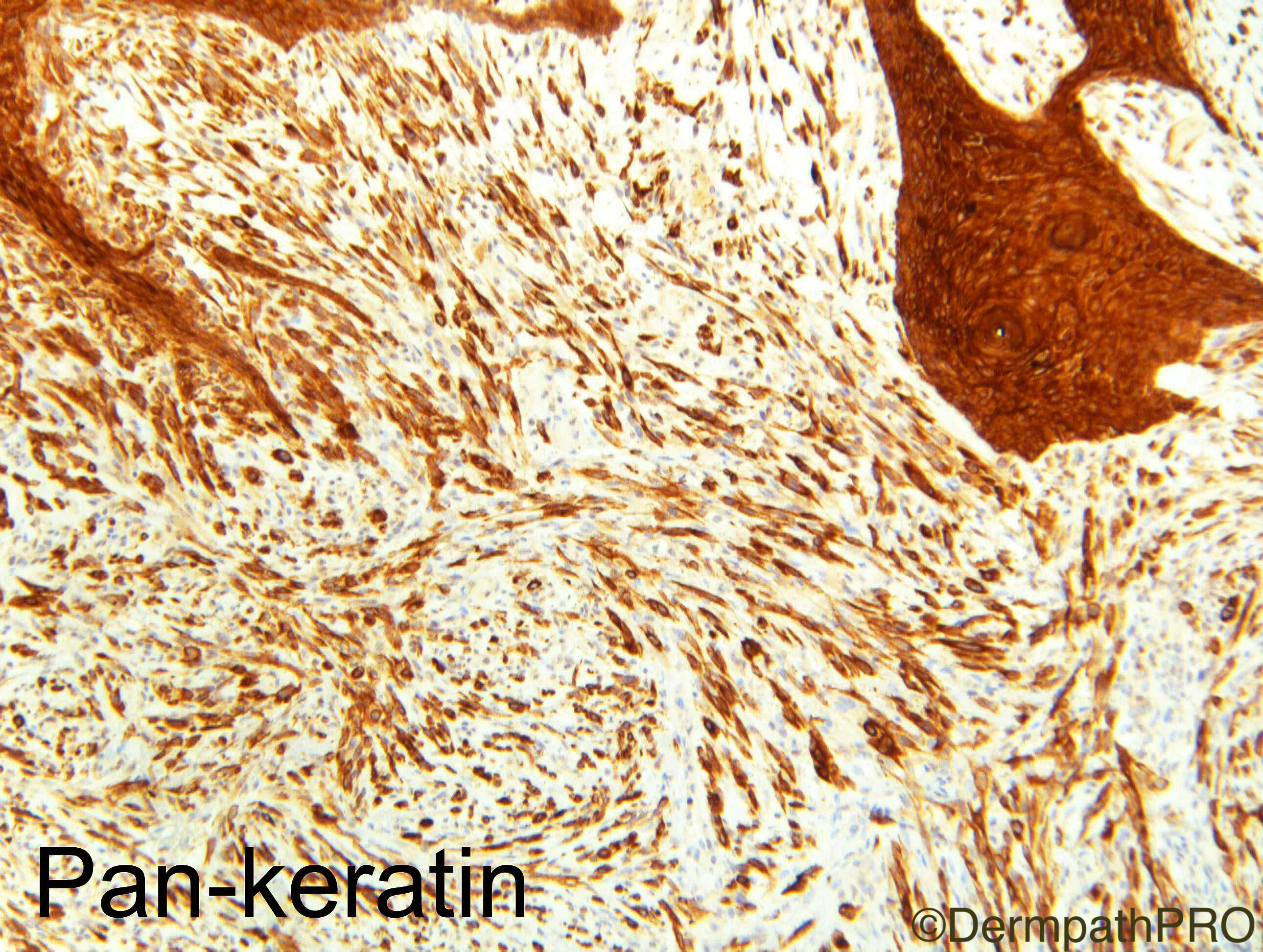
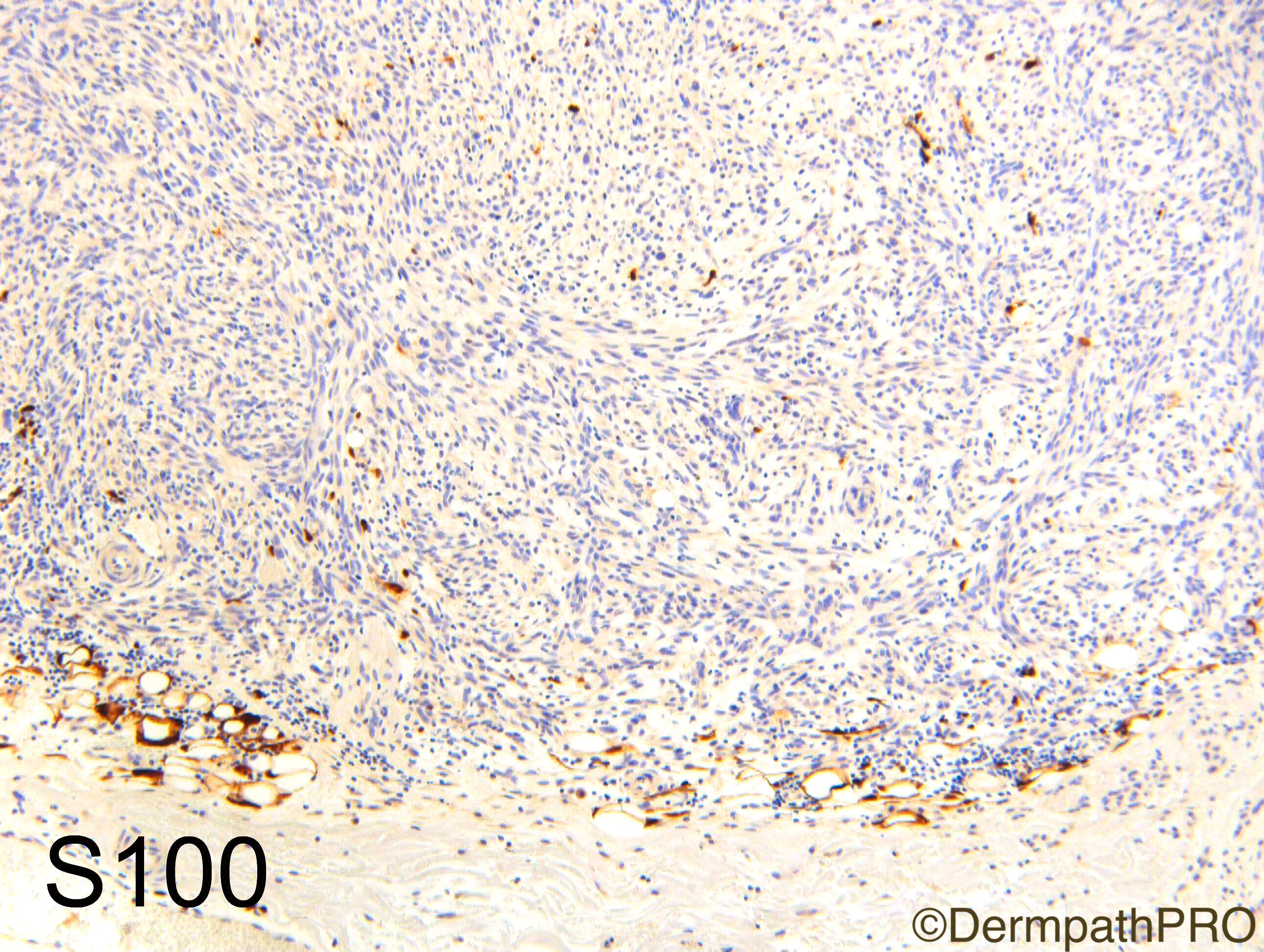
Thanks for the question about p53 which was the point of the case.  Saleem Taibjee drew my attention to it's use for margins in AFX/AFX-type cutaneous sarcoma (the latter my preferred designation for lesions that are either dermal and not circumscript or in which there is clear-cut invasion of subcutis - I am not a fan of so-called "pleomorphic dermal sarcoma" designation on several levels). p53 when diffusely strong can help with assessing margins for the "superficial spreading variant" that I have seen several times and on one or two occasions recurring (this is not in the text-books). However CD10 (a much maligned antibody IMHO) has been doing this job for me for a long time quite adequately actually and is far more sensitive, I also believe diffuse CD10 is pretty specific for AFX (we've collected over a 100 cases). In this case CD10 was ++40% (mainly interstitial, presumed reactive, cells).  To repeat my approach to this DDx I use a very limited panel S100 only (for melanoma), Pan-keratin & p63 (for carcinoma) and CD10 (diffuse supports AFX/AFX-type cutaneous sarcoma). I'm experimenting with p53 right now but will probably drop it soon I suspect.  I only add CD30 and vascular markers as required by morphology although a spindle cell variant of angiosarcoma can be a catch it is exceedingly rare.  I don't worry about cutaneous leiomyosarcomas at all at the treatment & prognosis for them is the same as AFX/AFX-type sarcoma.

Share this comment


Link to comment
share_externally

Arti Bakshi

Posted

Thanks Richard, very useful! I remember seeing a case of 'superficial spreading' AFX in your collection and the degree of horizontal spread of the tumour is easily missed.

Share this comment


Link to comment
share_externally


Join the conversation

You can post now and register later. If you have an account, sign in now to post with your account.

Guest
Add a comment...

×   Pasted as rich text.   Paste as plain text instead

  Only 75 emoji are allowed.

×   Your link has been automatically embedded.   Display as a link instead

×   Your previous content has been restored.   Clear editor

×   You cannot paste images directly. Upload or insert images from URL.

Loading...

×
×
  • Create New...