Jump to content
In this section we have spot diagnoses posted on a daily basis since June 2010, now over 4000! You can review the archived cases and read the suggested diagnoses by users and the final comment by the contributors.
Case are uploaded each week day by 10 am UK time with the correct diagnosis will generally be posted at 8 pm UK time. Why not view the most recent spot diagnosis and proffer a diagnosis?

Case Number : Case 1615 - 2 September Posted By: Guest

Please read the clinical history and view the images by clicking on them before you proffer your diagnosis.
Submitted Date :
   (0 reviews)

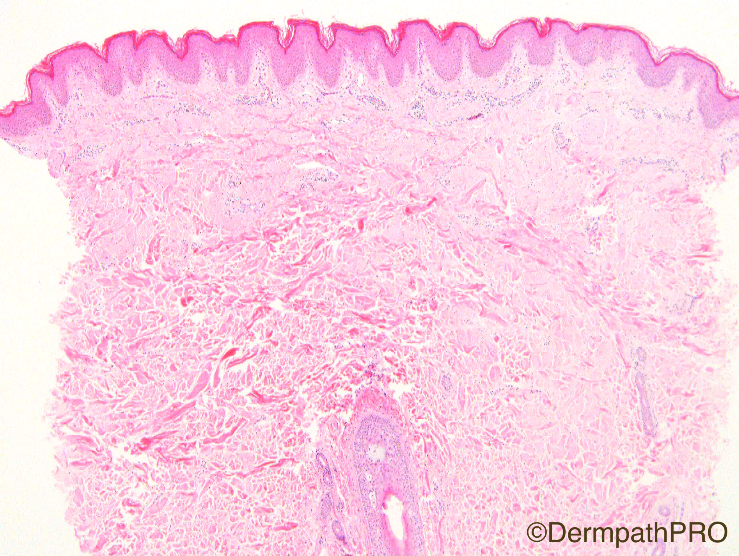
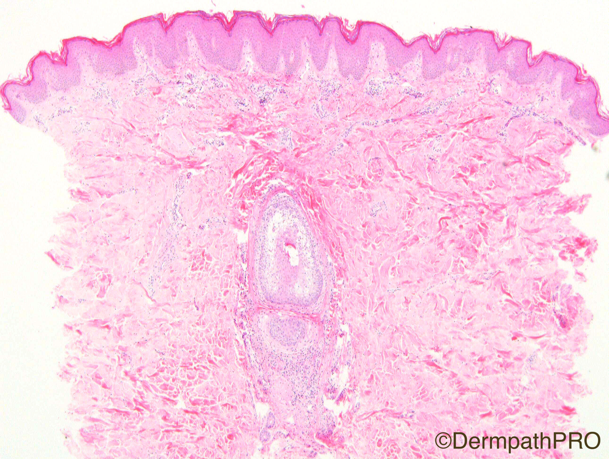
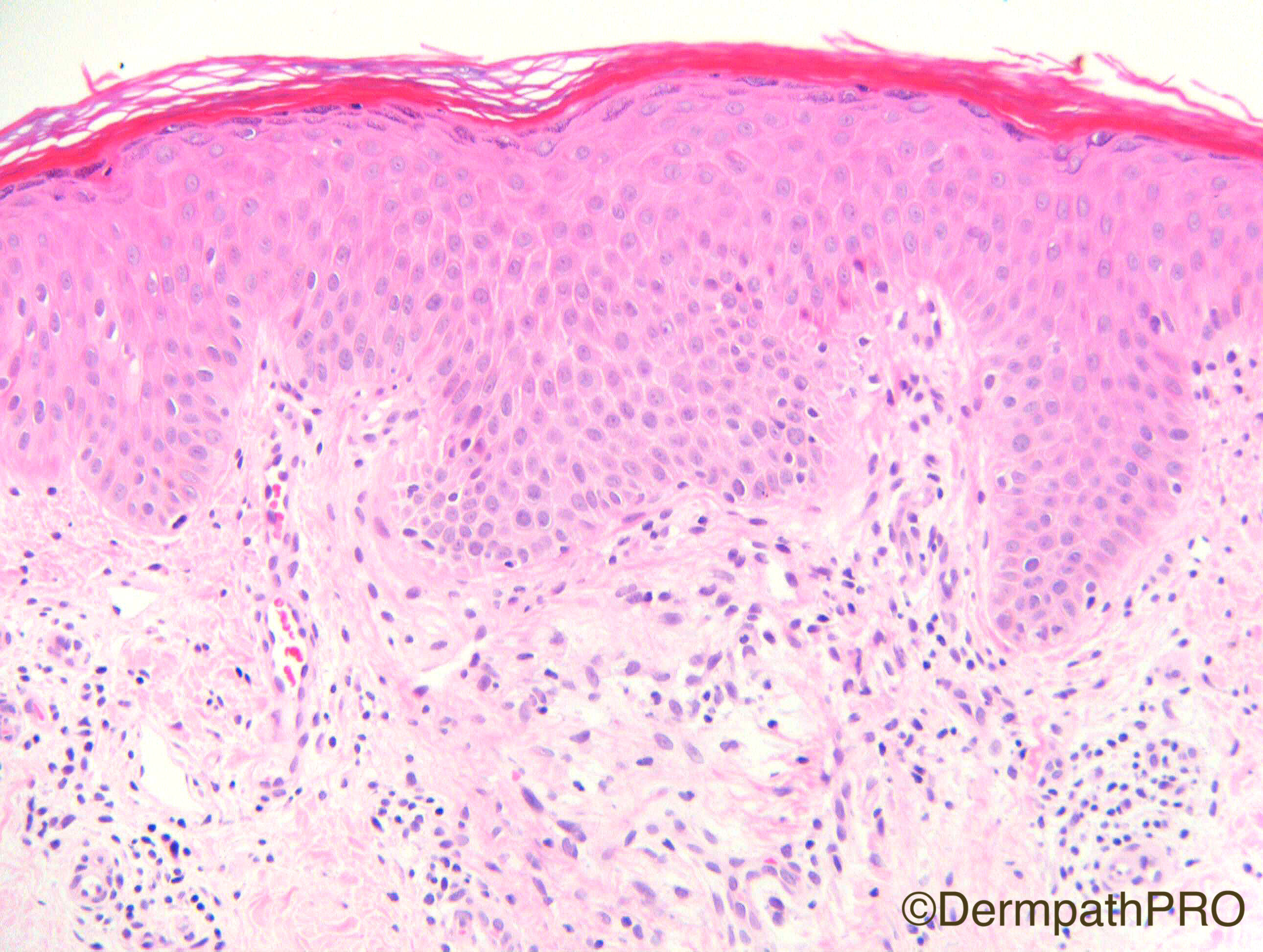
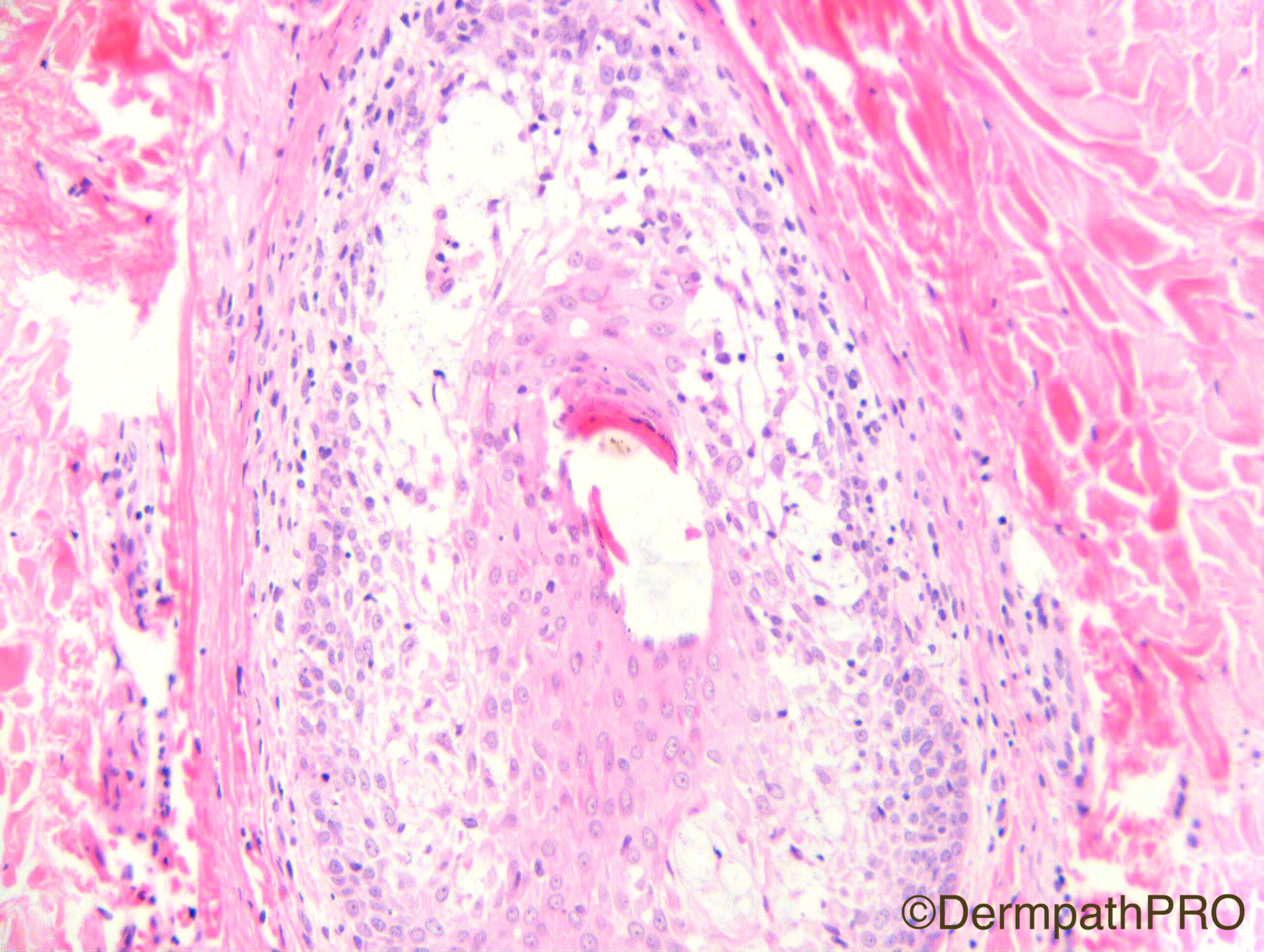
M20. Eczematous changes in skin 7 years. Symmetrical & flexural but also folliculocentric. Predominantly folliculocentric in antecubital and popliteal fossae and trunk. No facial changes. ?follicular atopic eczema.

Case Posted by Dr Richard Carr


  • Like 1
  Report Record

User Feedback


Dr. Mona Abdel-Halim

Posted

Follicular mucinosis.
Do not see sufficient evidence of follicular MF, may be it is just primary follicular mucinosis. Will like to take biopsies from different lesions to be sure.

Share this comment


Link to comment
share_externally

vincenzo polizzi

Posted

I'm thinking of a secondary follicular mucinosis, developing in atopic dermatitis.

Share this comment


Link to comment
share_externally

Guest Michelle Liang

Posted

Follicular mucinosis which can be primary or secondary to CTCL. I would be concerned of underlying CTCL given its multicentricity and his age (no longer a child).

Share this comment


Link to comment
share_externally

Sasi Attili

Posted

Interesting. There is mild acanthosis associated with compact hyperkeratosis alternating with orthokeratosis. Mild epidermal spongiosis is also evident, associated with a mild-moderate perivascular lymphocytic infiltrate in the upper dermis. There is obviously a follicular spongiosis with lymphocyte exocytosis and follicular mucin. However the lymphocytes aren't atypical. I think the mucin is secondary. I think this is just follicular mucinosis on the background of chronic atopic dermatitis. 

Share this comment


Link to comment
share_externally

Dr. Richard Carr

Posted

The histology does indeed show mild compact hyperkeratosis, mild acanthosis, a lack of epidermal spongiosis, prominent follicular mucinosis with exocytosis of some mildly convoluted lymphocytes.  The cells in the epithelium were positive for CD3, CD5 and mainly CD4 (compared with CD8) with some loss of CD2 and CD7. Gene re-arrangement studies were weakly polyclonal for TRG and non-amplifying for TRB studies. We are suspicious that this will be a case of secondary follicular mucinosis to CTCL/mycosis fungoides rather than idiopathic but follow-up will be required for a final conclusion. Thank you to Werner Kempf and Hesham El-Daly who were consulted on this case and the latter for arranging additional studies.  Dr Kempf indicated that there was a report that oral minocycline can be effective in idiopathic follicular mucinosis.

Share this comment


Link to comment
share_externally


Join the conversation

You can post now and register later. If you have an account, sign in now to post with your account.

Guest
Add a comment...

×   Pasted as rich text.   Paste as plain text instead

  Only 75 emoji are allowed.

×   Your link has been automatically embedded.   Display as a link instead

×   Your previous content has been restored.   Clear editor

×   You cannot paste images directly. Upload or insert images from URL.

Loading...

×
×
  • Create New...