Jump to content
In this section we have spot diagnoses posted on a daily basis since June 2010, now over 4000! You can review the archived cases and read the suggested diagnoses by users and the final comment by the contributors.
Case are uploaded each week day by 10 am UK time with the correct diagnosis will generally be posted at 8 pm UK time. Why not view the most recent spot diagnosis and proffer a diagnosis?

Case Number : Case 1625 - 16 September Posted By: Guest

Please read the clinical history and view the images by clicking on them before you proffer your diagnosis.
Submitted Date :
   (0 reviews)

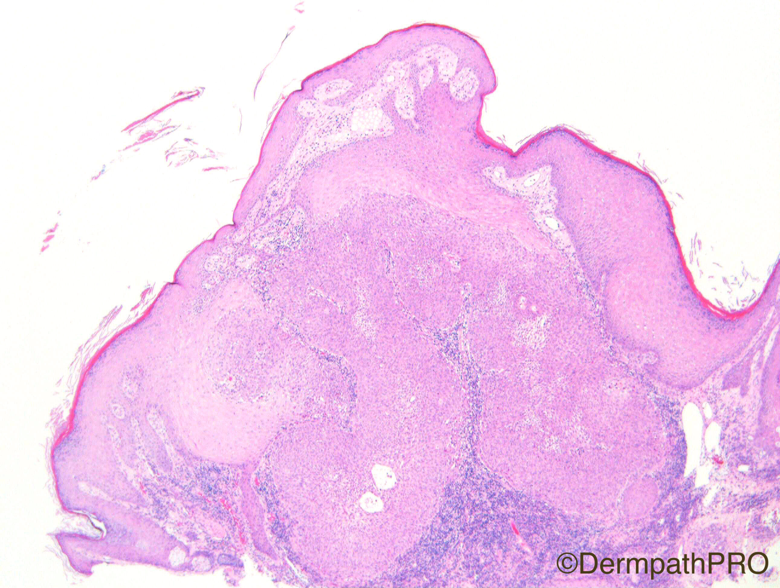
M80. Cheek. ?AK, ?SEBK

Case Posted by Dr Richard Carr

  Report Record

User Feedback


vincenzo polizzi

Posted

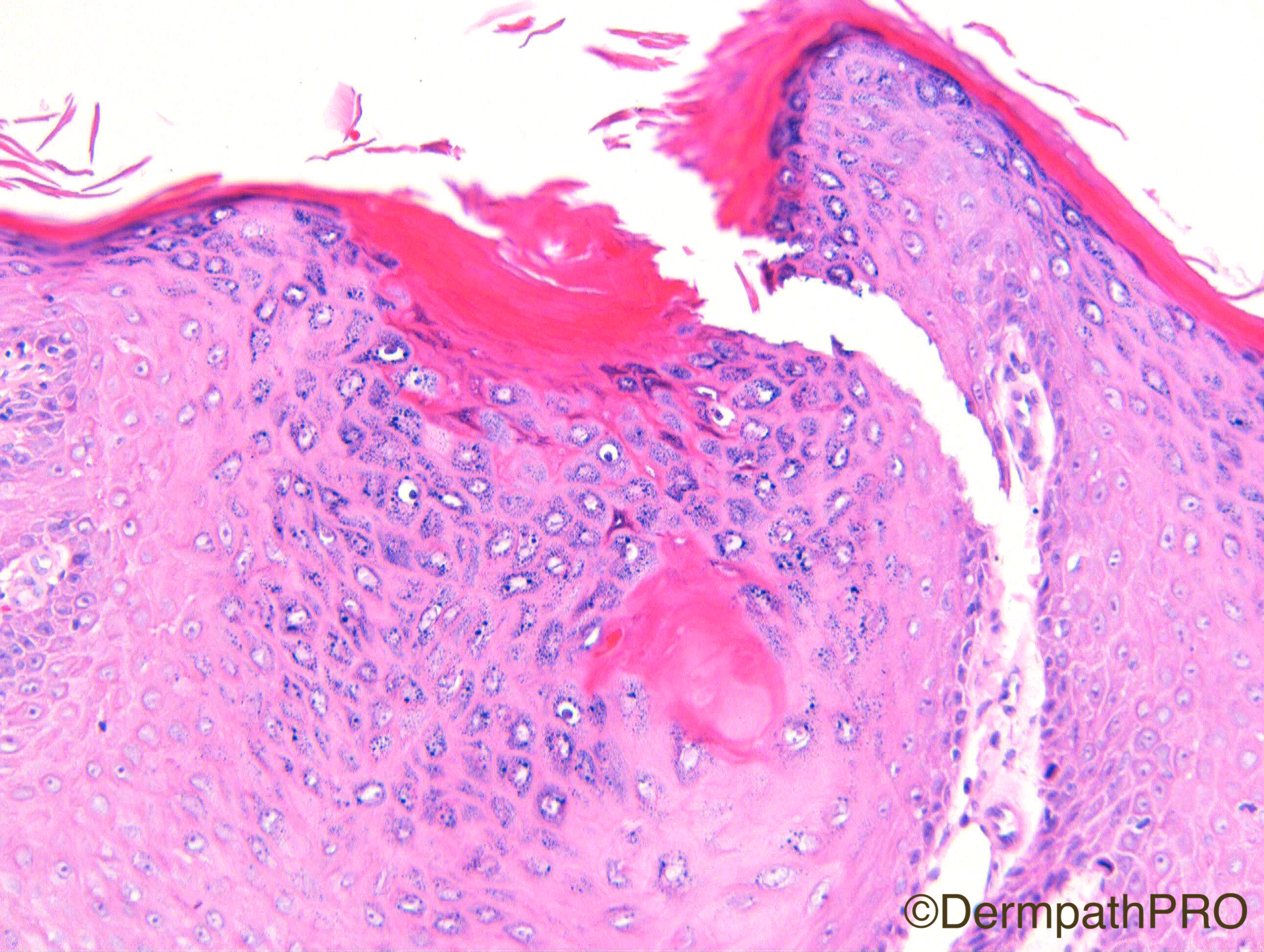
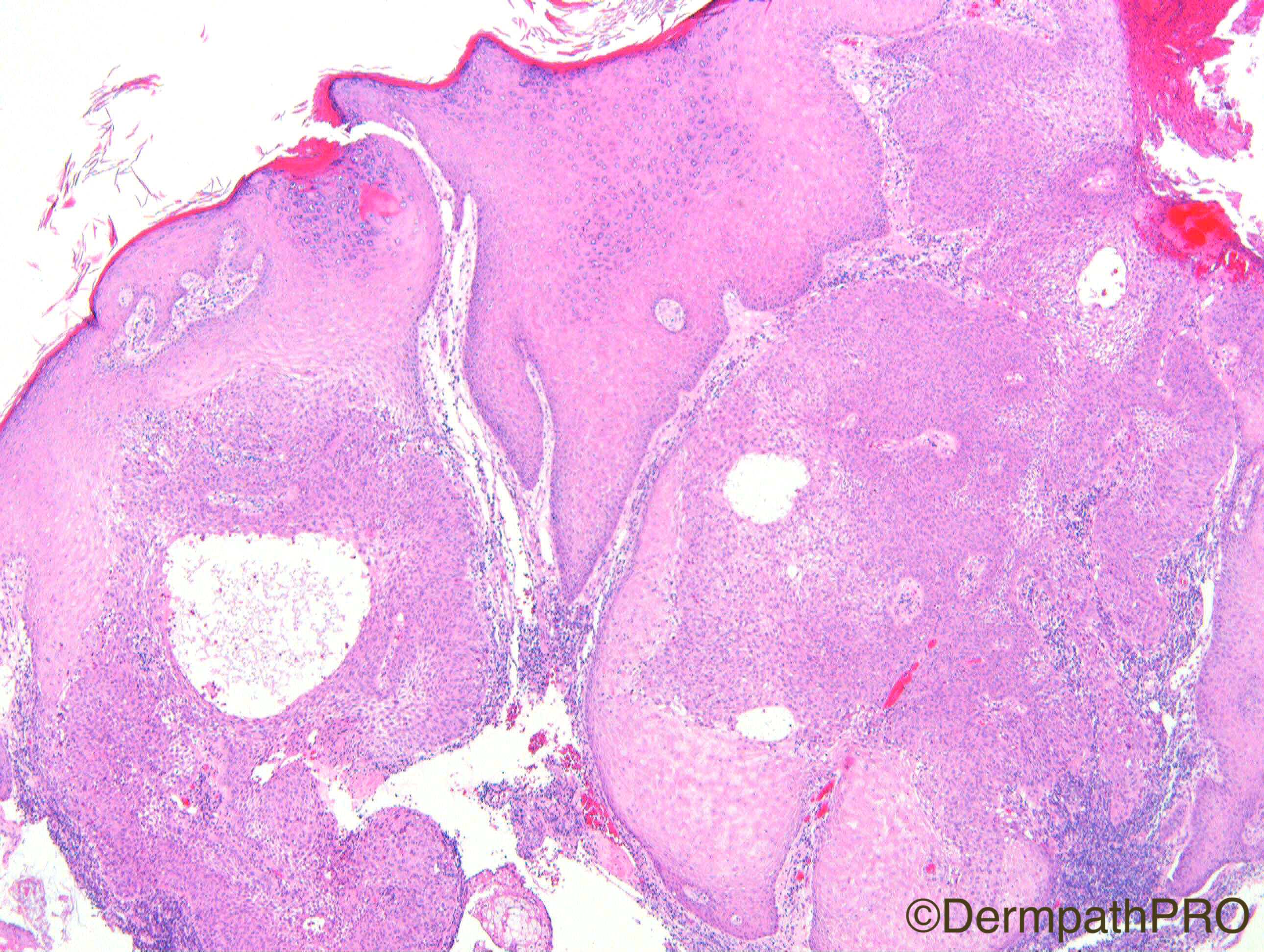
Difficult case for me. I was thinking of an atypical irritated seborrheic keratosis, but atypia seems over the hyper plastic reactive range and there are much features typical of infundibular and trichilemmal differentiation. The border with non-atypical epidermis is sharp, remarking clonal nature of atypical one...so I favour Follicular SCC with trichilemmal and infundibular differentiation.

Share this comment


Link to comment
share_externally

Arti Bakshi

Posted

Agree completely with Vicenzo's comment. The same differentials came to my mind ie inverted follicular keratosis and follicular SCC. There is intraepithelial mucin, which can be seen in both.

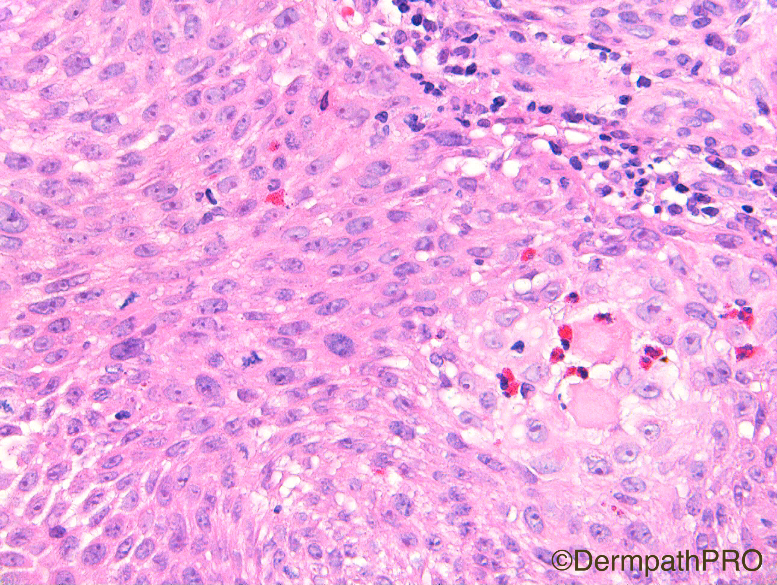
I too would favour Follicular SCC (insitu) due to the degree of cytological atypia and mitoses in figs 6 and 7.

Share this comment


Link to comment
share_externally

Dr. Mona Abdel-Halim

Posted

Me too, was working my mind between IFK and Follicular SCC. Favour follicular SCC.

Share this comment


Link to comment
share_externally

Nitin Khirwadkar

Posted

Agree with differentials. Favour follicular SCC.

Share this comment


Link to comment
share_externally

Raul Perret

Posted

Agree with the comments of my colleagues. SCC follicular, Did you consider it in situ Richard?

Share this comment


Link to comment
share_externally

Guest Arash Daryakarr

Posted

me too agree with colleagues. there is an endophytic growth of squamous neoplasm with sharp borders with adjacent epidermis bearing atypias ,mitoses and mucin deposition.

follicular in situ SCC seems a good idea.

what is acidophilic material surrounded by eosinophils in 7th image?something important?

Share this comment


Link to comment
share_externally

Dr. Richard Carr

Posted

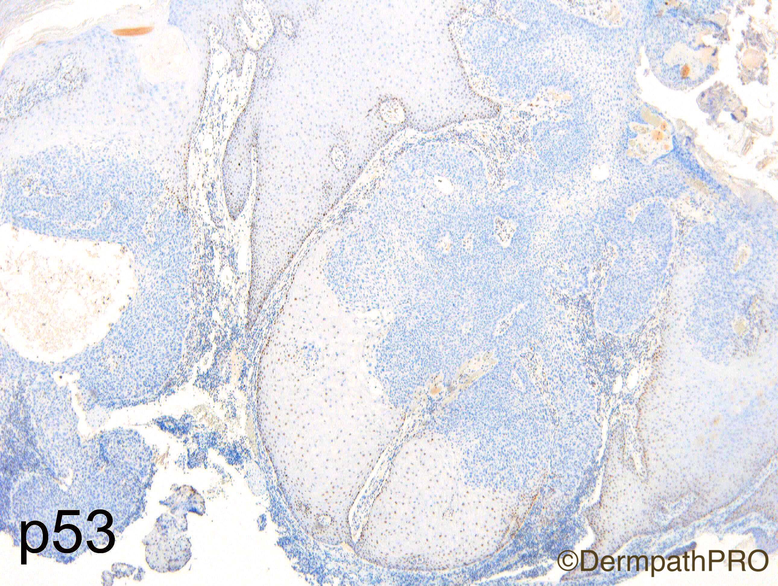
Thanks for excellent discussion. I have arranged for some IHC to get posted (soon hopefully).

Share this comment


Link to comment
share_externally

Dr. Richard Carr

Posted

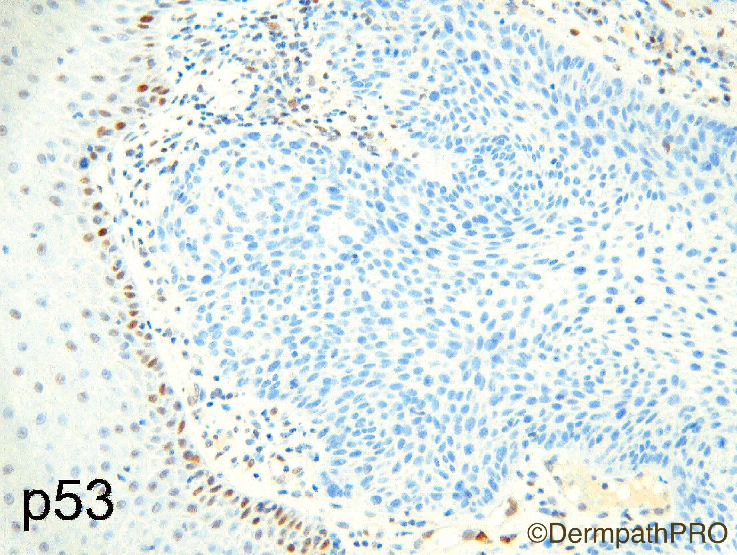
Well done Vincenzo.  You perfectly summarised the case as a differential between follicular SCC v's inverted follicular keratosis / irritated seborrheic keratosis. Thanks for reminding me of Case 1032 which also discusses the differential in depth.  This case was indeed reported as follicular SCC with pushing only borders and for practical purposes regarded as in situ (resembling IFK but with atypia too much for a benign lesion - supported by a null p53 - done retrospectively).  CD34 was also negative as expected. The acidophilic material in image 7 is just cross-cutting of the large "glassy" tricholemmal keratinocytes.  Eosinophils, neutrophils etc. can be attracted to squamous lesions such as this (including keratoacanthoma).

Share this comment


Link to comment
share_externally


Join the conversation

You can post now and register later. If you have an account, sign in now to post with your account.

Guest
Add a comment...

×   Pasted as rich text.   Paste as plain text instead

  Only 75 emoji are allowed.

×   Your link has been automatically embedded.   Display as a link instead

×   Your previous content has been restored.   Clear editor

×   You cannot paste images directly. Upload or insert images from URL.

Loading...

×
×
  • Create New...