Jump to content
In this section we have spot diagnoses posted on a daily basis since June 2010, now over 4000! You can review the archived cases and read the suggested diagnoses by users and the final comment by the contributors.
Case are uploaded each week day by 10 am UK time with the correct diagnosis will generally be posted at 8 pm UK time. Why not view the most recent spot diagnosis and proffer a diagnosis?

Case Number : Case 1630 - 23 September Posted By: Guest

Please read the clinical history and view the images by clicking on them before you proffer your diagnosis.
Submitted Date :
   (0 reviews)

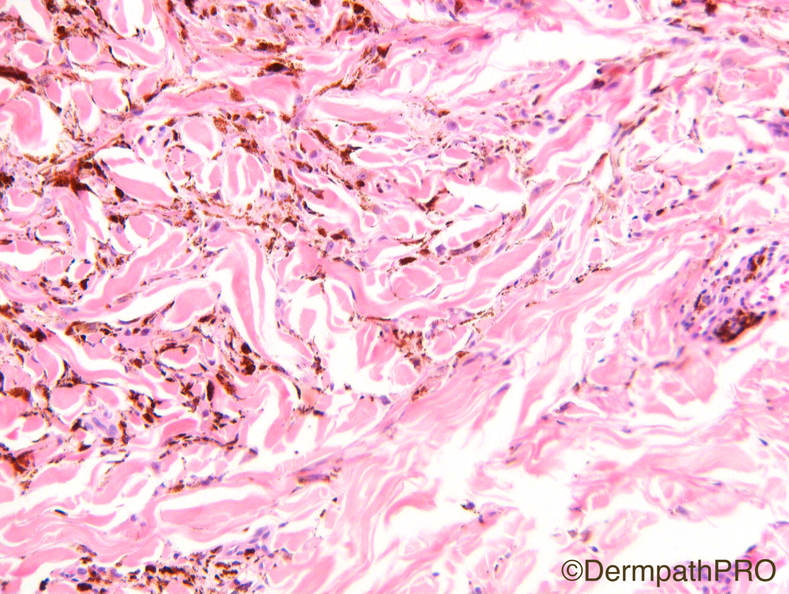
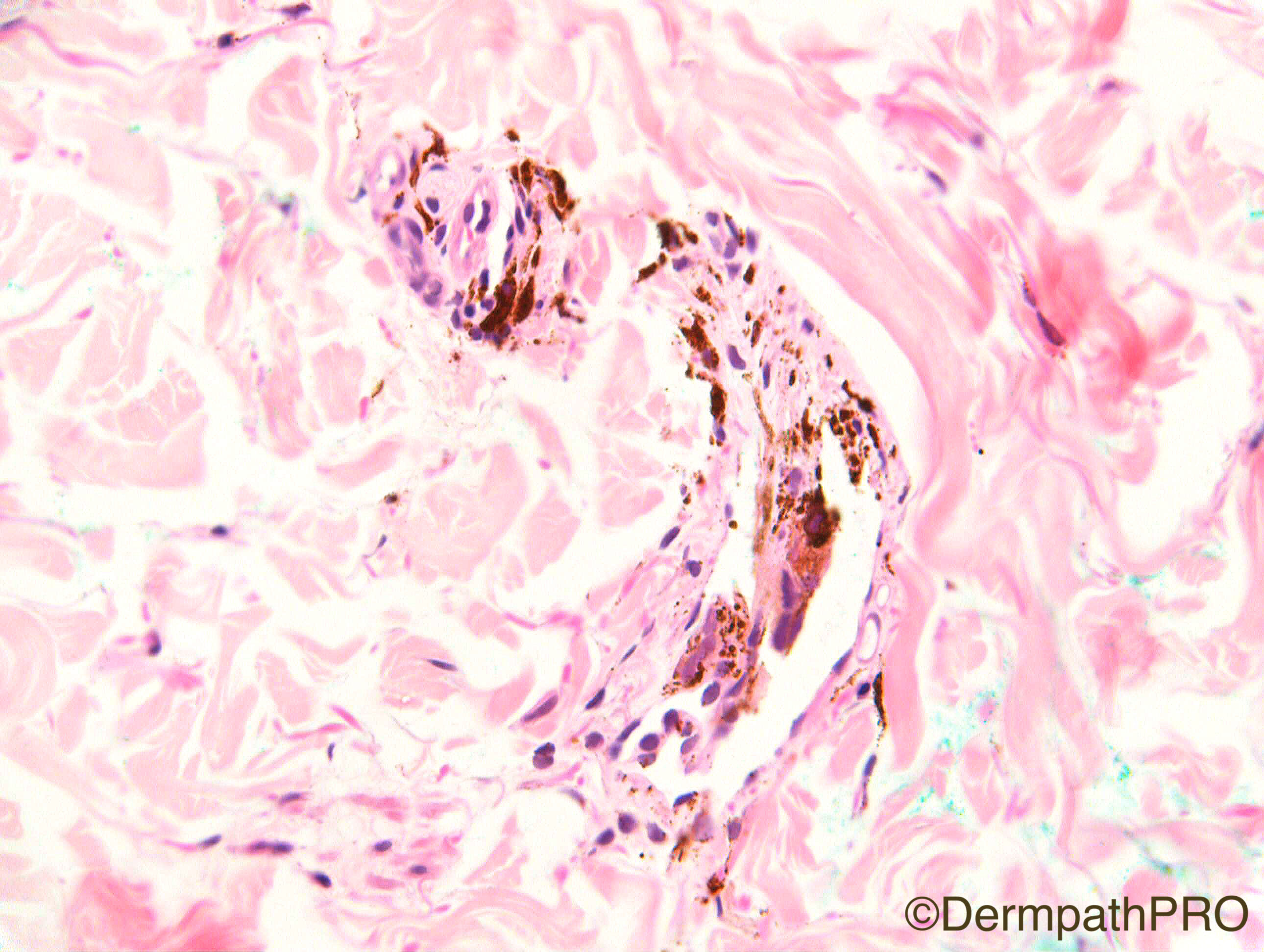
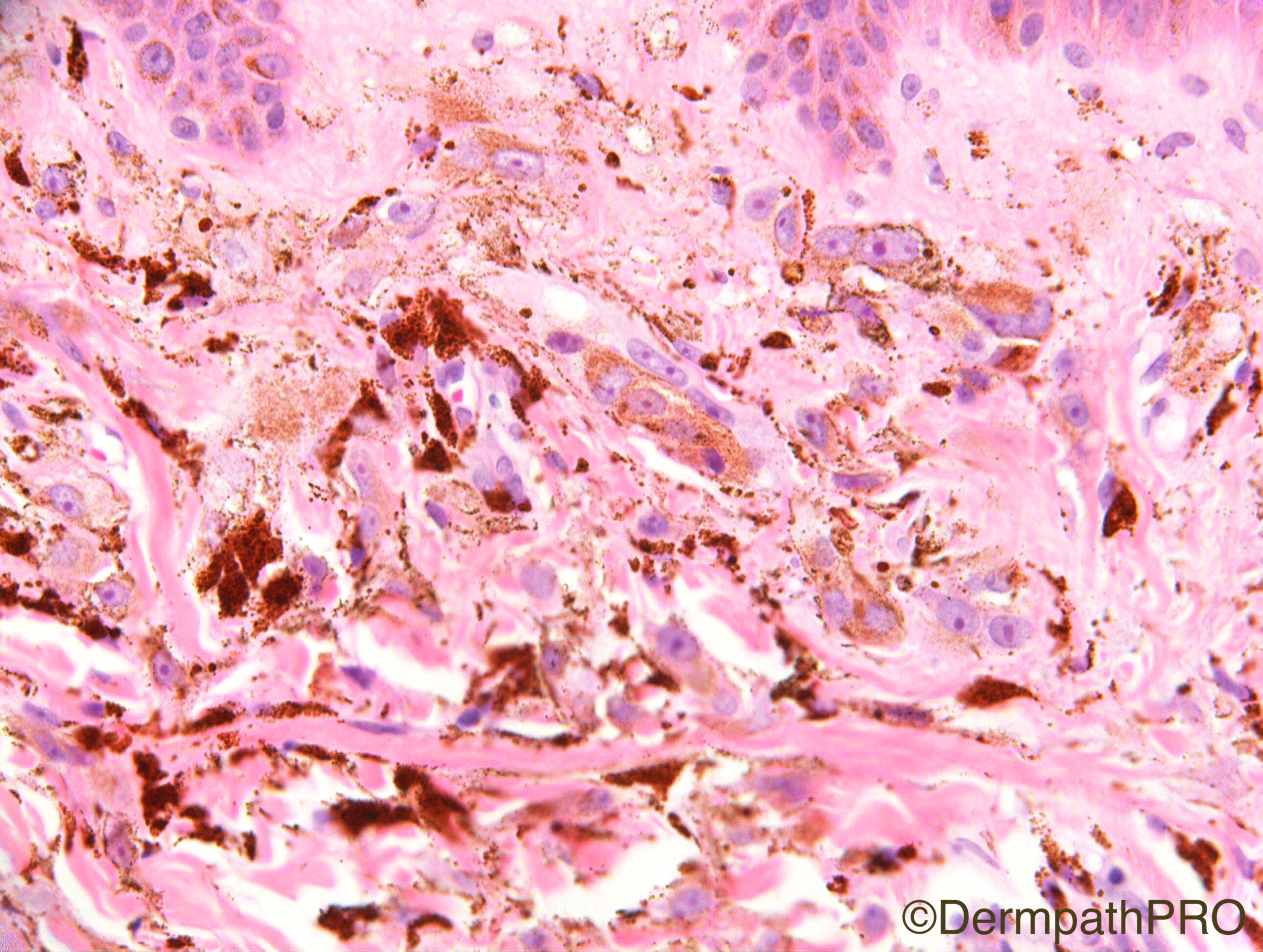
F45 Mole on back

Case Posted by Dr Richard Carr

  Report Record

User Feedback


Admin_Dermpath

Posted

Here is Dr Richard Carr's interesting Spot Diagnosis Case of the Day to test you before the weekend. Remember, when you figured this interesting case out take a look at the 'Video Case of the Week' which has just been updated.


Cheers, Geoff Cross

Share this comment


Link to comment
share_externally

vincenzo polizzi

Posted

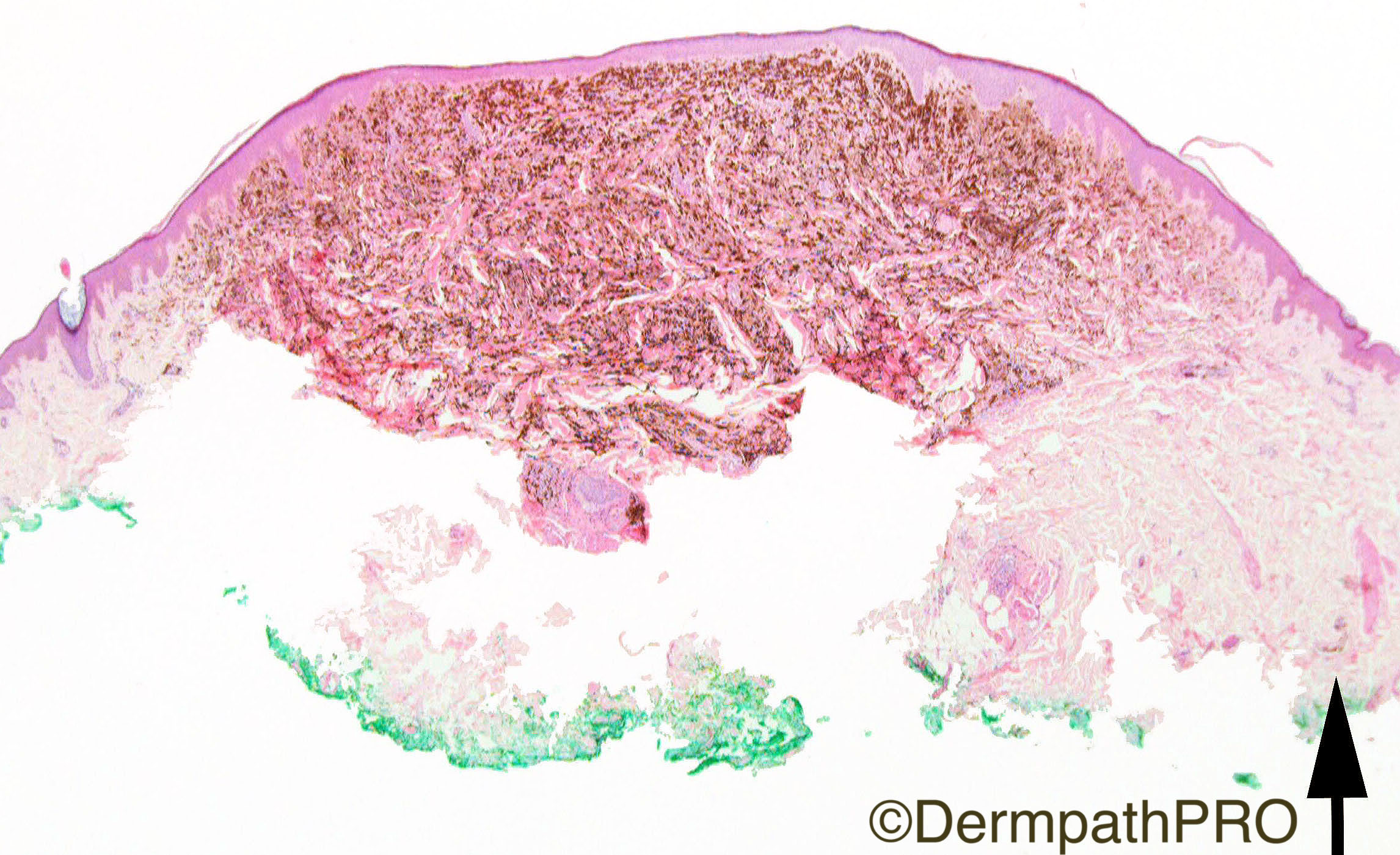
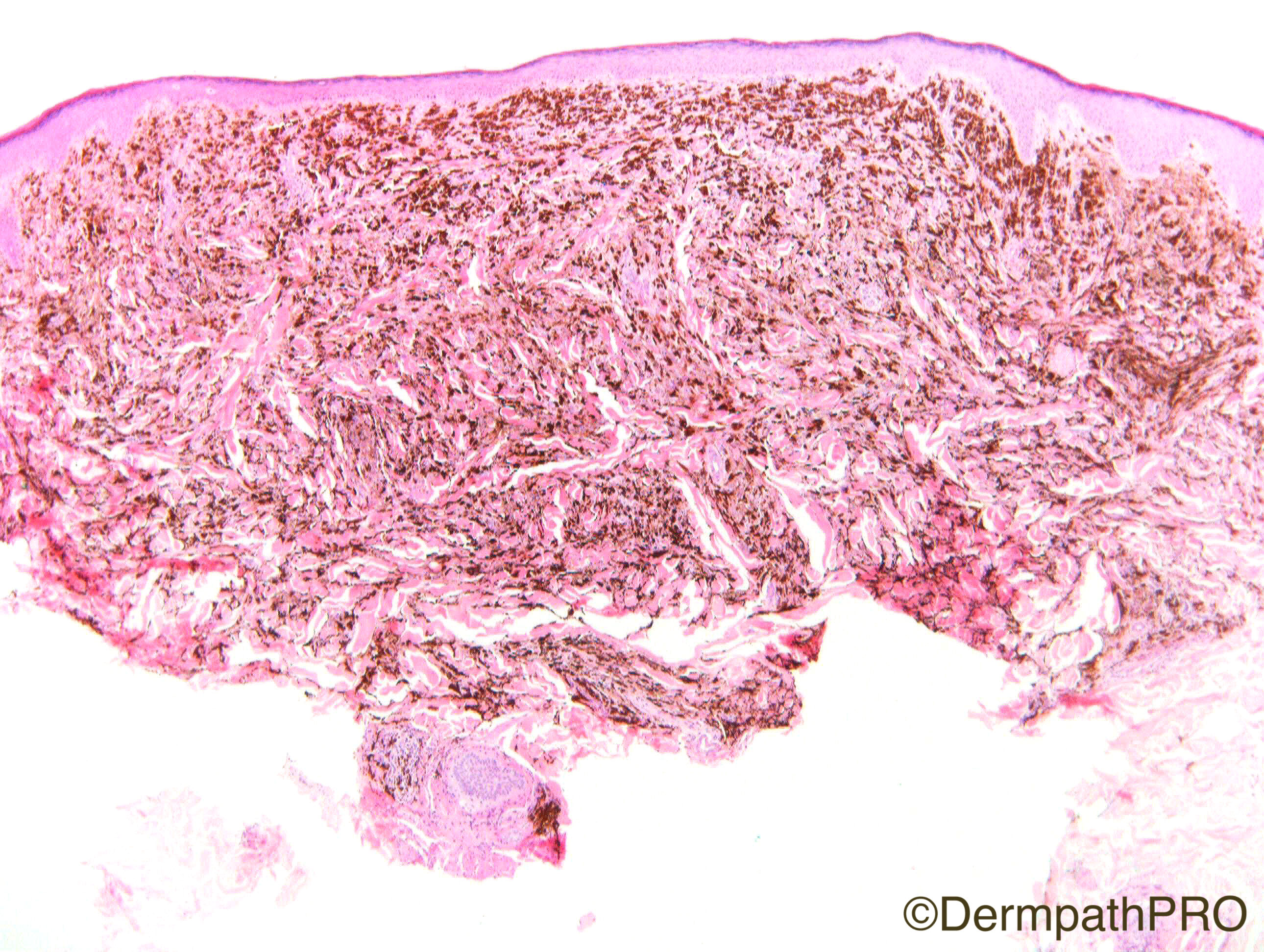
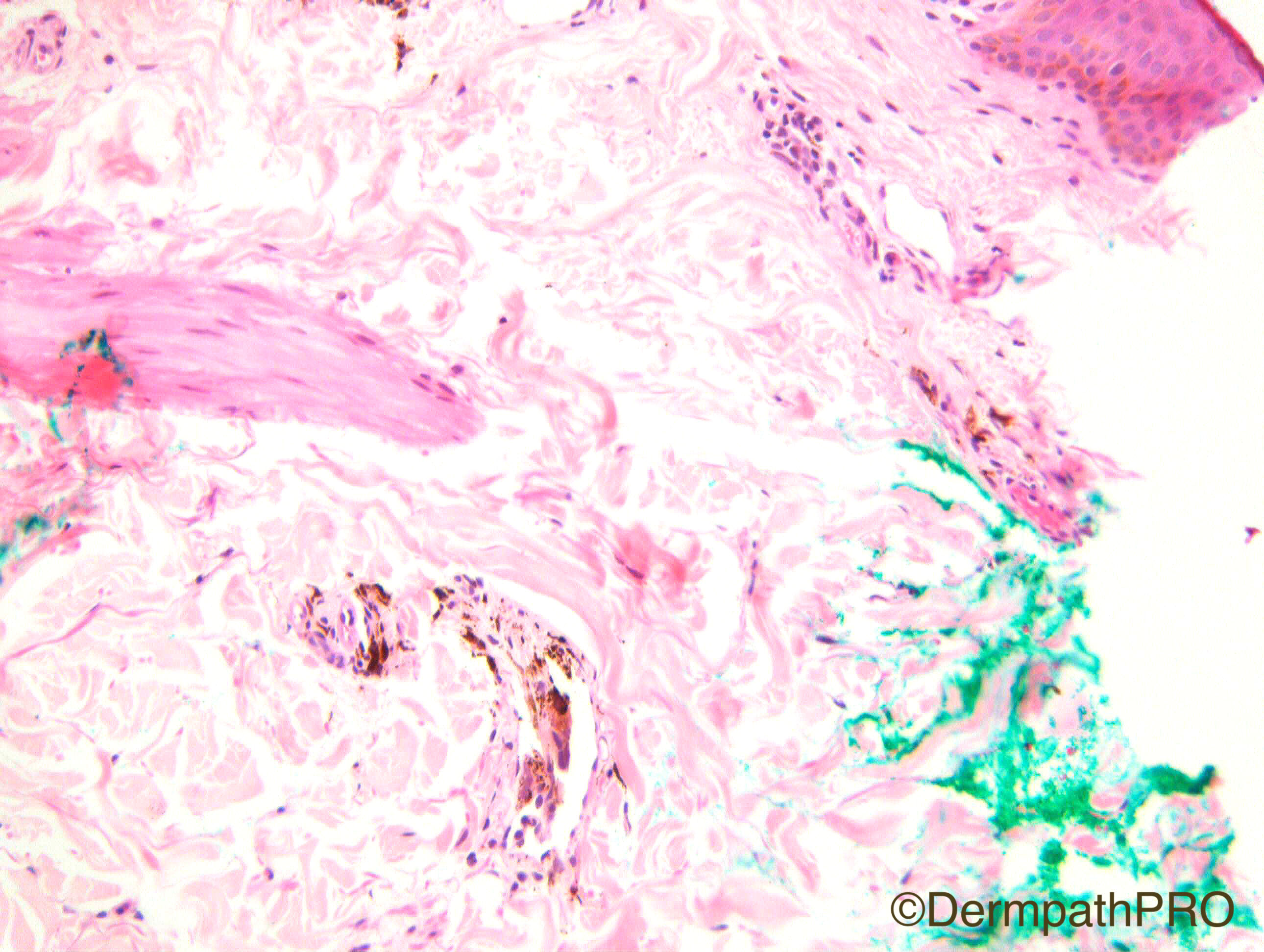
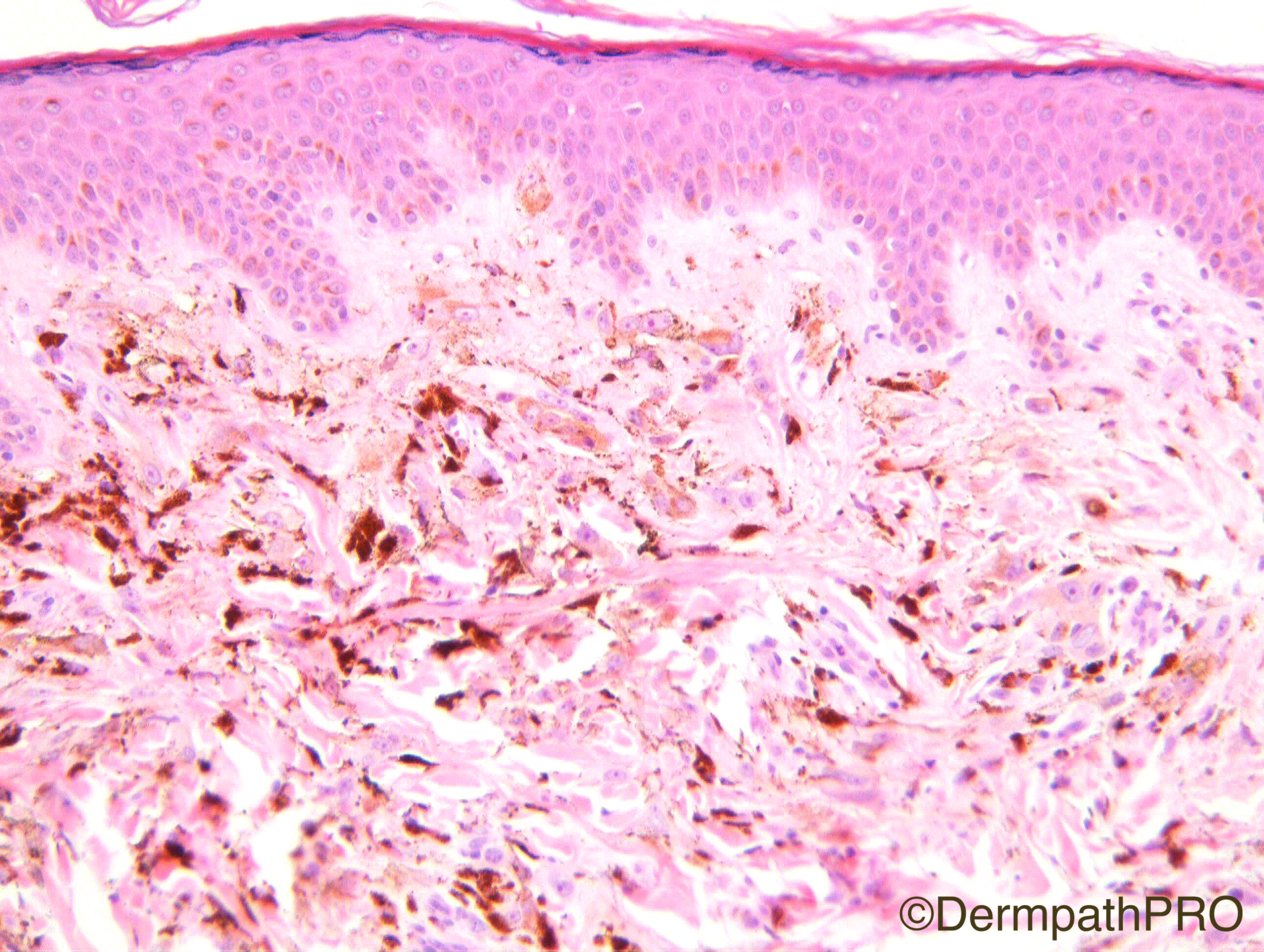
What about a Pigmented Epithelioid Melanocytoma?   There are the typical large epithelioid cells with abundant clear or lightly pigmented cytoplasm, and large vesicular nuclei with prominent nucleoli; look like "fried eggs". These cells are most abundant at the periphery of the lesion. The pattern is infiltrative, between collagen bandles, mainly to the periphery. And some lymph space is infiltrated.

Share this comment


Link to comment
share_externally

Dr. Mona Abdel-Halim

Posted

Agree with Vincenzo, I think it is pigmented epithelioid melanocytoma. I read that some consider epithelioid blue nevus and pigmented epithelioid melanocytoma two entities in a clinical and pathological spectrum, with the lymphatic invasion, I am tempted more to describe it as pigmented epithelioid melanocytoma. Waiting to be enlightened by the experts !!

Share this comment


Link to comment
share_externally

Raul Perret

Posted

Looks like a heavily and homogeneously pigmented melanocytic lesion with deep extension and tropism for adnexae. There are some cells that look banal (dendritic blue nevus cells) others have worrisome features (macrocariosis with irregular nuclear borders, giant nucleoli, ample dusty like pigmented cytoplasm), no mitosis, no necrosis. Should bleach (must be systematic on heavily pigmented lesions). As vincenzo mentioned I would favour pigmented epithelioid melanocytoma, the absence of necrosis and brisk mitotic activity make favour it over the so called malignant blue nevus. By the way, association with carney complex in some cases should be mentioned in the report (from my point of view)

Share this comment


Link to comment
share_externally

Dr. Richard Carr

Posted

Forgot to thank Dr Mark Atkinson for kindly referring this case.  I have asked some international colleagues from a melanocytic slide club that I organise to partake in this case as Mona asked what the experts think!  Hopefully we'll get a few of them responding (a few have already to me privately).

Share this comment


Link to comment
share_externally

Raul Perret

Posted

Forgot to mention, Richard did you perform cd31/d2-40 on this lesion? My impression only based on morphology is that melanocytic cells are not inside the lumen of the vessel but that this is rather vascular pseudoinvasion. Blue nevi are usually infiltrative lesions and rather adnexotropic/neurotropic. From my optic I would still have called this PEM even if that part was not present.

Share this comment


Link to comment
share_externally

Nitin Khirwadkar

Posted

Would favour a PEM. Waiting for the answer.


Richard, what were the margins? Did this case have a SLNB? Thanks

Share this comment


Link to comment
share_externally

Arti Bakshi

Posted

Agree with pigmented epithelioid melanocytoma.

PEM occurring in a setting of Carney's complex or combined melanocytoma are considered benign. But histologically similar leisons, which are sporadic and not combined with other naevi are potentially metastatic to regional lymph nodes (although these too supposedly have a good long term outcome as distant metastasis exceedingly uncommon).  

Share this comment


Link to comment
share_externally

Dr. Richard Carr

Posted

These were some responses of colleagues in the melanocytic slide club:

Gerardo Ferrara (Benevento, Italy): PEM

Arnaud de la Fouchardiere (Lyon, France): I agree with PEM give the junction is epithelioid and so are the dermal cells. There are no large nests in the dermis. The lateral and deep border is ill defined.  The nuclear detail is very suggestive with large nucleoli in an oval nucleus. MelanA in red should show a diffuse mesh displayed with not that many cells and junctional cells.

Alan Spatz (Montreal, Canada): I also agree with PEM, The website is truly fantastic, congratulations

David Slater (Sheffield, UK): Agreed PEM. The neuropathologistes I worked with had a run of these in the spinal column.

David Elder (Pennysylvania, USA): I agree also with the diagnosis of PEM


I reported it along similar lines choosing to categorise it as a heavily pigmented variant of (atypical) Spitz tumour, uncertain malignancy, probably benign with minimal risk for metastasis (with a discussive comment that I don't doubt some pathologists would push it into the category of pigmented epithelioid melanocytoma).  I noted that it followed neurovascular bundles focally up to excision margins and therefore advised a modest re-excision and follow-up. I did raise the possibility of Carney's in the report. No SLNB as far as I am aware  but in my report I favoured a likely benign course give the lack of dermal mitotic activity. I don't remember doing a vascular marker but not sure it would change my interpretation in either case with respect to biological behaviour (likely benign). I found this case difficult so I am impressed that you all made the same relatively confident diagnosis!

You have got me again today. Regards to all.

Share this comment


Link to comment
share_externally


Join the conversation

You can post now and register later. If you have an account, sign in now to post with your account.

Guest
Add a comment...

×   Pasted as rich text.   Paste as plain text instead

  Only 75 emoji are allowed.

×   Your link has been automatically embedded.   Display as a link instead

×   Your previous content has been restored.   Clear editor

×   You cannot paste images directly. Upload or insert images from URL.

Loading...

×
×
  • Create New...