Jump to content
In this section we have spot diagnoses posted on a daily basis since June 2010, now over 4000! You can review the archived cases and read the suggested diagnoses by users and the final comment by the contributors.
Case are uploaded each week day by 10 am UK time with the correct diagnosis will generally be posted at 8 pm UK time. Why not view the most recent spot diagnosis and proffer a diagnosis?

Case Number : Case 1631 - 26 September Posted By: Guest

Please read the clinical history and view the images by clicking on them before you proffer your diagnosis.
Submitted Date :
   (0 reviews)

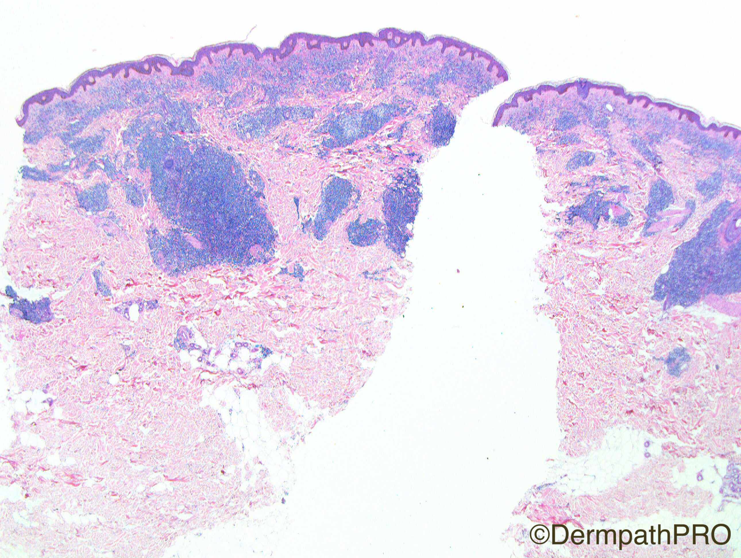
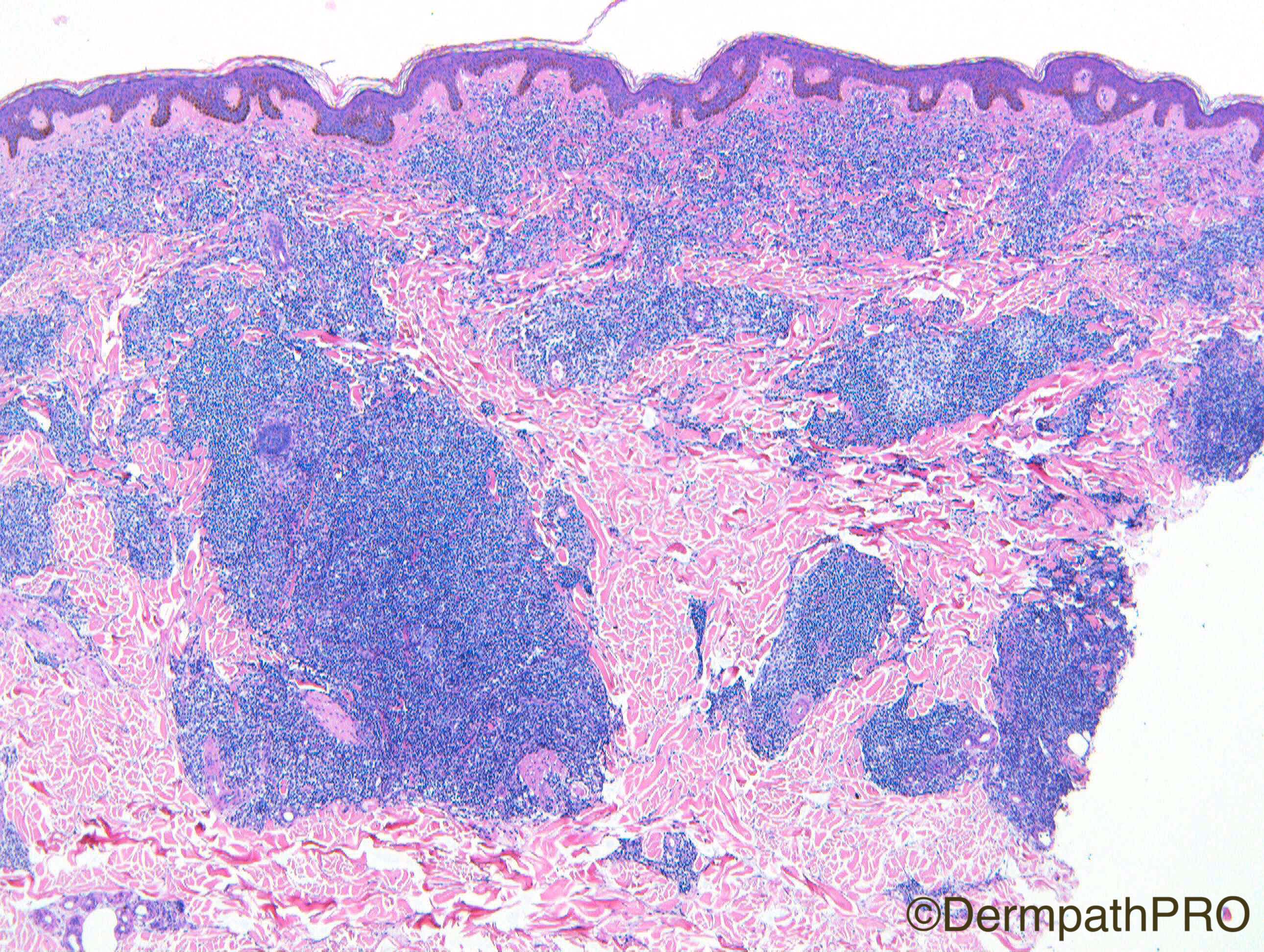
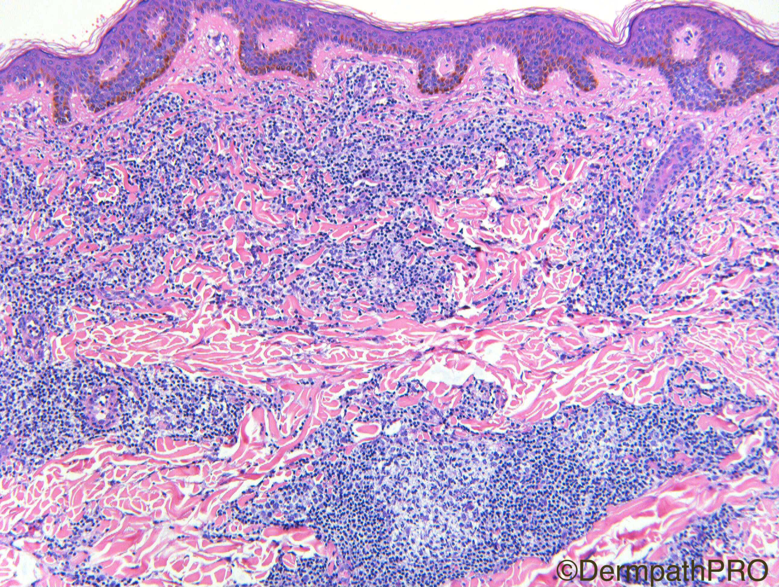
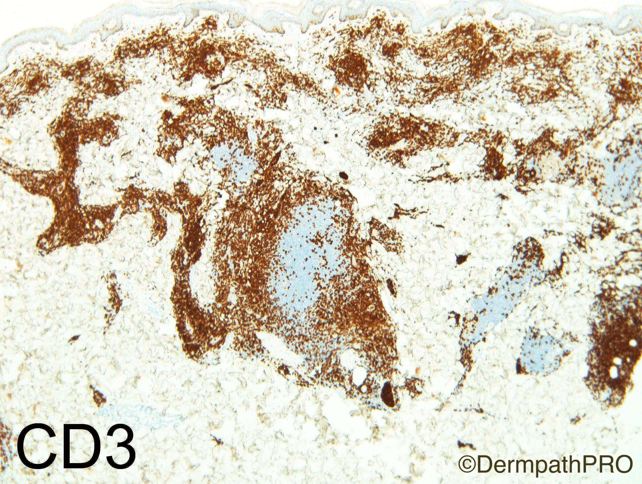
F60. Arm. ?GA, ?Sarcoid, ?B cell lymphoma

Case Posted by Dr Richard A Carr


  Report Record

User Feedback


Admin_Dermpath

Posted

Today we have a special 'extra' case from Dr Richard Carr, there will be extra images posted at 6pm UK time to aid your diagnosis.


Cheers, Geoff

Share this comment


Link to comment
share_externally

vincenzo polizzi

Posted

It is a cellular spindle cell neoplasm, polypoid, with patternless arrangement of the cells but regularly distributed thick vessels. Something looking like spindle cell lipoma but mammary type myofibroblastoma too. So I'm thinking of a cellular angiofibroma. Unusual site.

Share this comment


Link to comment
share_externally

Dr. Mona Abdel-Halim

Posted

I think we saw this case before, cellular angiofibroma.

Share this comment


Link to comment
share_externally

Admin_Dermpath

Posted

Give me 5 minutes and I will replace case and matching images


Cheers, Geoff

Share this comment


Link to comment
share_externally

Admin_Dermpath

Posted

Done, new case images uploaded.


Cheers, Geoff

Share this comment


Link to comment
share_externally

Raul Perret

Posted

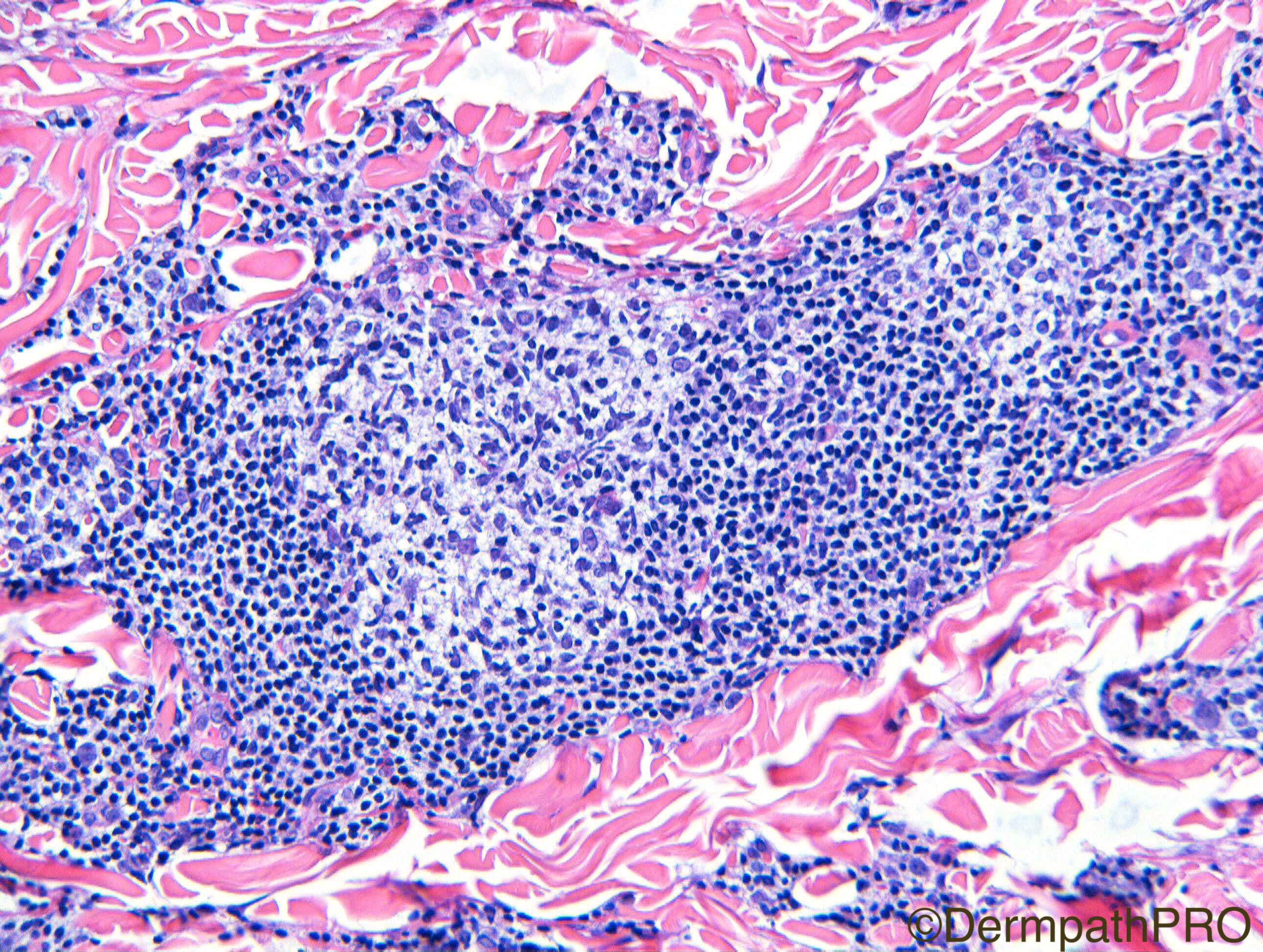
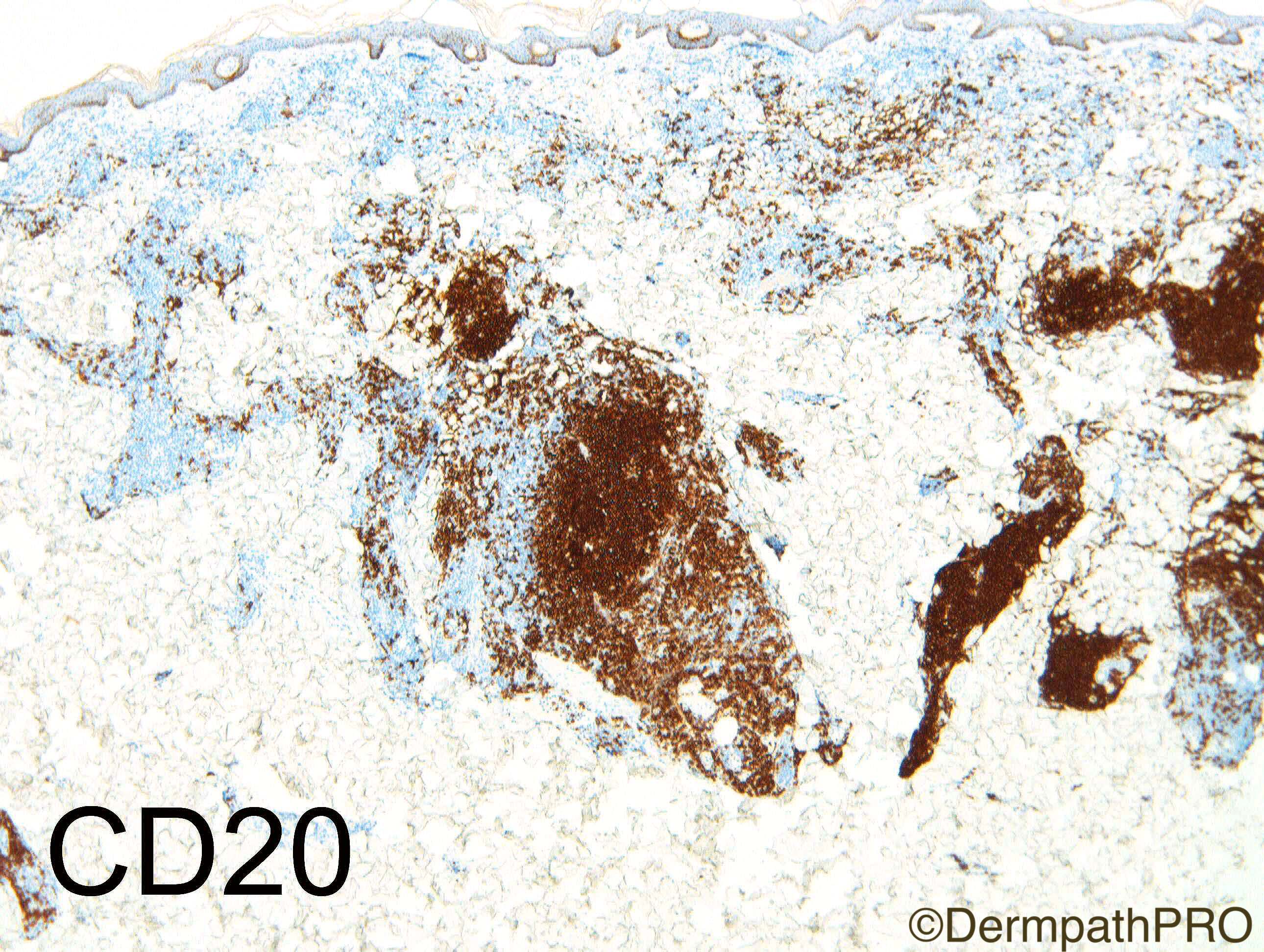
Dermal based nodular lymphoid proliferation with tropism for adnexae. The follicle highlighted doesnt show tingible body macrophages or zonation, there are some atypical cells present, some of larger size. There are some plasma cells too. Favour b cell lymphoma, immunohistochemistry is a must (cd20, cd3, cd10, bcl-2, bcl-6, cd138, kappa, lambda, ki67) but the morphology makes favour FCL over MZL for correlation with the clinical setting.

Share this comment


Link to comment
share_externally

Dr. Mona Abdel-Halim

Posted

Agree with Raul, waiting for the immunos.

Share this comment


Link to comment
share_externally

vincenzo polizzi

Posted

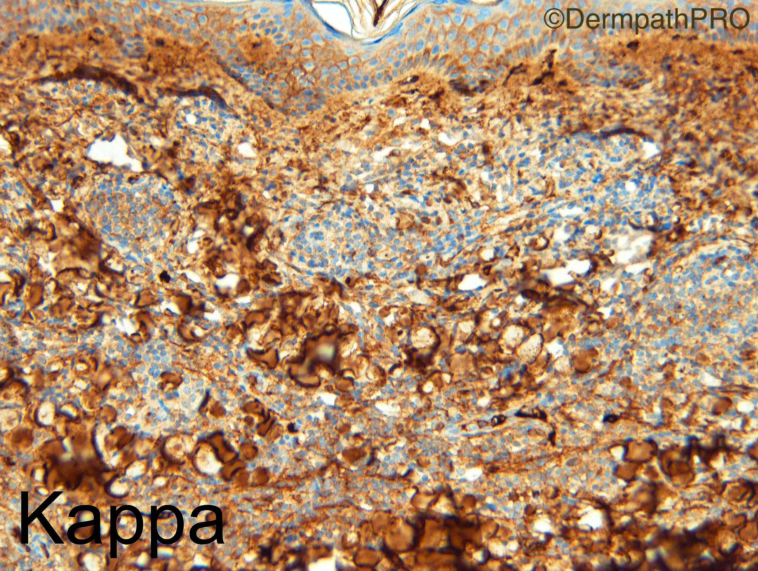
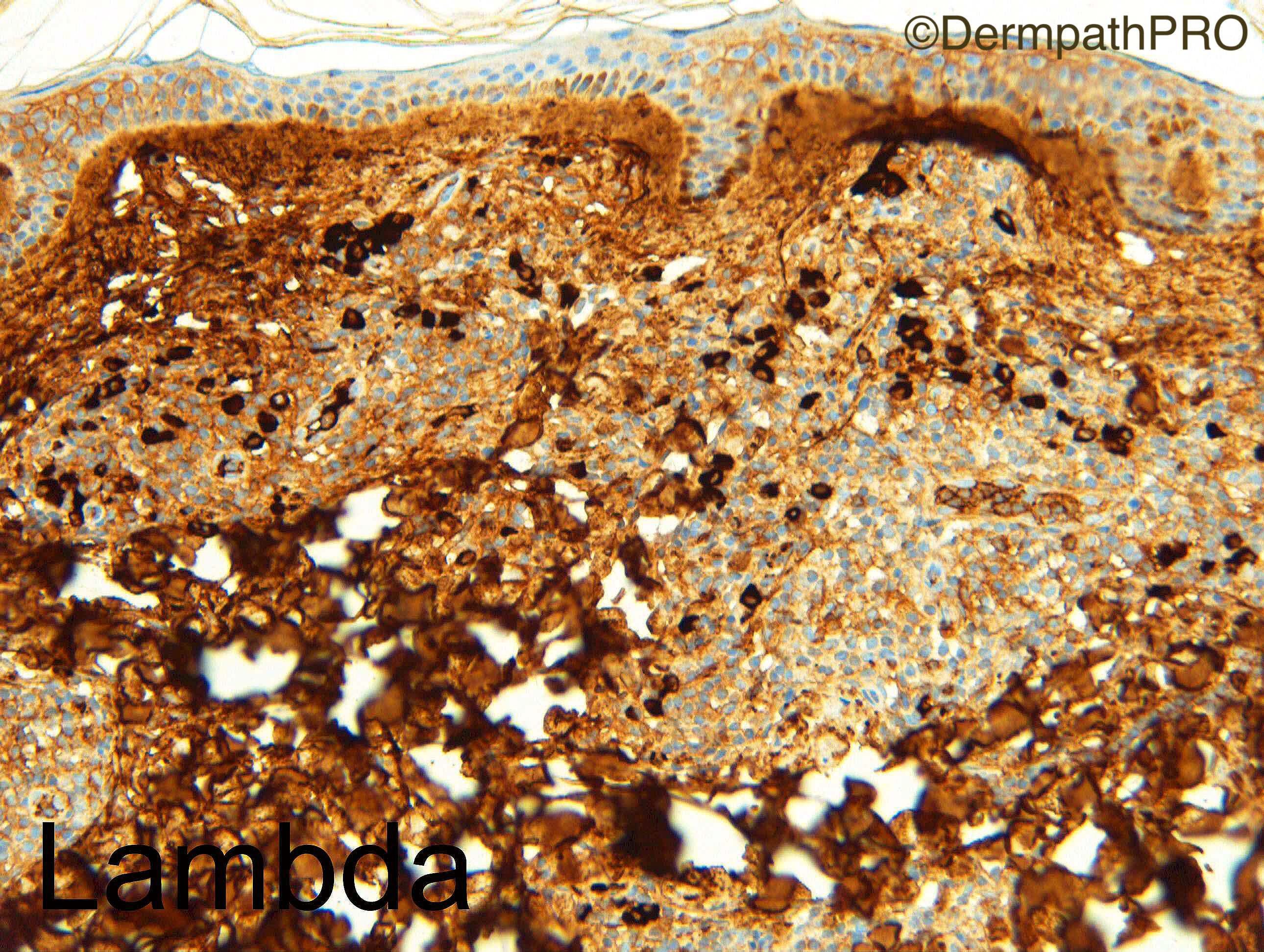
Medium sized centrocytoid cells are found beyond the germinal centre boundaries, a clue to a primary cutaneous FCL, so agree with Raul comment. But I'm finding this case particularly difficult to differentiate by a MZL and, above all, by a benign lymphoid hyperplasia. Peripheral plasma cells are few, so could be difficult k/lambda restriction interpretation...The last two fig make me favour a follicular lymphoma.

Share this comment


Link to comment
share_externally

vincenzo polizzi

Posted

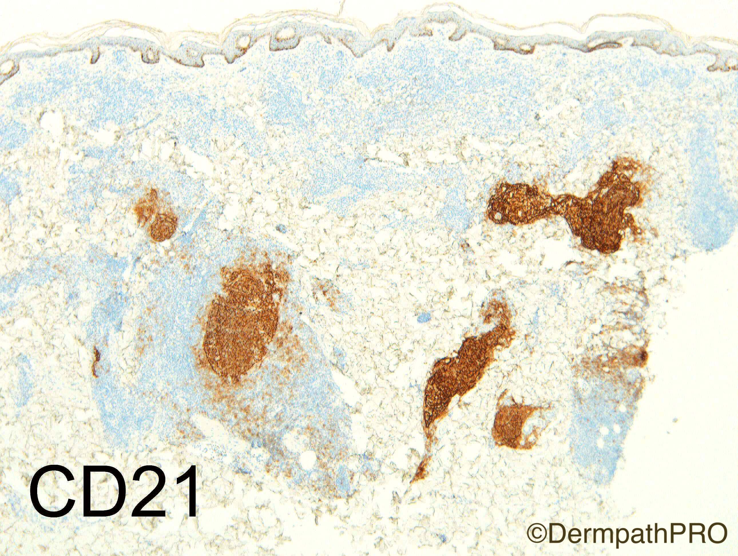
Light c.r.  fit more with MZL but, may be I'm wrong, for me CD21 doesn't show a colonized follicular center, instead an early low grade FCL with plasmocytic differentiation. What do you think about?

Share this comment


Link to comment
share_externally

Dr. Mona Abdel-Halim

Posted

Same confusion Vincenzo !!!
Light chain restriction favor MZL, CD21 does not show colonization. My instinct is still favouring FCL especially that a small population of monoclonal plasma cells can be seen in FCL . Would love to see Bcl2 and Bcl6, CD10 and Ki67.

Share this comment


Link to comment
share_externally

Raul Perret

Posted

funny things in this case is that the plasma cells seem to be lambda restricted and cd21 highlights irregularly shaped follicles. I would still perform the rest of the immunos as you guys mentioned and still favour FCL.

Share this comment


Link to comment
share_externally

Nitin Khirwadkar

Posted

Agree with the differentials. The monocytoid cells in the inter follicular regions haven't carried the FDC networks. Want to look at CD43 and BCL6. Would favour a marginal zone lymphoma.

Share this comment


Link to comment
share_externally

Dr. Richard Carr

Posted

Good discussion. I interpreted these as reactive GCs (I'm guessing therefore Ki67 was very high, Bcl2 negative, sorry did not photograph). I thought the peripheral plasma cells and immature plasmacytoid cells & monotcytoid cells in the superficial dermis showing light chain restriction was quite typical of marginal zone lymphoma in this case but of course supported by Lambda light chain restriction. PCR showed weak clonal bands in a polyclonal background - I did not think this altered the interpretation of a likely marginal zone lymphoma in this case.

Share this comment


Link to comment
share_externally

Alice Roberts

Posted

We have switched to ISH for kappa/lambda expression study... For us it provides a cleaner more easily interpreted stain

Share this comment


Link to comment
share_externally

Dr. Richard Carr

Posted

Agree - the NHS is still in the dark ages though!

Share this comment


Link to comment
share_externally


Join the conversation

You can post now and register later. If you have an account, sign in now to post with your account.

Guest
Add a comment...

×   Pasted as rich text.   Paste as plain text instead

  Only 75 emoji are allowed.

×   Your link has been automatically embedded.   Display as a link instead

×   Your previous content has been restored.   Clear editor

×   You cannot paste images directly. Upload or insert images from URL.

Loading...

×
×
  • Create New...