Jump to content
In this section we have spot diagnoses posted on a daily basis since June 2010, now over 4000! You can review the archived cases and read the suggested diagnoses by users and the final comment by the contributors.
Case are uploaded each week day by 10 am UK time with the correct diagnosis will generally be posted at 8 pm UK time. Why not view the most recent spot diagnosis and proffer a diagnosis?

Case Number : Case 1632 - 27 September Posted By: Guest

Please read the clinical history and view the images by clicking on them before you proffer your diagnosis.
Submitted Date :
   (0 reviews)

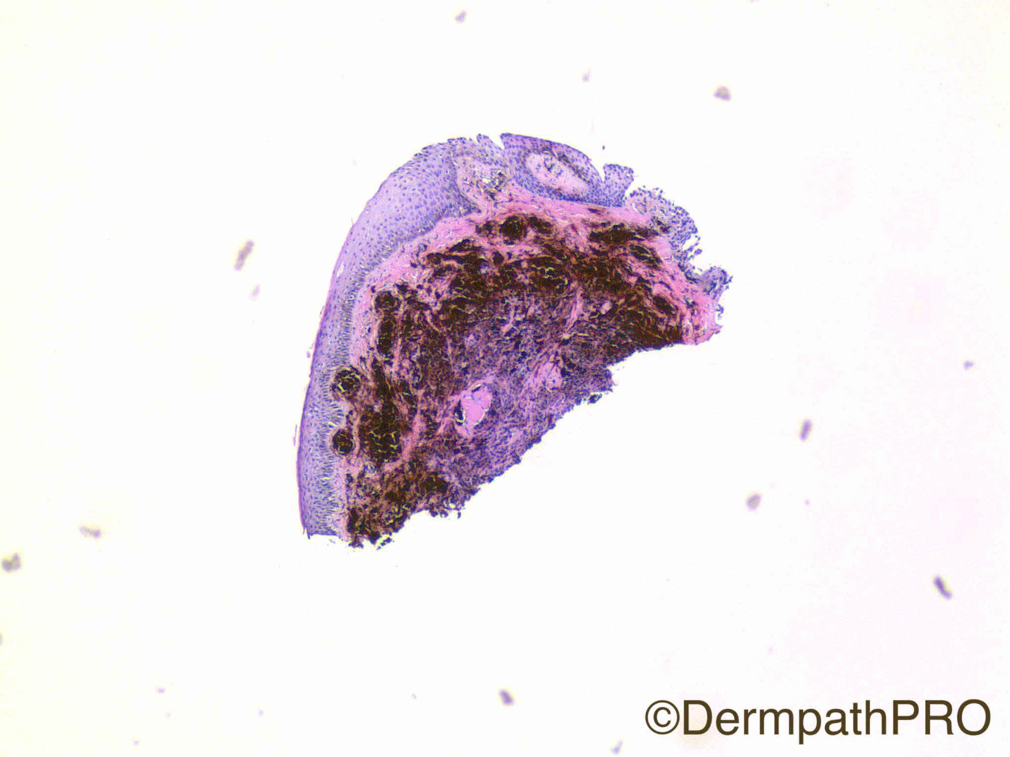
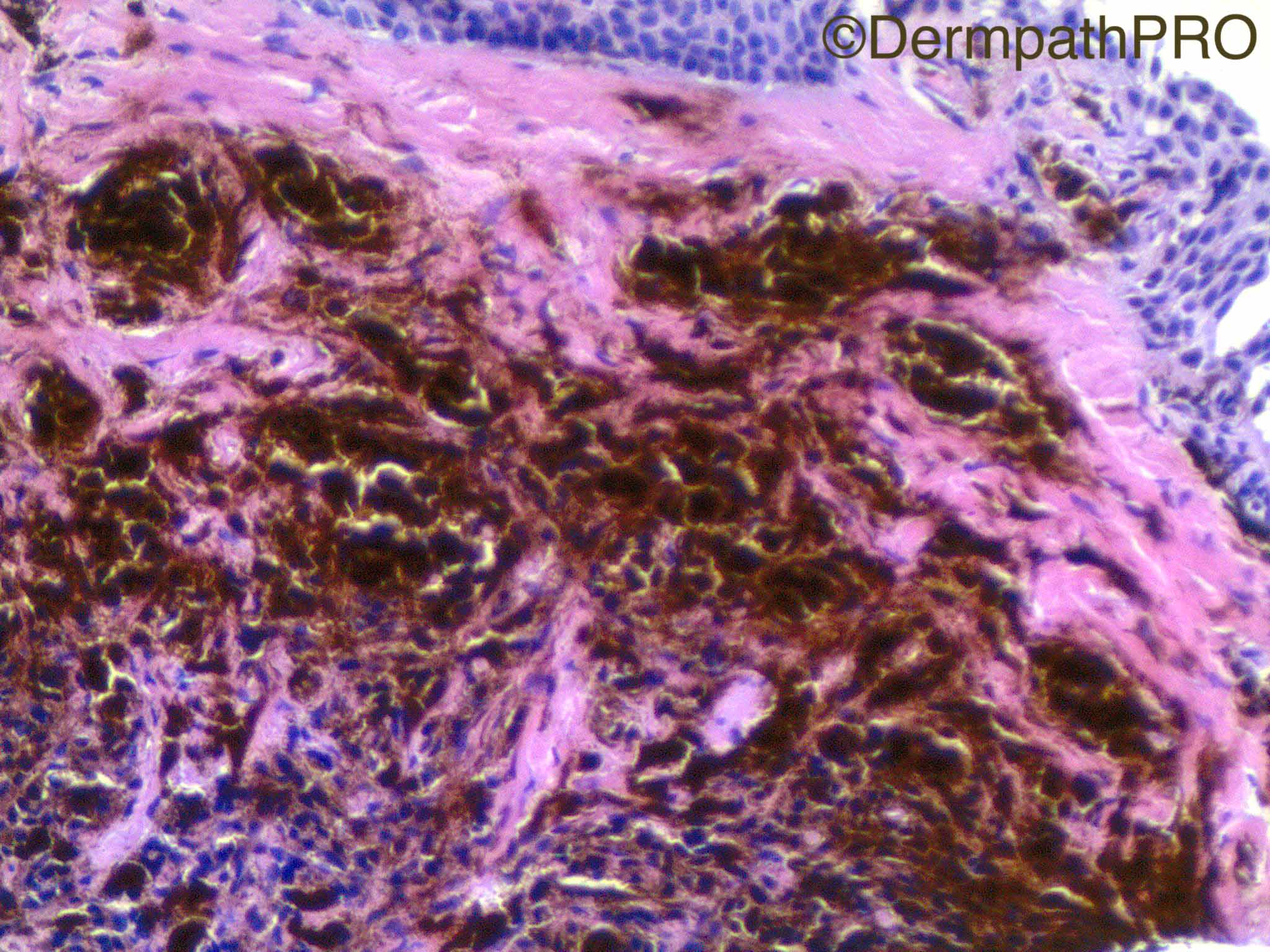
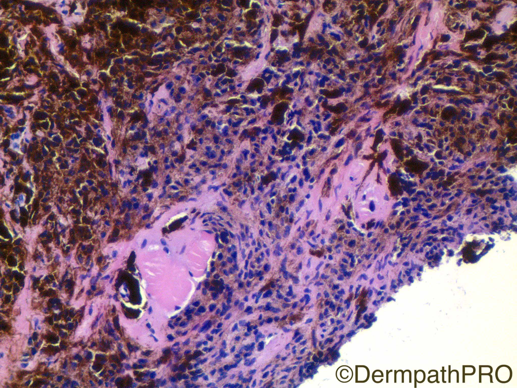
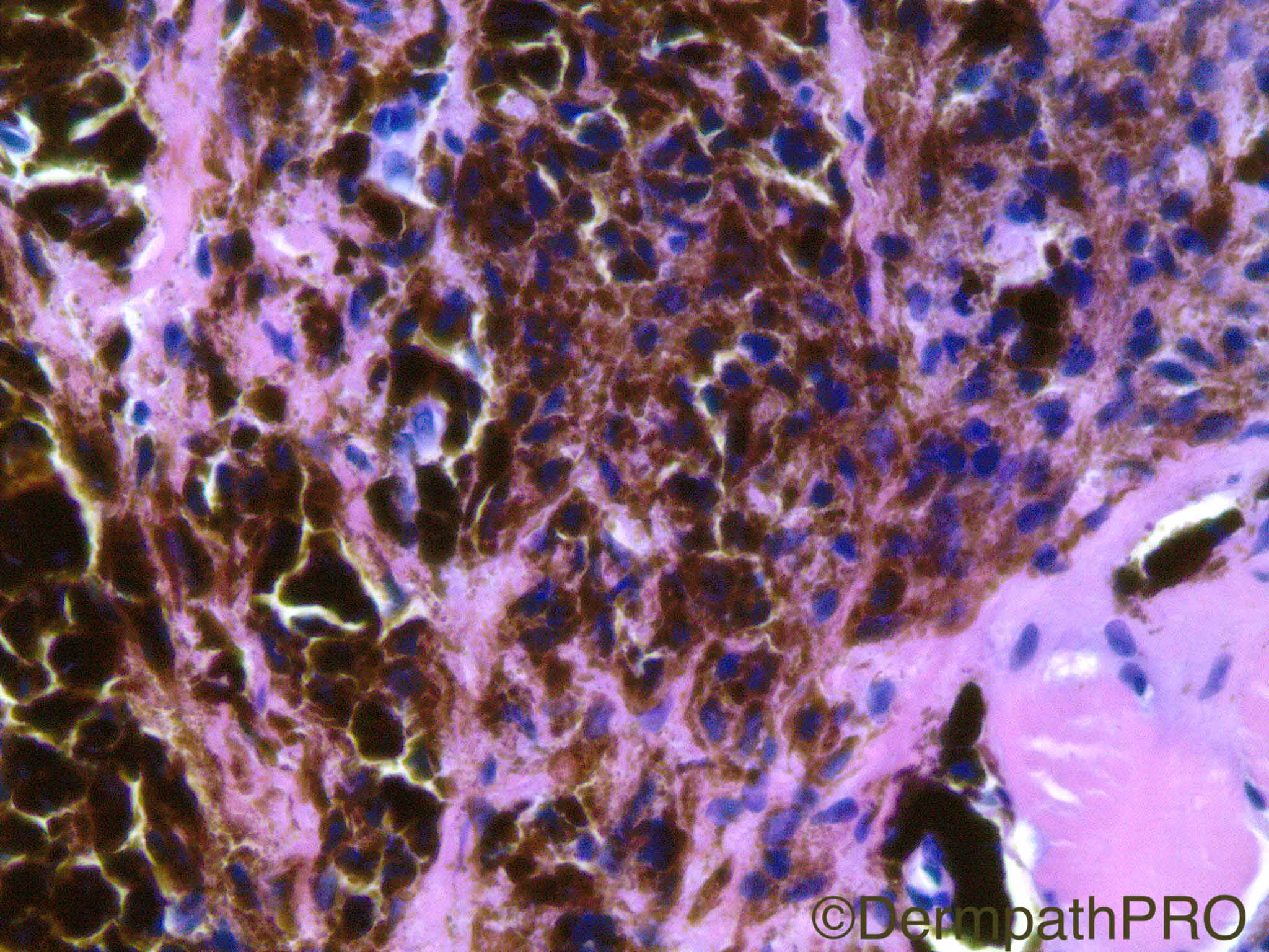
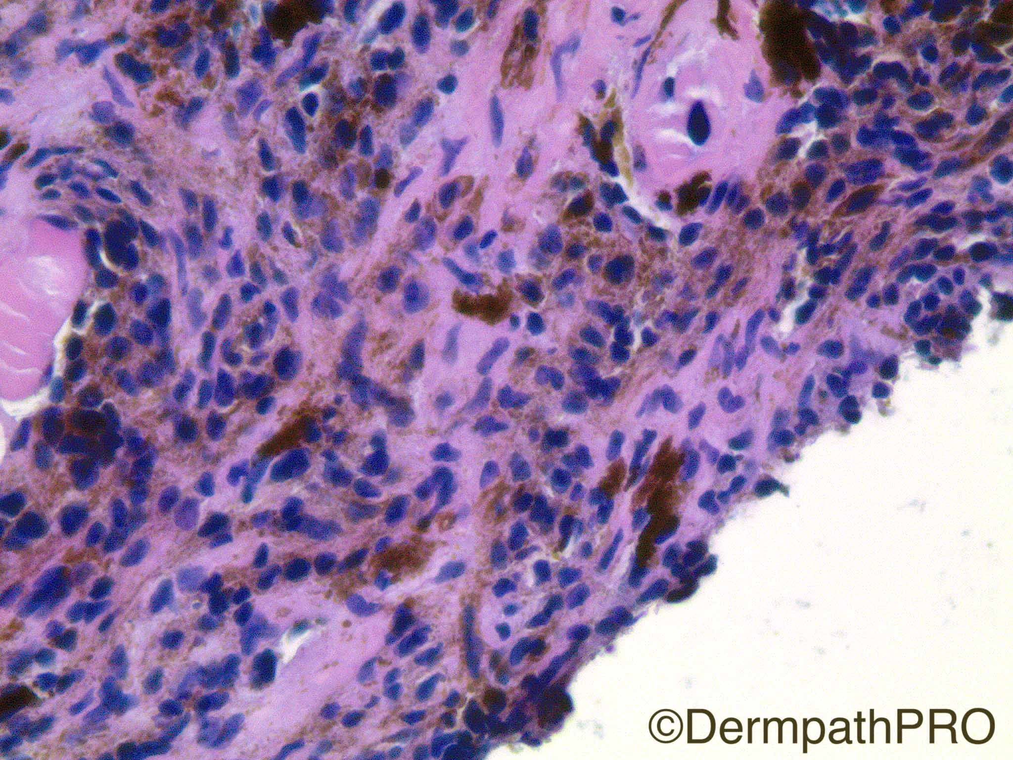
41 year old male with darkly pigmented left lower eyelid lesion.

Case Posted by Dr Uma Sundram


  Report Record

User Feedback


Admin_Dermpath

Posted

Another interesting case by Dr Uma Sundram.


When you have worked this one out please take a look at a new addition to the Difficult Opinion Cases (just follow the link on home page) a wonderful case by Maraya Bittencourt.


Cheers, Geoff

Share this comment


Link to comment
share_externally

Raul Perret

Posted

I would have called this lesion hyperpigmented blue nevus. I think we could argue if this is just a hyperpigmented subepithelial nevus (considering the rounded subepithelial areas seen in the second picture that are not in my experience seen in BN) but the overall architecture is for me consistent with BN. The topography is quite infrequent for any melanocytic neoplasm so should be careful and talk to the ophtalmologist just in case any other clinically relevant data.

Share this comment


Link to comment
share_externally

vincenzo polizzi

Posted

Another tricky case. Roughly looks like a PEM , but completely devoid of epithelioid cells and with a spindle mildly pigmented deep component, consistent with a blue nevus. So am thinking of a combined Nevus, PEM at the top and blue at the bottom. The site is odd as Raul's comments.

Share this comment


Link to comment
share_externally

Nitin Khirwadkar

Posted

Agree with comments above. Favour a blue / cellular blue naevus. Problem here is an odd site, incompletely excised lesion , further management?

Share this comment


Link to comment
share_externally

Dr. Mona Abdel-Halim

Posted

Thinking combined nevus. Incompletely excised.. same concern as Nitin, further management?

Share this comment


Link to comment
share_externally

I also think this is a combined nevus (BN + Intradermal common nevus).

Share this comment


Link to comment
share_externally

vincenzo polizzi

Posted

Or an odd deep penetrating nevus?...this is a partial superficial biopsy so we don't see the deepest component.

Share this comment


Link to comment
share_externally

Uma Sundram

Posted

This is a difficult case and I appreciate all of your opinions. We called this a combined nevus, IDN + BN, with heavy pigmentation complicating the assessment. I called the opthalmologist. The lesion is small and removed by the procedure, essentially. The patient refused further therapy at this site (surgically challenging) and both patient and clinician are comfortable with following clinically for recurrence (if any).

Share this comment


Link to comment
share_externally


Join the conversation

You can post now and register later. If you have an account, sign in now to post with your account.

Guest
Add a comment...

×   Pasted as rich text.   Paste as plain text instead

  Only 75 emoji are allowed.

×   Your link has been automatically embedded.   Display as a link instead

×   Your previous content has been restored.   Clear editor

×   You cannot paste images directly. Upload or insert images from URL.

Loading...

×
×
  • Create New...