Jump to content
In this section we have spot diagnoses posted on a daily basis since June 2010, now over 4000! You can review the archived cases and read the suggested diagnoses by users and the final comment by the contributors.
Case are uploaded each week day by 10 am UK time with the correct diagnosis will generally be posted at 8 pm UK time. Why not view the most recent spot diagnosis and proffer a diagnosis?

Case Number : Case 1634 - 29 September Posted By: Guest

Please read the clinical history and view the images by clicking on them before you proffer your diagnosis.
Submitted Date :
   (0 reviews)

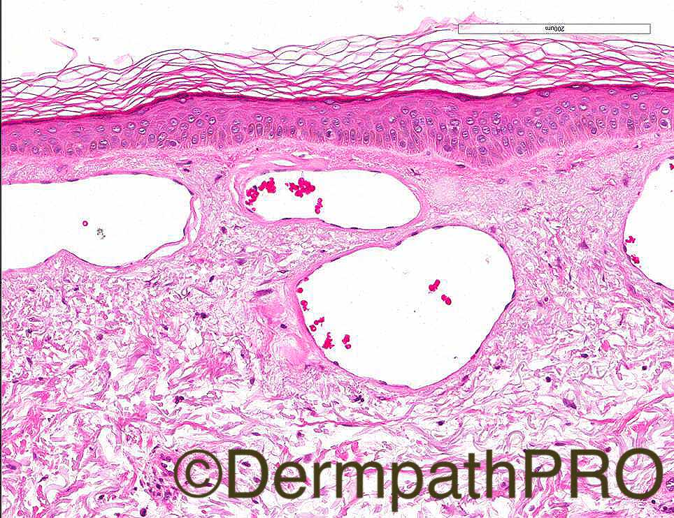
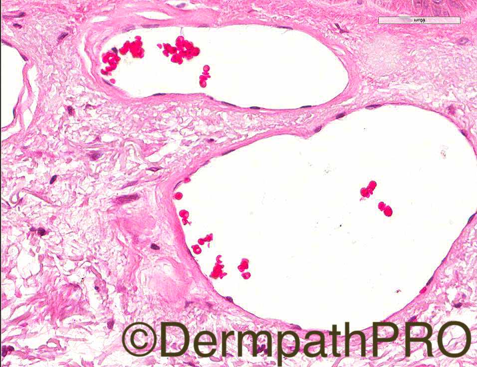
76/F slowly progressive asymptomatic rash since 2 years.  O/E- blanchable telangiectatic macules symmetrically distributed on limbs and trunk.  
Image 3 is PAS and Image 4 is Laminin.  Case courtesy of Dr Nitin Khirwadkar

Case Posted by Dr Arti Bakshi


  Report Record

User Feedback


Admin_Dermpath

Posted

Here is another great case from Dr Arti Bakshi. 


When you have worked this out take a trip over to the home page and check out the MIAMI HOT videos from last year's Understanding Dermatopathology Course which was hosted in the amazing but too hot for me Miami Beach. These videos are only available for the remainder of September to grab a fast flight to Florida and enjoy the great training and cases from some of the top Dermatopathologists in the World.


Cheers, Geoff

Share this comment


Link to comment
share_externally

vincenzo polizzi

Posted

Odd clinical background for a Multinucleate cell angiohistiicytoma. But this is my merely thought. Probably I'm wrong.

Share this comment


Link to comment
share_externally

Raul Perret

Posted

This is a really nice and rare case. When I first saw the pictures and in correlation with the clinical history I thought of generalized essential telangiectasia, but I could not make fit the vessel wall deposits. And then reading about the differentials of GET (that is actualy a diagnosis of exclusion) I found this entity called cutaneous collagenous vasculopathy. It shares the same clinical history as GET but there are vessell wall deposits and actually seems to be underreported (maybe because the deposits can be as delicate as in this case). Here a nice article

Share this comment


Link to comment
share_externally

Dr. Mona Abdel-Halim

Posted

I also thought of cutaneous collagenous vasculopathy

Share this comment


Link to comment
share_externally

vincenzo polizzi

Posted

I didn't know this entity, but after having read the article suggested by Raul ( Thanks Raul!! ), I completely agree with above.

Share this comment


Link to comment
share_externally

Raul Perret

Posted

I didn't know this entity, but after having read the article suggested by Raul ( Thanks Raul!! ), I completely agree with above.


You are welcome

Share this comment


Link to comment
share_externally

Arti Bakshi

Posted

Well done Raul and Mona!

Yes, this a rather lovely example of cutaneous collagenous vasculopathy, which my colleague, Nitin, diagnosed a few years back.The only one that I have seen, but agree that these may be under reported.

Thanks for the reference Raul, here is another one

https://www.ncbi.nlm.nih.gov/pubmed/25079204

Share this comment


Link to comment
share_externally

Great case. I read an article some time ago, but I had not seen one since then.

Share this comment


Link to comment
share_externally


Join the conversation

You can post now and register later. If you have an account, sign in now to post with your account.

Guest
Add a comment...

×   Pasted as rich text.   Paste as plain text instead

  Only 75 emoji are allowed.

×   Your link has been automatically embedded.   Display as a link instead

×   Your previous content has been restored.   Clear editor

×   You cannot paste images directly. Upload or insert images from URL.

Loading...

×
×
  • Create New...