Jump to content
In this section we have spot diagnoses posted on a daily basis since June 2010, now over 4000! You can review the archived cases and read the suggested diagnoses by users and the final comment by the contributors.
Case are uploaded each week day by 10 am UK time with the correct diagnosis will generally be posted at 8 pm UK time. Why not view the most recent spot diagnosis and proffer a diagnosis?

Case Number : Case 1878 - 09 August - Dr Uma Sundram Posted By: Guest

Please read the clinical history and view the images by clicking on them before you proffer your diagnosis.
Submitted Date :
   (0 reviews)

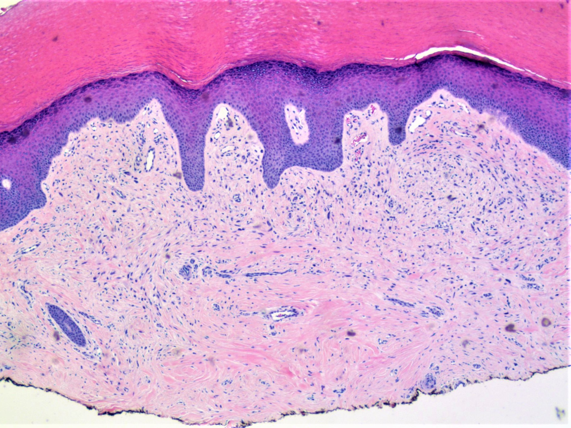
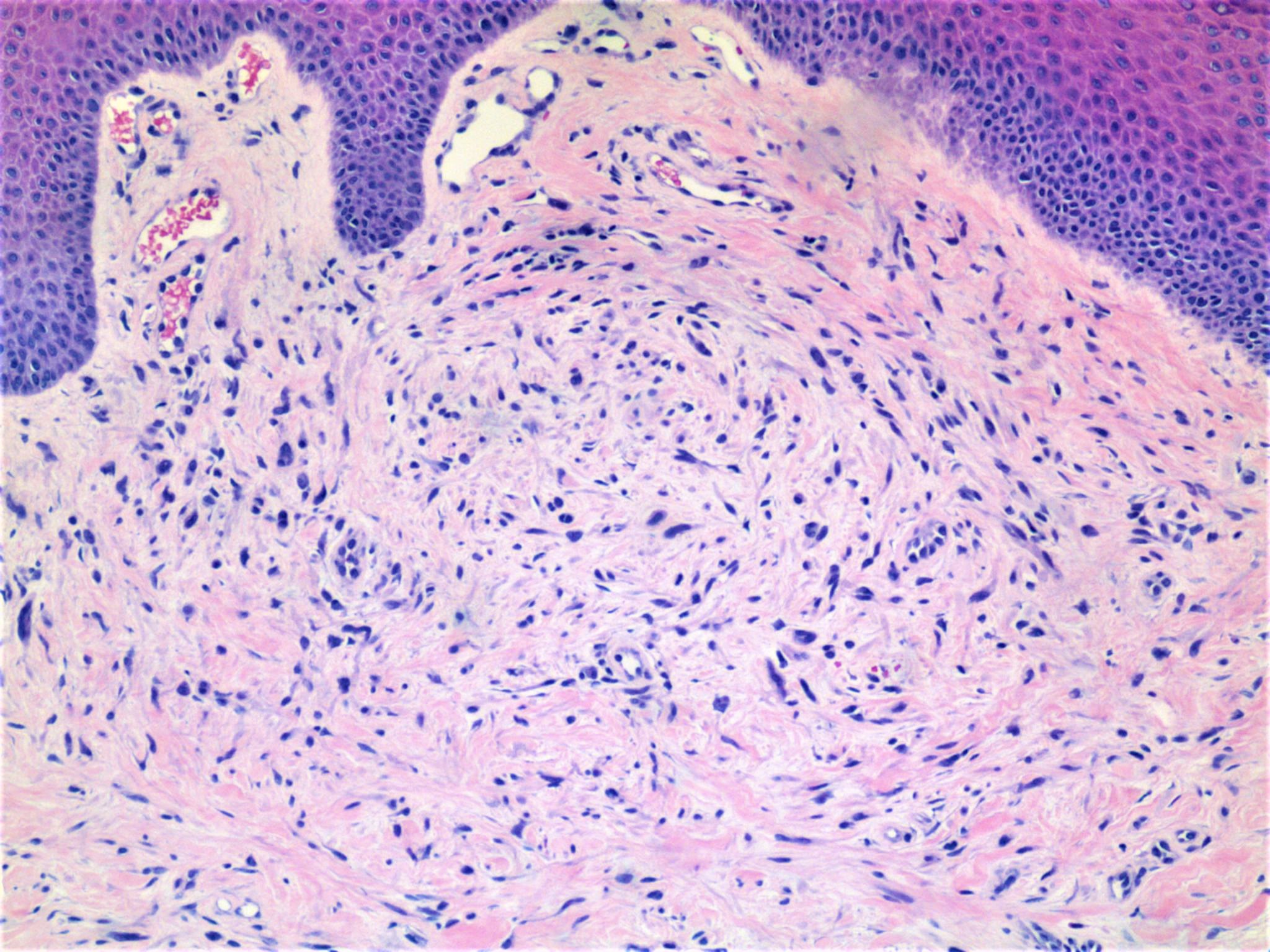
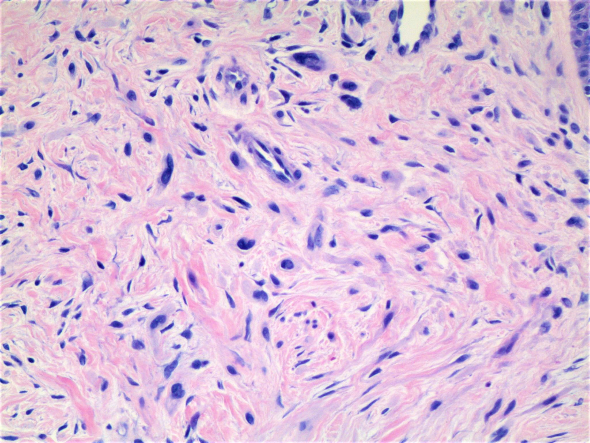
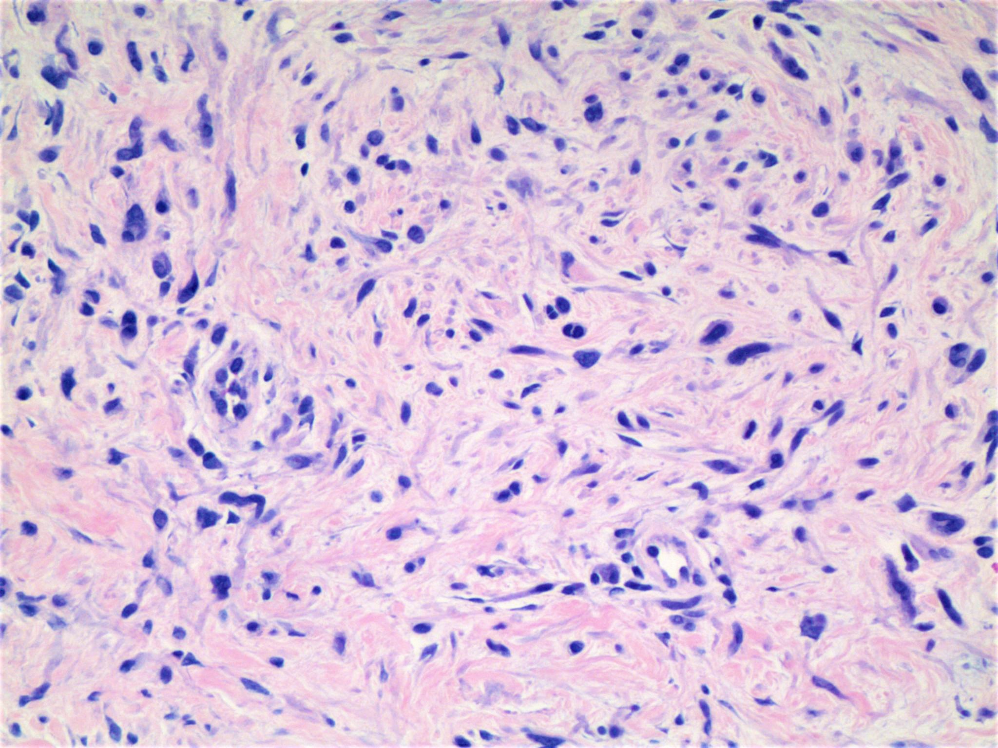
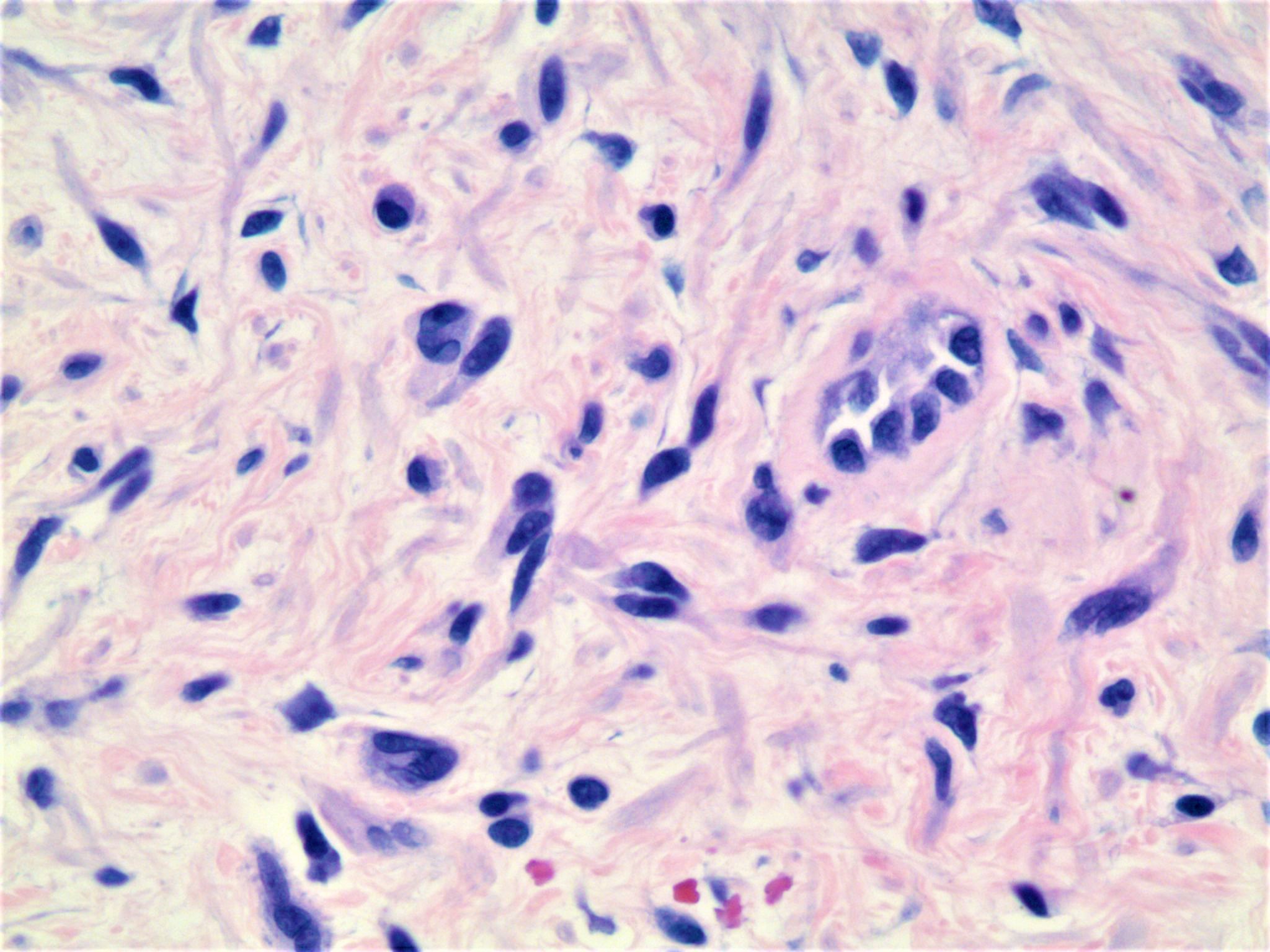
60 year old woman with papule on left lateral leg.


  Report Record

User Feedback


Dr. Richard Carr

Posted

I'm struggling. I find this sort of spindle cell lesion tricky and invariably end up doing a few immunos. My differential including: perineurioma, neurofibroma, hypopigmented blue n., desmoplastic naevus.

Share this comment


Link to comment
share_externally

Dr. Mona Abdel-Halim

Posted

Looks neural to me too, perinurioma?

Share this comment


Link to comment
share_externally

Nitin Khirwadkar

Posted

Agree with the differentials proposed by Richard. Favour a neural tumour ? Perineurioma

Share this comment


Link to comment
share_externally

vincenzo polizzi

Posted

I was thinking of mutinucleated cell angiohistiocytoma, but I'm afraid I'm wrong, if no other thought of this lesion...

I looked for alternative diagnosis ( as perineurioma or some  smooth muscle proliferation ), but this lesion has a typical plaque-like silhouette, and typical angulated multinucleate cells, so favour MCA.

Share this comment


Link to comment
share_externally

Some of the cells looks like mast cells.

Just to widen the differentials , the following are added with different priorities;Giant cell collagenoma (GCC) ,multinucleate cell angiohistiocytoma , circumscribed storiform collagenoma, pleomorphic fibroma/lipoma, regressive forms of dermatofibroma, solitary myofibroma,  giant cell fibroblastoma , neurofibroma, giant cell angiofibroma .

Share this comment


Link to comment
share_externally

Arti Bakshi

Posted

pleomorphic fibroma if other d/d (as above) excluded by immunos.

Share this comment


Link to comment
share_externally

Uma Sundram

Posted

My apologies for the late post. We did call this an unusual neurofibroma. Sox10 and s100 were positive but other melanocytic markers were negative. 

Share this comment


Link to comment
share_externally


Join the conversation

You can post now and register later. If you have an account, sign in now to post with your account.

Guest
Add a comment...

×   Pasted as rich text.   Paste as plain text instead

  Only 75 emoji are allowed.

×   Your link has been automatically embedded.   Display as a link instead

×   Your previous content has been restored.   Clear editor

×   You cannot paste images directly. Upload or insert images from URL.

Loading...

×
×
  • Create New...